Image-Based Metrics in Ultrasound for Estimation of Global Speed-of-Sound
Pith reviewed 2026-05-22 23:52 UTC · model grok-4.3
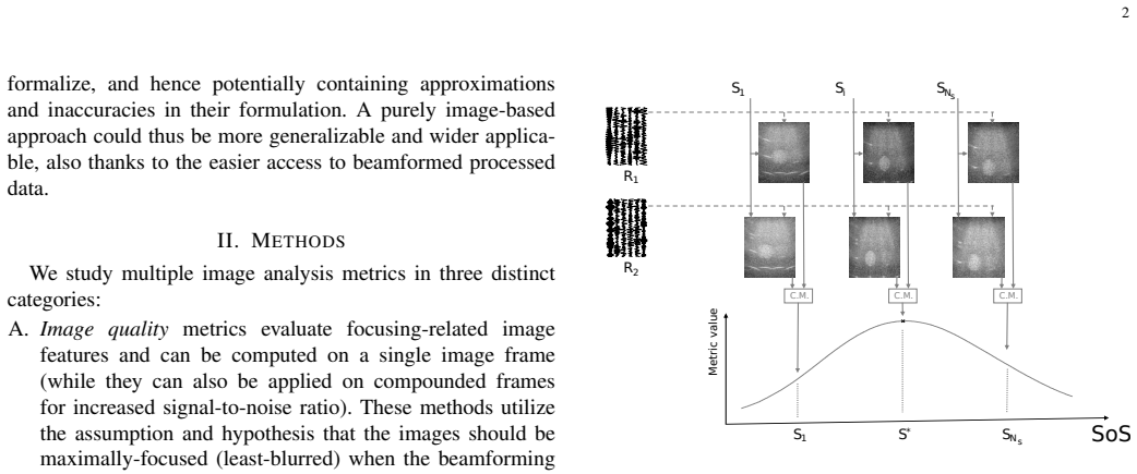
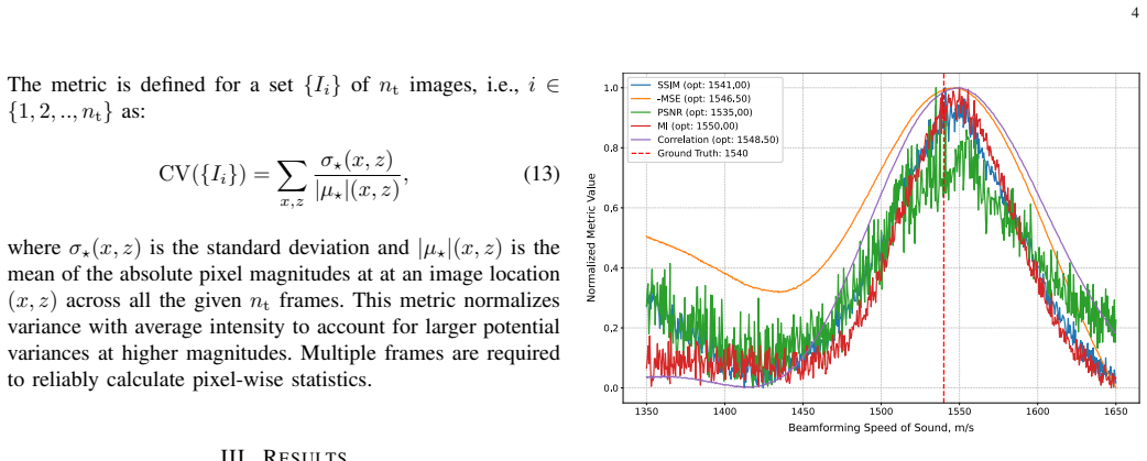
The pith
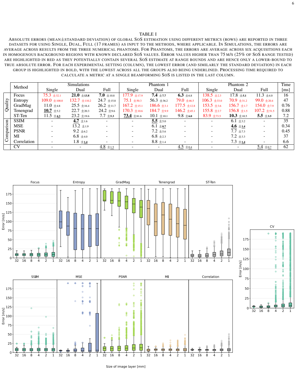
Common image metrics on beamformed ultrasound scans estimate global speed of sound to within 8 m/s without raw channel data.
A machine-rendered reading of the paper's core claim, the machinery that carries it, and where it could break.
Core claim
Differential image comparison metrics estimate global speed-of-sound from post-beamformed ultrasound images with errors consistently under 8 m/s even when applied to a single pair of frames or to relatively small image patches; mutual information and correlation metrics prove especially robust for this task, and the approach supports an in vivo demonstration of speed-of-sound-based breast density classification while operating solely on conventional beamformed or B-mode data.
What carries the argument
Differential image comparison metrics that quantify similarity or variation between frames to sense global speed-of-sound mismatch
If this is right
- The metrics function on post-beamformed and B-mode images, removing any requirement for raw channel data.
- Mutual information and correlation remain accurate on small patches, enabling focal or localized speed-of-sound estimation.
- Errors stay under 8 m/s when only a single pair of frames is available, supporting use with limited acquisitions.
- An in vivo breast density classification task demonstrates direct clinical applicability of the speed-of-sound estimates.
- Some single-frame quality metrics achieve 5-8 m/s accuracy once multiple frames are compounded.
Where Pith is reading between the lines
- Clinical scanners that cannot export raw data could still apply these metrics to correct the constant speed-of-sound assumption used in beamforming.
- Patch-wise application suggests a route toward spatially varying speed-of-sound maps rather than a single global value.
- The low computational cost may permit real-time speed-of-sound correction during live scanning sessions.
- Testing on additional tissue types and pathologies would clarify how far the metrics isolate speed-of-sound effects from other image degradations.
Load-bearing premise
The image metrics respond primarily to global speed-of-sound mismatch rather than to local tissue heterogeneity or beamforming artifacts.
What would settle it
Applying the metrics to images that contain known speed-of-sound mismatch plus controlled added heterogeneity or artifacts and finding that the error is dominated by those other factors instead of the mismatch would falsify the central assumption.
Figures
read the original abstract
Accurate speed-of-sound (SoS) estimation is crucial for ultrasound image formation, yet conventional systems often rely on an assumed value for imaging. We propose to leverage conventional image analysis techniques and metrics as a novel and simple approach to estimate tissue SoS. We study eleven metrics in three categories for assessing image quality, image similarity and multi-frame variation, by testing them in numerical simulations and phantom experiments, as well as testing in an in vivo scenario. Among single-frame image quality metrics, conventional Focus and a proposed metric variation on Tenengrad present satisfactory accuracy (5-8\,m/s on phantoms), but only when the metrics are applied after compounding multiple frames. Differential image comparison metrics were more successful overall with errors consistently under 8\,m/s even applied on a single pair of frames. Mutual information and correlation metrics were found to be robust in processing relatively small image patches, making them suitable for focal estimation. We present an in vivo study on breast density classification based on SoS, to showcase clinical applicability. The studied metrics do not require access to raw channel data as they can operate on post-beamformed and/or B-mode data. These image-based methods offer a computationally efficient and data-accessible alternative to existing physics- and model-based approaches for SoS estimation.
Editorial analysis
A structured set of objections, weighed in public.
Referee Report
Summary. The paper proposes using eleven conventional image analysis metrics (categorized as image quality, similarity, and multi-frame variation) to estimate global speed-of-sound (SoS) from post-beamformed or B-mode ultrasound data. It reports that differential metrics such as mutual information and correlation achieve consistent errors under 8 m/s even on single frame pairs in numerical simulations and phantom experiments, with robustness on small patches, and demonstrates an in vivo application for breast density classification based on the resulting SoS estimates.
Significance. If the metrics can be shown to isolate global SoS mismatch, the approach offers a computationally lightweight, data-accessible alternative to physics- or model-based SoS estimation methods that does not require channel data, potentially enabling simpler integration into clinical systems and supporting applications such as tissue characterization.
major comments (3)
- [Abstract and Results] The abstract and results sections report specific error figures (5-8 m/s for focus/Tenengrad after compounding; <8 m/s for differential metrics on single pairs) but supply no description of the optimization procedure, search strategy, or statistical fitting that converts metric values into an SoS estimate; without this, the accuracy claims cannot be evaluated or reproduced.
- [Methods and Results (phantom/simulation experiments)] The phantom and simulation protocols (described in the methods and results) do not include controls or ablation tests to establish that the chosen metrics vary predominantly with global SoS mismatch rather than with local tissue heterogeneity, speckle statistics, or residual beamforming artifacts; this assumption is load-bearing for attributing the reported errors to SoS estimation.
- [Results (in vivo study)] The in vivo breast-density classifier (results section) applies the SoS estimates without reporting how the metric-to-SoS mapping is performed on clinical data, any ground-truth validation, or assessment of confounding factors such as varying breast composition within the imaged region.
minor comments (1)
- [Abstract] The abstract states that eleven metrics were studied but does not enumerate them; a brief listing or reference to a table would improve clarity.
Simulated Author's Rebuttal
We thank the referee for the detailed and constructive report. We address each major comment below with clarifications and proposed revisions. We agree that additional methodological transparency is required and will update the manuscript accordingly.
read point-by-point responses
-
Referee: [Abstract and Results] The abstract and results sections report specific error figures (5-8 m/s for focus/Tenengrad after compounding; <8 m/s for differential metrics on single pairs) but supply no description of the optimization procedure, search strategy, or statistical fitting that converts metric values into an SoS estimate; without this, the accuracy claims cannot be evaluated or reproduced.
Authors: We agree the conversion procedure requires explicit description. SoS estimation is performed via exhaustive grid search: each metric is evaluated over a discrete range of candidate SoS values (1400–1600 m/s at 1 m/s steps) on the beamformed or B-mode data, and the value that optimizes the metric (maximum for focus/Tenengrad/MI/correlation; minimum for variance-based metrics) is selected as the estimate. No statistical fitting or regression is applied. We will expand the Methods section with a dedicated subsection detailing the search strategy, range, step size, and selection rule to enable full reproducibility. revision: yes
-
Referee: [Methods and Results (phantom/simulation experiments)] The phantom and simulation protocols (described in the methods and results) do not include controls or ablation tests to establish that the chosen metrics vary predominantly with global SoS mismatch rather than with local tissue heterogeneity, speckle statistics, or residual beamforming artifacts; this assumption is load-bearing for attributing the reported errors to SoS estimation.
Authors: The simulation and phantom setups use media with spatially uniform SoS (known constant values in simulation; calibrated homogeneous phantoms), which isolates global mismatch as the primary variable while holding speckle statistics and beamforming parameters fixed. This design provides implicit control. However, we did not perform explicit ablation experiments that independently vary local heterogeneity or residual artifacts. We will add a paragraph in the Discussion acknowledging this limitation and justifying the attribution based on the controlled uniform-media protocols. revision: partial
-
Referee: [Results (in vivo study)] The in vivo breast-density classifier (results section) applies the SoS estimates without reporting how the metric-to-SoS mapping is performed on clinical data, any ground-truth validation, or assessment of confounding factors such as varying breast composition within the imaged region.
Authors: The metric-to-SoS mapping on clinical data follows the identical grid-search optimization used in the phantom experiments. No independent ground-truth SoS measurements or detailed intra-breast composition maps were available for the retrospective clinical dataset, and potential confounders (e.g., local density variations) were not quantified. The in vivo component is presented strictly as a proof-of-concept illustration of clinical applicability rather than a validated diagnostic tool. We will revise the Results and Discussion sections to state the mapping procedure explicitly, note the absence of ground truth, and discuss the confounding factors as study limitations. revision: partial
Circularity Check
No significant circularity; empirical evaluation against known ground truth
full rationale
The paper evaluates eleven image metrics empirically on simulations and phantoms where SoS is controlled and known, reporting errors directly against those values. No derivation chain, self-definitional equations, fitted parameters renamed as predictions, or load-bearing self-citations are present. The in-vivo application is presented as a showcase rather than a derived result. This is a standard empirical validation study with independent external benchmarks.
Axiom & Free-Parameter Ledger
Reference graph
Works this paper leans on
-
[1]
S. A. Goss, R. L. Johnston, and F. Dunn. Compilation of empirical ultrasonic properties of mammalian tissues. II. The Journal of the Acoustical Society of America , 68(1):93–108, 1980
work page 1980
-
[2]
M. E. Anderson and G. E. Trahey. The direct estimation of sound speed using pulse-echo ultrasound. The Journal of the Acoustical Society of America, 104:3099–3106, 1998
work page 1998
-
[3]
C. D. Bezek and O. Goksel. Analytical estimation of beamforming speed-of-sound using transmission geometry. Ultrasonics, 134, 2023
work page 2023
-
[4]
D. Napolitano, C.-H. Chou, G. McLaughlin, T.-L. Ji, L. Mo, D. DeBuss- chere, and R. Steins. Sound speed correction in ultrasound imaging. Ultrasonics, 44:e43–e46, 2006
work page 2006
-
[5]
H. C. Shin, R. Prager, H. Gomersall, N. Kingsbury, G. Treece, and A. Gee. Estimation of average speed of sound using deconvolution of medical ultrasound data. Ultrasound in Medicine & Biology, 36(4):623– 636, 2010
work page 2010
-
[6]
C. Yoon, Y . Lee, J. H. Chang, T. Song, and Y . Yoo. In vitro estimation of mean sound speed based on minimum average phase variance in medical ultrasound imaging. Ultrasonics, 51(7):795–802, 2011
work page 2011
-
[7]
S. J. Park, J. Lee, W. Y . Lee, and Y . Yoo. Mean sound speed estimation with focusing quality evaluation for medical ultrasound imaging. In 2011 IEEE International Ultrasonics Symposium , pages 2205–2208, 2011
work page 2011
-
[8]
X. Qu, T. Azuma, J. T. Liang, and Y . Nakajima. Average sound speed estimation using speckle analysis of medical ultrasound data. International Journal of Computer Assisted Radiology and Surgery , 7(6):891–899, 2012
work page 2012
-
[9]
H. Hasegawa and R. Nagaoka. Initial phantom study on estimation of speed of sound in medium using coherence among received echo signals. Journal of Medical Ultrasonics , 46:297–307, 2019
work page 2019
-
[10]
C.-C. Shen and K.-L. Tu. Ultrasound DMAS beamforming for estima- tion of tissue speed of sound in multi-angle plane-wave imaging. Applied Sciences, 10(18):6298, 2020
work page 2020
- [11]
-
[12]
B. R. Chintada, R. Rau, and O. Goksel. Phase-aberration correction in shear-wave elastography imaging using local speed-of-sound adaptive beamforming. Frontiers in Physics, 9:690385, 2021
work page 2021
- [13]
-
[14]
Amelia Carolina Sparavigna. Entropy in image analysis. Entropy, 21(5):502, 2019
work page 2019
-
[15]
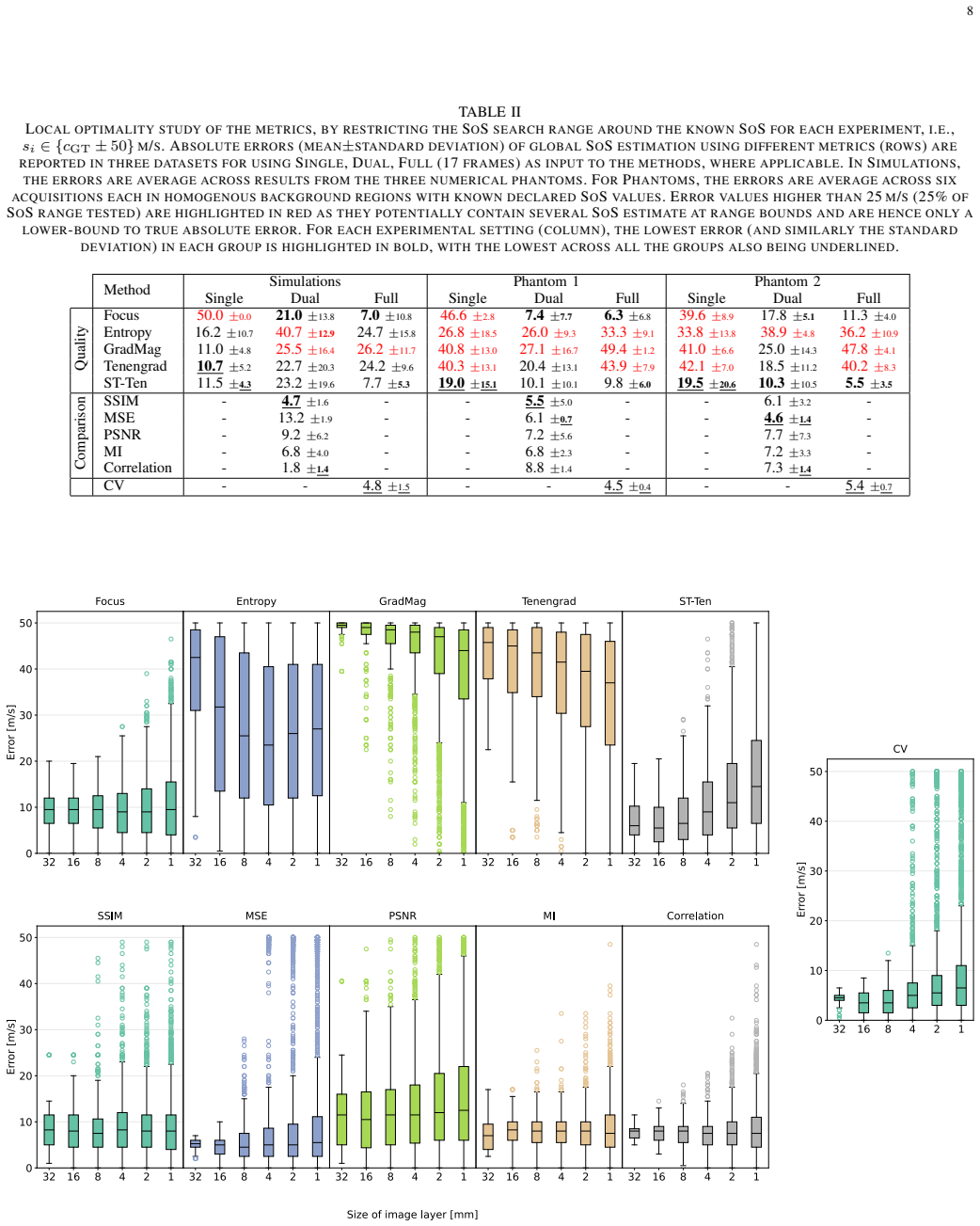
E. Krotkov. Focusing. International Journal of Computer Vision , 1(3):223–237, 1988. 8 TABLE II LOCAL OPTIMALITY STUDY OF THE METRICS , BY RESTRICTING THE SOS SEARCH RANGE AROUND THE KNOWN SOS FOR EACH EXPERIMENT , I.E., si ∈ {cGT ± 50} M/S. A BSOLUTE ERRORS (MEAN ±STANDARD DEVIATION ) OF GLOBAL SOS ESTIMATION USING DIFFERENT METRICS (ROWS ) ARE REPORTED ...
work page 1988
-
[16]
Z. Wang, A. C. Bovik, H. R. Sheikh, and E. P. Simoncelli. Image quality assessment: From error visibility to structural similarity. IEEE Transactions on Image Processing , 13(4):600–612, April 2004
work page 2004
-
[17]
Q. Huynh-Thu and M. Ghanbari. Scope of validity of PSNR in image/video quality assessment. Electronics Letters , 44(13):800–801, 2008
work page 2008
-
[18]
D. Xiao, P. De la Torre, and A. C. H. Yu. Real-time speed-of-sound estimation in vivo via steered plane wave ultrasound. IEEE Transactions on Ultrasonics, Ferroelectrics, and Frequency Control , 71(6):673–686, 2024
work page 2024
-
[19]
B. E. Treeby and B. T. Cox. K-wave: MATLAB toolbox for the simulation and reconstruction of photoacoustic wave fields. J. Biomed. Opt., 15(2):021314, 2010
work page 2010
-
[20]
R. Rau, D. Schweizer, V . Vishnevskiy, and O. Goksel. Speed-of-sound imaging using diverging waves. Int. J. Comput. Assist. Radiol. Surg. , 16(7):1201–1211, Jul 2021
work page 2021
discussion (0)
Sign in with ORCID, Apple, or X to comment. Anyone can read and Pith papers without signing in.