REVIEW 5 major objections 6 minor 34 references
Diffusion Bridge Models for 3D Medical Image Translation
T0 review · 5 major / 6 minor · reviewed 2026-08-16 · deepseek-v4-flash
Pith's one-line read A diffusion bridge model can learn to translate between T1-weighted MRI and DTI fractional-anisotropy images in 3D, generating synthetic FA scans that match real data in downstream Alzheimer's and sex classification.
desk verdict A clean but incremental 3D application of the authors' own diffusion bridge method; the evaluation doesn't yet prove the headline claim that synthetic FA recovers diffusion-specific microstructure. read the letter →
The pith
A machine-rendered reading of the paper's core claim, the machinery that carries it, and where it could break.
The reading
What carries the argument
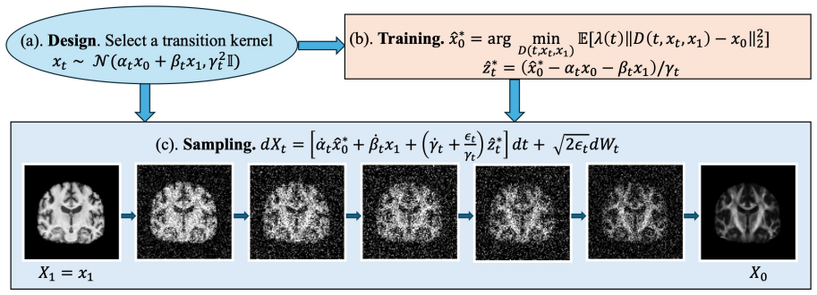
The object that carries the argument is the diffusion bridge process: a stochastic process $p_t$ defined for $t\in[0,1]$ by a Gaussian transition kernel that interpolates between the source and target distributions and satisfies boundary conditions forcing $p_1=\pi_1$ and $p_0=\pi_0$. The paper uses the simplified parameterization $\alpha_t=1-t$, $\beta_t=t$, $\gamma_t^2=4\gamma_{\max}^2 t(1-t)$, trains a denoiser $\hat{x}_\theta^0$ to approximate the conditional expectation $\mathbb{E}[x_0|x_t,x_1]$, and then samples by simulating an SDE (or its deterministic ODE limit) with 40 steps. The denoiser is a 3D adaptation of the ADM UNet, and sampling noise is controlled by a parameter $\eta$ that interpolates between stochastic and deterministic generation.
What would settle it
Measure MS-SSIM, PSNR, and voxelwise correlation between real and synthetic FA restricted to a white-matter skeleton (for example the corpus callosum and corticospinal tract) instead of the whole brain. If scores in those regions are near chance while whole-brain scores remain above 0.9, the claim that the model preserves white-matter integrity is refuted. A complementary check is to run tractography on synthetic FA and compare streamline counts or tract-averaged FA against values from real FA.
Extended reading notes
Core claim
On its own terms, the paper's central discovery is that the conditional distribution $\pi_{0|1}(x_0|x_1)$ between T1w and FA images can be learned with a diffusion bridge whose transition kernel is $p_{t|0,1}(x_t|x_0,x_1)=\mathcal{N}((1-t)x_0+t x_1,\,4\gamma_{\max}^2t(1-t)I)$, using $\gamma_{\max}=0.125$ and a neural-network denoiser trained to estimate $\mathbb{E}[x_0|x_t,x_1]$. Across 167 test subjects, synthetic FA images reach MS-SSIM close to 1, PSNR above 30 dB, and MMD below 0.005. In downstream tasks, synthetic FA reaches 88.0 percent accuracy for Alzheimer's disease classification, matching real FA exactly, and 83.2 percent for sex classification versus 86.8 percent on real FA; synthetic T1 reaches 91.7 percent for AD, matching real T1. The authors interpret these results as evidence that the translation preserves white matter integrity and neurobiologically relevant anatomical detail.
Load-bearing premise
The load-bearing premise is that T1-weighted images carry enough information about water diffusion directionality to reconstruct fractional anisotropy, so that a meaningful conditional distribution $\pi_{0|1}(x_0|x_1)$ exists. If that premise fails, the high similarity scores could reflect background dominance and coarse anatomical overlap rather than genuine recovery of white-matter microstructure.
Editorial extensions
If this is right
- DTI-FA maps can be synthesized from T1w scans alone, so cohorts with only structural MRI could be augmented with diffusion-derived biomarkers without additional scanning time.
- Synthetic FA images can substitute for real data when training downstream classifiers: AD classification accuracy is identical (88.0 percent) whether training on real or synthetic FA.
- Translation works in both directions, and translating FA to T1 can even slightly improve AD classification accuracy (synthetic T1 91.7 percent versus source FA 88.0 percent), suggesting the translation step can enhance task-relevant structure.
- Deterministic ODE sampling gives reproducible synthetic images, which is useful for clinical or multi-site settings where exact replication matters.
- The approach may reduce the DTI acquisition bottleneck in resource-limited settings, with caveats for pathologies beyond MCI and dementia.
Reading between the lines
- An implication the paper leaves implicit is that whole-brain MS-SSIM may be inflated by background zeros, so a stricter test would measure similarity only inside white-matter masks or along known tracts; high scores there would be stronger evidence that true microstructure is recovered.
- If T1-to-FA translation holds in general, it would mean T1 contrast encodes latent white-matter-integrity information, which would motivate joint T1-diffusion models and could change how acquisition protocols are designed.
- A testable extension is to generate the full diffusion tensor or orientation distribution function instead of just FA, enforcing positive definiteness, and to check whether tractography from synthetic data reproduces known streamline patterns.
- The downstream evaluation is limited to binary tasks on one dataset; applying the same synthetic images to voxelwise FA regression or multi-site generalization would reveal which anatomical regions the model actually reproduces.
Signed reviews
Editorial analysis
A structured set of objections, weighed in public.
Referee Report
Summary. The paper applies a diffusion bridge model (SDB, from the authors' prior work) to paired 3D T1-weighted MRI and DTI-FA images from ADNI, translating in both directions. The authors report whole-volume MS-SSIM, PSNR, and MMD values for generated images, and evaluate synthetic data through sex and Alzheimer's disease classification. The central claim is that the generated FA images are quantitatively and practically comparable to real FA images, so that DTI acquisition could be augmented or partially replaced by T1w-based synthesis.
Significance. If established, the result would be practically valuable: DTI acquisition is time-consuming, and reliable T1-to-FA synthesis could support dataset augmentation and dementia research. The manuscript gives useful implementation details, including the bridge-process parameterization, the sampling algorithm, and extensive tables of model configurations, and the deterministic ODE sampler is a reproducible choice. However, the current evidence does not support the strong clinical and microstructural claims. The whole-volume metrics are dominated by background and template-registered gross anatomy, the downstream classification table lacks the critical control conditions, no comparison is made against GAN/VAE or atlas-warping baselines, and all results are point estimates from a single split. The central claim is defensible in principle, but the evaluation needs substantial additional work before the paper can be accepted.
major comments (5)
- [§IV-D-3 / Table III] The downstream evaluation does not isolate diffusion-specific information. For AD classification, training on real T1w images already yields 91.7% accuracy, which is higher than the 88.0% obtained from synthetic FA, and the rows compare classifiers trained and tested on the same modality. To support the claim that synthetic FA can substitute for real FA, the paper should report cross-domain evaluations (train on synthetic and test on real, and vice versa) and include a T1-only control matched on architecture, preprocessing, and test subjects. Without these, the comparable AD accuracy may reflect macrostructural atrophy visible in T1w rather than recovered white-matter microstructure.
- [§IV-C / Fig. 3 / Fig. 4] Whole-volume MS-SSIM, PSNR, and MMD are dominated by background and by coarse, template-registered anatomy; the paper itself notes in Fig. 3 that outer slices score higher because there is just less brain. Since all images are registered to a common template, a trivial baseline that warps a population mean FA atlas into each T1w image could produce high global similarity. To substantiate the abstract and conclusion claims about preserving white matter pathway integrity, the authors should report metrics restricted to a brain mask and to the white-matter skeleton or tract-specific FA (e.g., TBSS-style analysis), together with the atlas-warping baseline.
- [§IV-B / §IV-D] No comparison is made against GAN- or VAE-based translation methods, even though the introduction motivates diffusion bridges by the training instability of GANs and the blurriness of VAEs. The relevant CycleGAN baseline of Gu et al. [8] is cited but not evaluated. Because the central claim is that the diffusion bridge approach is preferable for this task, the authors should add at least one strong baseline (e.g., CycleGAN or a VAE-based synthesizer) evaluated with the same data, preprocessing, and metrics.
- [§IV-D-3] All classification results are point estimates from a single train/validation/test split with a fixed seed, and no confidence intervals or significance tests are reported. With roughly 167 test subjects, differences such as 88.0 vs. 88.0 for AD and 74.3 vs. 83.2 for sex are not interpretable without uncertainty quantification. The paper should report bootstrap confidence intervals or repeated-seed/repeated-split results and, for paired subjects, a McNemar test.
- [§III-A] The problem formulation assumes that a learnable conditional distribution π_{0|1}(x0|x1) exists between T1w and FA images, but FA reflects water diffusion directionality that T1 contrast does not directly measure. This information-theoretic premise is not tested. A concrete negative control would be to compare the bridge output against a model that maps T1w anatomy only through a population FA atlas; if that baseline matches the reported whole-volume scores, the premise would be unsupported. I recommend adding such a baseline.
minor comments (6)
- [§II, Eq. (1)] The fractional anisotropy formula appears to be missing the standard normalization factor of sqrt(3/2); please verify the equation against the standard definition.
- [Fig. 2 caption] The caption states that the table presents four types of images but then lists only three (true T1, synthetic FA, and true FA); please clarify what the fourth type is.
- [§IV-C] The MMD results depend on the choice of kernel and bandwidth, but the manuscript only cites the MONAI implementation; please specify the kernel and its parameters for reproducibility.
- [§IV-D-3] The phrase 'a small but significant drop' is used without any statistical test; if significance testing is added, please revise this wording accordingly.
- [Table III] The columns 'Training data', 'Test data', and 'Sampler' are ambiguous; please clarify, for example, whether the classifier is trained on synthetic images and tested on synthetic images of the same type, or whether transfer between synthetic and real images is being measured.
- [Eqs. (7)-(9) / Algorithm 1] The notation is inconsistent: Eq. (9) uses x_T while Algorithm 1 uses x_N for the source endpoint; please unify the notation.
Circularity Check
No significant circularity: the diffusion-bridge derivation and evaluations are self-contained on held-out ADNI data.
full rationale
The paper's claimed derivation chain is not circular. The bridge process (Eq. 4) with alpha_t = 1-t, beta_t = t, gamma_t^2 = 4*gamma_max^2*t*(1-t), and the training objective in Eq. (6) define a conditional denoiser for pi_{0|1}(x0|x1); these equations do not encode the evaluation metrics or downstream labels. The reported MS-SSIM/PSNR/MMD numbers and sex/AD classification accuracies are computed on a held-out test split (167 subjects) that is independent of model training, so they are not forced by construction. The only notable self-citation is to the authors' prior SDB paper [13] for the coefficient schedule and gamma_max=0.125; that value is inherited, not fitted to the ADNI data in this paper, and no fitted constant is renamed as a prediction. The paper itself flags that outer-slice MS-SSIM is inflated by background ('there is just less brain in the outer images', Fig. 3), and the absence of a T1-only classifier baseline is an experimental-design concern, not a circularity. Per the scoring rules, this is an honest non-finding: no circularity. Score 0.
Assumptions & free parameters
free parameters (1)
- gamma_max =
0.125
assumptions (5)
- standard math The stochastic interpolant/bridge SDE in Equation (5) is a valid representation of the conditional distribution pi_{0|1} when the boundary conditions on alpha, beta, and gamma hold.
- domain assumption A neural network F_theta of the 3D UNet architecture can approximate the conditional expectation in Equation (6) well enough for high-fidelity translation.
- domain assumption Paired T1w and dMRI images are accurately aligned after nonlinear registration to the T1w space.
- domain assumption T1-weighted images contain sufficient information to predict fractional anisotropy.
- domain assumption ADNI participants and the 7:1.5:1.5 split provide a representative enough setting for the reported generalization claims.
Cite this review
Pith. "Pith review of Diffusion Bridge Models for 3D Medical Image Translation." pith.science (2026). https://pith.science/paper/ZZ3GMHTH
@misc{pith2026250415267,
  author       = {Pith},
  title        = {Pith review of: Diffusion Bridge Models for 3D Medical Image Translation},
  year         = {2026},
  howpublished = {\url{https://pith.science/paper/ZZ3GMHTH}},
  note         = {Machine review of arXiv:2504.15267}
}
read the original abstract
Diffusion tensor imaging (DTI) provides crucial insights into the microstructure of the human brain, but it can be time-consuming to acquire compared to more readily available T1-weighted (T1w) magnetic resonance imaging (MRI). To address this challenge, we propose a diffusion bridge model for 3D brain image translation between T1w MRI and DTI modalities. Our model learns to generate high-quality DTI fractional anisotropy (FA) images from T1w images and vice versa, enabling cross-modality data augmentation and reducing the need for extensive DTI acquisition. We evaluate our approach using perceptual similarity, pixel-level agreement, and distributional consistency metrics, demonstrating strong performance in capturing anatomical structures and preserving information on white matter integrity. The practical utility of the synthetic data is validated through sex classification and Alzheimer's disease classification tasks, where the generated images achieve comparable performance to real data. Our diffusion bridge model offers a promising solution for improving neuroimaging datasets and supporting clinical decision-making, with the potential to significantly impact neuroimaging research and clinical practice.
Figures


Figures from the paper (1 more)

Reference graph
Works this paper leans on
-
[8]
X. Gu, H. Knutsson, M. Nilsson, and A. Eklund, “Generating diffusion MRI scalar maps from T1 weighted images using generative adver- sarial networks,” in Image Analysis: 21st Scandinavian Conference, SCIA 2019, Norrköping, Sweden, June 11–13, 2019, Proceedings 21 . Springer, 2019, pp. 489–498
work page 2019
-
[1]
Machine learning for medical image translation: A systematic review,
J. McNaughton, J. Fernandez, S. Holdsworth, B. Chong, V . Shim, and A. Wang, “Machine learning for medical image translation: A systematic review,” Bioengineering, vol. 10, no. 9, p. 1078, 2023
work page 2023
-
[2]
J. Chen, S. Chen, L. Wee, A. Dekker, and I. Bermejo, “Deep learning based unpaired image-to-image translation applications for medical physics: a systematic review,” Physics in Medicine & Biology, vol. 68, no. 5, p. 05TR01, 2023
work page 2023
-
[3]
T1w MRI to T2W MRI image synthesis using SSIM-CycleGAN,
M. Meharban, M. Sabu, and T. Santhanakrishnan, “T1w MRI to T2W MRI image synthesis using SSIM-CycleGAN,” in 2024 11th Interna- tional Conference on Advances in Computing and Communications (ICACC). IEEE, 2024, pp. 1–6
work page 2024
-
[4]
PET image reconstruction using deep image prior,
K. Gong, C. Catana, J. Qi, and Q. Li, “PET image reconstruction using deep image prior,” IEEE transactions on medical imaging , vol. 38, no. 7, pp. 1655–1665, 2018
work page 2018
-
[5]
MCMT-GAN: multi-task coherent modality transferable GAN for 3D brain image synthesis,
Y . Huang, F. Zheng, R. Cong, W. Huang, M. R. Scott, and L. Shao, “MCMT-GAN: multi-task coherent modality transferable GAN for 3D brain image synthesis,” IEEE Transactions on Image Processing , vol. 29, pp. 8187–8198, 2020
work page 2020
-
[6]
Multiscale Metamorphic VAE for 3D Brain MRI Synthesis
J. Kapoor, J. H. Macke, and C. F. Baumgartner, “Multiscale metamorphic V AE for 3D brain MRI synthesis,” arXiv preprint arXiv:2301.03588, 2023
work page Pith review arXiv 2023
-
[7]
Dual-cycle constrained bijective V AE-GAN for tagged- to-cine magnetic resonance image synthesis,
X. Liu, F. Xing, J. L. Prince, A. Carass, M. Stone, G. El Fakhri, and J. Woo, “Dual-cycle constrained bijective V AE-GAN for tagged- to-cine magnetic resonance image synthesis,” in 2021 IEEE 18th International Symposium on Biomedical Imaging (ISBI). IEEE, 2021, pp. 1448–1452
work page 2021
Show all 34 references
-
[9]
Fast denoising diffusion probabilistic models for medical image-to-image generation,
H. Jiang, M. Imran, L. Ma, T. Zhang, Y . Zhou, M. Liang, K. Gong, and W. Shao, “Fast denoising diffusion probabilistic models for medical image-to-image generation,” arXiv preprint arXiv:2405.14802 , 2024
2024 arXiv
-
[10]
A multimodal comparison of latent denoising diffusion probabilistic models and generative adversarial networks for medical image syn- thesis,
G. Müller-Franzes, J. M. Niehues, F. Khader, S. T. Arasteh, C. Haar- burger, C. Kuhl, T. Wang, T. Han, T. Nolte, S. Nebelung et al. , “A multimodal comparison of latent denoising diffusion probabilistic models and generative adversarial networks for medical image syn- thesis,”...
2023
-
[11]
I 2SB: Image-to-image Schrödinger bridge,
G.-H. Liu, A. Vahdat, D.-A. Huang, E. A. Theodorou, W. Nie, and A. Anandkumar, “I 2SB: Image-to-image Schrödinger bridge,” arXiv preprint arXiv:2302.05872, 2023
2023 arXiv
-
[12]
Denoising diffusion bridge models,
L. Zhou, A. Lou, S. Khanna, and S. Ermon, “Denoising diffusion bridge models,” arXiv preprint arXiv:2309.16948 , 2023
2023 arXiv
-
[13]
Exploring the design space of diffusion bridge models via stochasticity control,
S. Zhang, Y . Cheng, X. Kong, and G. V . Steeg, “Exploring the design space of diffusion bridge models via stochasticity control,” arXiv preprint arXiv:2410.21553, 2024
2024 arXiv
-
[14]
A practical Alzheimer’s disease classifier via brain imaging- based deep learning on 85,721 samples
B. Lu, H.-X. Li, Z.-K. Chang, L. Li, N.-X. Chen, Z.-C. Zhu, and H.-X. Zhou, “A practical Alzheimer’s disease classifier via brain imaging- based deep learning on 85,721 samples.” Journal of Big Data , vol. 9, no. 1, p. 101, 2022
2022
-
[15]
Brain age analysis and dementia classification using convolutional neural networks trained on diffusion MRI: Tests in Indian and North American Cohorts,
T. Chattopadhyay, N. A. Joshy, S. S. Ozarkar, K. Buwa, Y . Feng, E. Laltoo, S. I. Thomopoulos, J. E. Villalon, H. Joshi, G. Venkatasub- ramanian, J. P. John, and P. M. Thompson, “Brain age analysis and dementia classification using convolutional neural networks trained on diff...
2024
-
[16]
Efficiently training vision transformers on structural MRI scans for Alzheimer’s disease detection,
N. J. Dhinagar, S. I. Thomopoulos, E. Laltoo, and P. M. Thompson, “Efficiently training vision transformers on structural MRI scans for Alzheimer’s disease detection,” in 2023 45th Annual International Conference of the IEEE Engineering in Medicine & Biology Society (EMBC). IE...
2023
-
[17]
NODDI and tensor-based microstructural indices as predictors of functional connectivity,
F. Deligianni, D. W. Carmichael, G. H. Zhang, C. A. Clark, and J. D. Clayden, “NODDI and tensor-based microstructural indices as predictors of functional connectivity,” Plos one , vol. 11, no. 4, p. e0153404, 2016
2016
-
[18]
Mean apparent propagator (MAP) MRI: a novel diffusion imaging method for mapping tissue microstructure,
E. Özarslan, C. G. Koay, T. M. Shepherd, M. E. Komlosh, M. O. ˙Irfano˘glu, C. Pierpaoli, and P. J. Basser, “Mean apparent propagator (MAP) MRI: a novel diffusion imaging method for mapping tissue microstructure,” NeuroImage, vol. 78, pp. 16–32, 2013
2013
-
[19]
Understanding disease progression and improving Alzheimer’s disease clinical trials: Recent highlights from the Alzheimer’s disease neuroimaging initiative,
D. P. Veitch, M. W. Weiner, P. S. Aisen, L. A. Beckett, N. J. Cairns, R. C. Green, D. Harvey, C. R. Jack Jr, W. Jagust, J. C. Morris et al., “Understanding disease progression and improving Alzheimer’s disease clinical trials: Recent highlights from the Alzheimer’s disease neu...
2019
-
[20]
Predicting dementia severity by merging anatomical and diffusion MRI with deep 3D convolutional neural networks,
T. Chattopadhyay, A. Singh, N. A. Joshy, S. I. Thomopoulos, T. M. Nir, H. Zheng, E. Nourollahimoghadam, U. Gupta, G. Ver Steeg, N. Ja- hanshad et al. , “Predicting dementia severity by merging anatomical and diffusion MRI with deep 3D convolutional neural networks,” in 18th In...
2023
-
[21]
3D grid-attention networks for interpretable age and Alzheimer’s disease prediction from structural MRI,
P. Lam, A. H. Zhu, I. B. Gari, N. Jahanshad, and P. M. Thompson, “3D grid-attention networks for interpretable age and Alzheimer’s disease prediction from structural MRI,” arXiv preprint arXiv:2011.09115 , 2020
2011 arXiv
-
[22]
Multi-site genetic analysis of diffusion images and voxelwise heritability analysis: A pilot project of the enigma–dti working group,
N. Jahanshad, P. V . Kochunov, E. Sprooten, R. C. Mandl, T. E. Nichols, L. Almasy, J. Blangero, R. M. Brouwer, J. E. Curran, G. I. de Zubicaray et al. , “Multi-site genetic analysis of diffusion images and voxelwise heritability analysis: A pilot project of the enigma–dti work...
2013
-
[23]
Diffusion MRI metrics and their relation to dementia severity: effects of harmonization approaches,
S. I. Thomopoulos, T. M. Nir, J. E. Villalon-Reina, A. Zavaliangos- Petropulu, P. Maiti, H. Zheng, E. Nourollahimoghadam, N. Jahanshad, and P. M. Thompson, “Diffusion MRI metrics and their relation to dementia severity: effects of harmonization approaches,” in 17th Internation...
2021
-
[24]
Deep normative tractometry for iden- tifying joint white matter macro-and micro-structural abnormalities in Alzheimer’s disease,
Y . Feng, B. Q. Chandio, J. E. Villalon-Reina, S. Benavidez, T. Chat- topadhyay, S. Chehrzadeh, E. Laltoo, S. I. Thomopoulos, H. Joshi, G. Venkatasubramanian et al., “Deep normative tractometry for iden- tifying joint white matter macro-and micro-structural abnormalities in Al...
2024
-
[25]
Stochastic interpolants: A unifying framework for flows and diffusions,
M. S. Albergo, N. M. Boffi, and E. Vanden-Eijnden, “Stochastic interpolants: A unifying framework for flows and diffusions,” arXiv preprint arXiv:2303.08797, 2023
2023 arXiv
-
[26]
Denoising diffusion probabilistic models,
J. Ho, A. Jain, and P. Abbeel, “Denoising diffusion probabilistic models,” Advances in neural information processing systems , vol. 33, pp. 6840–6851, 2020
2020
-
[27]
Denoising diffusion implicit models,
J. Song, C. Meng, and S. Ermon, “Denoising diffusion implicit models,” arXiv preprint arXiv:2010.02502 , 2020
2010 arXiv
-
[28]
Diffusion models beat GANs on image synthesis,
P. Dhariwal and A. Nichol, “Diffusion models beat GANs on image synthesis,” Advances in neural information processing systems, vol. 34, pp. 8780–8794, 2021
2021
-
[29]
A variational per- spective on solving inverse problems with diffusion models,
M. Mardani, J. Song, J. Kautz, and A. Vahdat, “A variational per- spective on solving inverse problems with diffusion models,” arXiv preprint arXiv:2305.04391, 2023
2023 arXiv
-
[30]
Pseudoinverse-guided diffusion models for inverse problems,
J. Song, A. Vahdat, M. Mardani, and J. Kautz, “Pseudoinverse-guided diffusion models for inverse problems,” in International Conference on Learning Representations , 2023
2023
-
[31]
Multiscale structural similarity for image quality assessment,
Z. Wang, E. P. Simoncelli, and A. C. Bovik, “Multiscale structural similarity for image quality assessment,” in The Thrity-Seventh Asilo- mar Conference on Signals, Systems & Computers, 2003, vol. 2. Ieee, 2003, pp. 1398–1402
2003
-
[32]
A kernel two-sample test,
A. Gretton, K. M. Borgwardt, M. J. Rasch, B. Schölkopf, and A. Smola, “A kernel two-sample test,” The Journal of Machine Learning Research, vol. 13, no. 1, pp. 723–773, 2012
2012
-
[33]
Ultimatesynth: MRI physics for pan-contrast ai,
R. Adams, W. Zhao, S. Hu, W. Lyu, K. M. Huynh, S. Ahmad, D. Ma, and P.-T. Yap, “Ultimatesynth: MRI physics for pan-contrast ai,” bioRxiv, pp. 2024–12, 2024
2024
-
[34]
Unsupervised MR har- monization by learning disentangled representations using information bottleneck theory,
L. Zuo, B. E. Dewey, Y . Liu, Y . He, S. D. Newsome, E. M. Mowry, S. M. Resnick, J. L. Prince, and A. Carass, “Unsupervised MR har- monization by learning disentangled representations using information bottleneck theory,” NeuroImage, vol. 243, p. 118569, 2021
2021
Reviewed August 16, 2026 · model on record in the stance chip above.
Discussion (0). Continue with ORCID to comment.