REVIEW 4 major objections 6 minor 20 references
Dynamic Arthroscopic Navigation System for Anterior Cruciate Ligament Reconstruction Based on Multi-level Memory Architecture
T0 review · 4 major / 6 minor · reviewed 2026-08-16 · deepseek-v4-flash
Pith's one-line read A three-level memory architecture lets a markerless arthroscopic tracker follow the femoral condyle continuously, cutting pixel error by about 45 percent over 1000 frames while running at 25.3 FPS.
desk verdict Plausible XMem adaptation for arthroscopic ACL navigation, but the evaluation's ground-truth reliability and internal arithmetic need serious work before the accuracy claims hold. read the letter →
The pith
A machine-rendered reading of the paper's core claim, the machinery that carries it, and where it could break.
The reading
What carries the argument
The central mechanism is the three-level memory store built on the Atkinson-Shiffrin model: arthroscopic sensory memory, a GRU updated every frame and gated by an image-quality factor; arthroscopic working memory, which stores high-resolution features every r frames together with viewpoint parameters; and arthroscopic long-term memory, which compresses features into anatomical prototypes tagged with region identifiers such as intercondylar notch or lateral condyle. Reading fuses all three levels with current-frame features through attention and viewpoint similarity, while writing uses consolidation, potentiation, and a phase-aware forgetting rule. This machinery supplies temporal continuity, viewpoint-aware retrieval, and anatomical priors, which together are claimed to produce the accuracy and memory-efficiency gains.
What would settle it
Recompute the pixel-error comparison on a set of frames independently annotated by at least two surgeons and report inter-annotator agreement, for example the mean distance between surgeon labels. If the average disagreement between annotators is comparable to or larger than the dynamic system's 5.3-pixel error, the claimed accuracy improvement over the static system is not established. A second check would be to run both systems on held-out sequences from a different hospital and verify that the roughly 45 percent gap persists.
Extended reading notes
Core claim
The paper's central claim is that a multi-level memory architecture, with sensory memory updated every frame, working memory holding viewpoint-tagged features, and long-term memory storing anatomical prototypes, is what lets the system track the femoral condyle across viewpoint changes, instrument occlusion, and tissue deformation. The authors argue that the static system fails during these events because it re-registers each frame independently, whereas the memory-based dynamic system reuses stored features and recovers tracking after disruptions. The quantitative evidence is the error comparison: 3.2 vs 4.1 pixels on 100-frame clips, 4.5 vs 7.8 on 500-frame clips, and 5.3 vs 12.6 on 1000-frame clips, with ablations showing that each memory level contributes and that removing long-term memory more than doubles memory use.
Load-bearing premise
The reported pixel errors and improvement percentages treat the positions annotated by three surgeons as ground truth, yet the paper does not report how much the surgeons agreed with each other or how many frames were annotated; if those labels are noisy, the error numbers and the 45 percent improvement would not be trustworthy.
Editorial extensions
If this is right
- Surgeons could receive continuous navigation without recalibration during viewpoint changes, because the tracker reuses stored features instead of re-registering each frame.
- The system meets real-time clinical demands, running at 25.3 FPS with 39.5 ms latency on a mainstream laptop GPU, comfortably above the 15 FPS threshold cited for arthroscopic surgery.
- Memory efficiency improves with sequence length, with 1.8 GB used over 1000 frames versus 2.7 GB for the static baseline, making long procedures feasible on standard hardware.
- Tracking failure rates drop under occlusion, illumination changes, and extreme viewpoint changes, with severe motion blur remaining the largest weakness at a 15.7 percent failure rate.
- The anatomy-tagged long-term memory should allow the system to re-identify the femoral condyle after the arthroscope returns to a previously seen viewpoint.
Reading between the lines
- Beyond the paper's data, a direct comparison against a general video object segmentation model fine-tuned on arthroscopy would isolate whether the gains come from the three-level memory structure itself or from the arthroscopy-specific additions such as viewpoint parameters, anatomical identifiers, and quality gating.
- Because the static baseline's error grows sharply with sequence length while the dynamic system's error grows slowly, the 45 percent improvement on 1000 frames likely understates the benefit on full 60-90 minute procedures, assuming the trend continues.
- The authors list tibial plateau tracking as future work; if the memory architecture generalizes, the same system could plausibly track both femur and tibia without markers, giving complete ACL reconstruction navigation.
- A practical deployment test on lower-resolution or compressed arthroscope feeds would be valuable, since motion blur is already the dominant failure mode and compression would add further degradation.
Signed reviews
Editorial analysis
A structured set of objections, weighed in public.
Referee Report
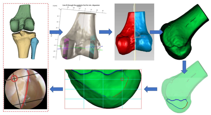
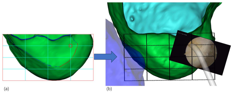
Summary. This manuscript extends the authors' previously published static markerless navigation system for ACL reconstruction by adding a dynamic tracking component based on a three-level memory architecture inspired by XMem and the Atkinson-Shiffrin memory model. The system performs automatic foreground segmentation of the femoral condyle, virtual-camera registration, multi-level memory tracking, and projection of a Bernard-Hertel grid onto arthroscopic views. Experiments on 20 arthroscopic video sequences are reported to show lower pixel error than the static baseline (5.3 vs. 12.6 pixels at 1000 frames), real-time operation at 25.3 FPS with 39.5 ms latency, reduced GPU memory, and lower tracking failure rates under occlusion, illumination change, motion blur, and viewpoint changes. Ablation and robustness analyses are also included.
Significance. If the reported results are reliable and reproducible, the system would be a practically useful contribution to arthroscopic navigation: it is markerless, runs in real time on mainstream hardware, and explicitly targets a clinical problem that is not well served by existing navigation systems. The authors deserve credit for reporting failure-rate analysis, ablation studies, and a candid limitations section. However, the central quantitative claim rests entirely on a self-referential comparison with the authors' own prior static system, and the evaluation protocol omits the annotation-reliability information needed to interpret the pixel-error numbers. Moreover, the headline improvement percentages are arithmetically inconsistent with the reported means. For these reasons the paper is best viewed as a promising engineering report whose performance claims require correction and strengthening before publication.
major comments (4)
- [Abstract and §4.2] The reported improvement percentages are inconsistent with the reported error means. For 1000-frame sequences, (12.6−5.3)/12.6 = 57.9%, not approximately 45%; for 500-frame sequences, (7.8−4.5)/7.8 = 42.3%, not approximately 35%; and for 100-frame sequences, (4.1−3.2)/4.1 = 22.0%, not approximately 19%. These are the paper's headline quantitative claims and must be corrected or the calculation explicitly explained.
- [§4.1–§4.2] The pixel-error evaluation is not interpretable without details of the manual reference annotations. The paper does not state how many frames were annotated, how the three surgeons' annotations were combined, what exactly is meant by 'femoral condyle position' (centroid, boundary, or anatomical landmark), or any inter-annotator agreement statistic. Because the improvements at 500 and 100 frames are only 3.3 px and 0.9 px, label noise of even a few pixels could change the conclusions. The authors should provide this information and confirm that identical reference frames and identical annotations were used for both the dynamic and static systems.
- [§4.4 and Fig. 12] The ablation study is reported only in prose and in a figure, with no numeric table in the text. Claims such as '62% higher tracking error,' '133% higher memory usage,' and '29% increase' cannot be checked from the material provided. Please include a table with per-condition tracking error, memory usage, and frame rate, including standard deviations and the number of sequences used.
- [§3.4 and §3.5] The multi-level memory architecture is described at a level of abstraction that prevents independent implementation. The training procedure, loss function, training data, network architecture for the feature extractor, GRU, and decoder, and key hyperparameters (e.g., α, β, γ, δ, r, and T_max) are not specified, and no code or model is released. Given that Section 2.2 identifies XMem as the source of the three-level memory structure, the authors should state precisely which components are borrowed and which are new, and provide enough implementation detail to reproduce the claimed gains.
minor comments (6)
- [Section numbering] The heading '3.4' is used twice, once for 'Multi-level Memory Architecture' and once for 'Bernard & Hertel Grid Projection', although the system overview in §3.1 describes the latter as Section 3.5; renumbering is needed.
- [§4.2, figure reference] The sentence 'Visual evidence supporting our quantitative findings is presented in Figure 3' appears to refer to the tracking comparison shown in Figure 10, not to the segmentation framework in Figure 3.
- [§3.4.1, citation] The statement 'Inspired by XMem [4]' cites the wrong reference; XMem is reference [20] in the bibliography.
- [Equations] Several equations contain typographical artifacts; for example, Eq. (34) is missing terminal punctuation and Eq. (12) is difficult to read as rendered. Please ensure all mathematical notation is legible and consistent.
- [Data Availability Statement] The Data Availability Statement says data are included in the article or supplementary material, but no supplementary file is provided; please clarify what data or code can be shared.
- [§4.5, terminology] The robustness section refers to 'moderate motion blur' when describing the condition with a 15.7% failure rate, while the preceding text calls the same condition 'severe motion blur'; the terminology should be made consistent.
Circularity Check
No circularity: the dynamic tracking gains are empirical measurements against the authors' prior static baseline, not consequences of the architecture's definitions.
full rationale
Walking the derivation chain, the central claim is an experimental comparison: the dynamic multi-level-memory system achieves lower pixel error than the previous static matching system on arthroscopic sequences. The accuracy values in Section 4.2 (e.g., 5.3±1.5 px vs 12.6±3.7 px on 1000-frame sequences) are measured against reference positions annotated by three surgeons, not computed from the memory equations. No parameter is fitted to reproduce the reported error values, and the ablation studies in Section 4.4 are removal experiments rather than inverse constructions. The B&H grid projection uses camera calibration and landmark-based least-squares fine-tuning (Equations 39-41), but that is calibration of a visualization overlay, not a prediction of tracking error. The only self-referential element is that the comparative baseline is the authors' own prior system, cited as [17] and mis-cited as [3] in Section 4.1; this makes the evaluation same-lab and potentially favorable to the new system, but it is not a circular reduction of the claim to its inputs. The paper itself flags limited clinical validation in Section 5.3, and Section 4.1 under-specifies the annotation protocol (no inter-annotator agreement, no number of annotated frames, no combination rule), which is an evidence-quality concern rather than circularity. There is also a citation-number inconsistency in Section 3.4.1, where XMem is cited as [4] although reference [4] is Tensho et al. and XMem is [20]; again, this is a bibliographic error, not a circular step. No quoted equation or passage reduces the claimed prediction to its own input, so no circular step can be exhibited.
Assumptions & free parameters
free parameters (6)
- HSV color thresholds L_H, U_H, L_S, U_S, L_V, U_V =
not reported (empirically set)
- Registration similarity weights alpha, beta, gamma =
not reported; constraint alpha+beta+gamma=1 with alpha >> beta > gamma
- Memory potentiation hyperparameters alpha, beta, gamma =
not reported
- Region importance weights omega_r for memory consolidation =
not reported
- Working-memory frame sampling interval r =
dynamically adjusted; no value
- Forgetting decay coefficient delta =
not reported
assumptions (5)
- standard math Perspective projection, camera intrinsic and distortion models, Levenberg-Marquardt optimization, moments, and IoU are standard and correct.
- domain assumption Cartilage of the femoral condyle can be isolated by HSV color thresholds with a whitish, bright, low-saturation appearance.
- domain assumption The articular margin boundary is the most reliable feature, and the weighting alpha >> beta > gamma in the similarity function is appropriate.
- domain assumption The Atkinson-Shiffrin three-level memory architecture, as implemented in XMem, remains effective when adapted to arthroscopic video with viewpoint and anatomical augmentations.
- domain assumption Semantic-SAM pretrained on arthroscopic image datasets is available and provides useful candidate masks.
invented entities (1)
-
Source
Cite this review
Pith. "Pith review of Dynamic Arthroscopic Navigation System for Anterior Cruciate Ligament Reconstruction Based on Multi-level Memory Architecture." pith.science (2026). https://pith.science/paper/WRUS7DL3
@misc{pith2026250419398,
  author       = {Pith},
  title        = {Pith review of: Dynamic Arthroscopic Navigation System for Anterior Cruciate Ligament Reconstruction Based on Multi-level Memory Architecture},
  year         = {2026},
  howpublished = {\url{https://pith.science/paper/WRUS7DL3}},
  note         = {Machine review of arXiv:2504.19398}
}
read the original abstract
This paper presents a dynamic arthroscopic navigation system based on multi-level memory architecture for anterior cruciate ligament (ACL) reconstruction surgery. The system extends our previously proposed markerless navigation method from static image matching to dynamic video sequence tracking. By integrating the Atkinson-Shiffrin memory model's three-level architecture (sensory memory, working memory, and long-term memory), our system maintains continuous tracking of the femoral condyle throughout the surgical procedure, providing stable navigation support even in complex situations involving viewpoint changes, instrument occlusion, and tissue deformation. Unlike existing methods, our system operates in real-time on standard arthroscopic equipment without requiring additional tracking hardware, achieving 25.3 FPS with a latency of only 39.5 ms, representing a 3.5-fold improvement over our previous static system. For extended sequences (1000 frames), the dynamic system maintained an error of 5.3 plus-minus 1.5 pixels, compared to the static system's 12.6 plus-minus 3.7 pixels - an improvement of approximately 45 percent. For medium-length sequences (500 frames) and short sequences (100 frames), the system achieved approximately 35 percent and 19 percent accuracy improvements, respectively. Experimental results demonstrate the system overcomes limitations of traditional static matching methods, providing new technical support for improving surgical precision in ACL reconstruction.
Figures


Figures from the paper (10 more)




Reference graph
Works this paper leans on
-
[1]
The influence of bony morphology on the ACL
Musahl V, et al. The influence of bony morphology on the ACL. Knee Surg Sports Traumatol Arthrosc. 2010;18(9):1158-1163
work page 2010
-
[2]
Shea KG, et al. The relationship between femoral tunnels created by the transtibial, anteromedial portal, and outside-in techniques and the anterior cruciate ligament footprint. Am J Sports Med. 2011;39(8):1841-1850
work page 2011
-
[3]
Advantages of computer-assisted ACL reconstruction
Mauch F, et al. Advantages of computer-assisted ACL reconstruction. Knee Surgery, Sports Traumatology, Arthroscopy. 2007;15(5):521-528
work page 2007
-
[4]
CT-based navigation for anterior cruciate ligament reconstruction
Tensho K, et al. CT-based navigation for anterior cruciate ligament reconstruction. The Knee. 2020;27(3):676-684
work page 2020
-
[5]
Meuffels DE, et al. Computer-assisted surgery is not more accurate or precise than conventional arthroscopic ACL reconstruction: a prospective randomized clinical trial. JBJS. 2012;94(17):1538-1545
work page 2012
-
[6]
Klos TV, et al. Computer-assisted anterior cruciate ligament reconstruction: an evidence- based approach of the first 15 years. Arthroscopy. 2013;29(9):1557-1573
work page 2013
-
[7]
Computer-assisted ACL reconstruction: clinical outcomes and complications
Yavari E, et al. Computer-assisted ACL reconstruction: clinical outcomes and complications. Journal of Knee Surgery. 2021;34(10):1089-1097
work page 2021
-
[8]
Virtual reality for ACL rehabilitation: current applications and future directions
Iribarren J, et al. Virtual reality for ACL rehabilitation: current applications and future directions. Journal of Clinical Medicine. 2023;12(5):1823
work page 2023
Show all 20 references
-
[9]
The role of navigation technology in anterior cruciate ligament reconstruction bone tunnel positioning
Zhang Z, et al. The role of navigation technology in anterior cruciate ligament reconstruction bone tunnel positioning. Journal of Robotic Surgery. 2025;19:90
2025
-
[10]
Mixed reality technology in orthopedic surgery: a review of the literature
Qiao J, et al. Mixed reality technology in orthopedic surgery: a review of the literature. Journal of Orthopaedic Surgery and Research. 2022;17(1):109
2022
-
[11]
Augmented reality in arthroscopic surgery: a systematic review
Kang X, et al. Augmented reality in arthroscopic surgery: a systematic review. Surgical Innovation. 2021;28(4):432-443
2021
-
[12]
Application of augmented reality in orthopedic surgery: current status and future directions
Cho HS, et al. Application of augmented reality in orthopedic surgery: current status and future directions. Archives of Orthopedic and Trauma Surgery. 2021;141(6):1057-1067
2021
-
[13]
Markerless tracking for computer-assisted orthopedic surgery: present and future
Xiang L, et al. Markerless tracking for computer-assisted orthopedic surgery: present and future. Frontiers in Bioengineering and Biotechnology. 2022;10:837104
2022
-
[14]
Computer-navigated versus conventional ACL reconstruction: a retrospective study
Foo WYX, et al. Computer-navigated versus conventional ACL reconstruction: a retrospective study. Journal of Orthopaedic Surgery. 2022;30(1):23094990221081494
2022
-
[15]
Long-term outcomes of computer-navigated versus conventional ACL reconstruction: a meta-analysis
Yavari E, et al. Long-term outcomes of computer-navigated versus conventional ACL reconstruction: a meta-analysis. The Knee. 2022;35:107-118
2022
-
[16]
Mixed reality in orthopedic surgery: a systematic review of applications and efficacy
Jud L, et al. Mixed reality in orthopedic surgery: a systematic review of applications and efficacy. BMC Musculoskeletal Disorders. 2023;24(1):59
2023
-
[17]
Marker-Less Navigation System for Anterior Cruciate Ligament Reconstruction with 3D Femoral Analysis and Arthroscopic Guidance
Wang, S.; Shi, W.; Yang, S.; Cui, J.; Guo, Q. Marker-Less Navigation System for Anterior Cruciate Ligament Reconstruction with 3D Femoral Analysis and Arthroscopic Guidance. Bioengineering 2025, 12, 464. https://doi.org/10.3390/bioengineering12050464
2025 doi
-
[18]
Segment anything,
A. Kirillov, E. Mintun, N. Ravi, H. Mao, C. Rolland, L. Gustafson, T. Xiao, S. Whitehead, A. C. Berg, W.-Y. Lo et al., “Segment anything,” arXiv preprint arXiv:2304.02643, 2023
2023 arXiv
-
[19]
Track anything: Segment anything meets videos,
J. Yang, M. Gao, Z. Li, S. Gao, F. Wang, and F. Zheng, “Track anything: Segment anything meets videos,” arXiv preprint arXiv:2304.11968, 2023
2023 arXiv
-
[20]
Xmem: Long-term video object segmentation with an atkinson-shiffrin memory model,
H. K. Cheng and A. G. Schwing, “Xmem: Long-term video object segmentation with an atkinson-shiffrin memory model, ” in Computer Vision – ECCV 2022: 17th European Conference, Tel Aviv, Israel, October 23–27, 2022, Proceedings, Part XXVIII. Springer, 2022, pp. 640–658
2022
Reviewed August 16, 2026 · model on record in the stance chip above.
Discussion (0). Continue with ORCID to comment.