REVIEW 3 major objections 6 minor 105 references
Search for structural differences in spike glycoprotein variants of SARS-CoV-2: Infrared Spectroscopy, Circular Dichroism and Computational Analysis
T0 review · 3 major / 6 minor · reviewed 2026-08-16 · deepseek-v4-flash
Pith's one-line read The paper claims that Omicron's S1 spike subunit shows less beta-sheet content, a more hydrophilic surface, and a stronger tendency to stay in the open, RBD-up conformation than Alpha or Gamma, helping to explain its higher ACE2 affinity.
desk verdict Useful comparative spectroscopic dataset, but the headline claims about Omicron's beta-sheet content and open-state stability are not supported by the paper's own uncertainties. read the letter →
The pith
A machine-rendered reading of the paper's core claim, the machinery that carries it, and where it could break.
The reading
What carries the argument
The investigative engine is a three-way comparison of the monomeric S1 subunit (the part of the spike containing the N-terminal domain and receptor-binding domain), with open versus closed state tracked by the NTD–RBD center-of-mass distance (threshold about 5 nm) and by the radius of gyration $R_g$. The spectroscopic workhorse is the amide I band (1590–1720 cm$^{-1}$), deconvoluted into $\beta$-sheet, $\alpha$-helix, random-coil, and $\beta$-turn components, with the $\beta$-sheet signal split into the characteristic low-frequency $\nu_\perp$ and high-frequency $\nu_\parallel$ modes; CD spectra deconvoluted with three algorithms over six reference sets give an independent secondary-structure estimate. The computational complement is 600 ns molecular dynamics with free-energy surfaces in $R_g$/RMSD space, plus non-polar-to-polar surface ratio maps that locate hydrophilic patches on the RBD and NTD. The key interpretive link is the amide I redshift: lower vibrational frequencies are read as stronger hydrogen bonding to water, which ties together the lower $\beta$-sheet content, the more hydrophilic surfaces, and the tendency to remain open.
What would settle it
A concrete test would be to run multiple independent 600 ns molecular dynamics replicas of Alpha, Gamma, and Omicron S1 from open starting states and count how often each closes; if Alpha or Gamma remain open as often as Omicron, or if Omicron frequently closes, the conformational claim fails. An experimental cross-check is small-angle X-ray scattering of the three isolated S1 subunits in buffer, which would show a larger radius of gyration for Omicron than for Alpha and Gamma if the open state is truly retained.
Extended reading notes
Core claim
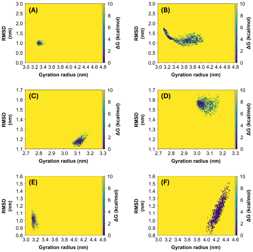
Combining attenuated total reflection infrared spectroscopy, circular dichroism, and molecular dynamics, the authors claim that the three variants share broadly similar secondary structure—about 27–28% random coil, 24–27% $\beta$-turn, 8–9% $\alpha$-helix—but Omicron stands apart in $\beta$-sheet content: 35±3% by IR, versus 39±2% for Alpha and 40±3% for Gamma, a gap the paper calls significant beyond error bars. The amide I maximum shifts from 1650 cm$^{-1}$ (Alpha) to 1648 cm$^{-1}$ (Gamma) to 1647 cm$^{-1}$ (Omicron), with all Omicron components slightly redshifted, which is interpreted as stronger C=O hydrogen bonding with water and therefore greater hydrophilicity. Surface polarity maps show larger hydrophilic areas on Omicron's RBD and NTD than on Alpha's, and MD simulations show that the open-state Omicron model keeps a radius of gyration near 4.3 nm over 600 ns, whereas Alpha and Gamma open-state models collapse to closed configurations. The paper identifies the open state with the RBD-up conformation of the full spike and concludes that Omicron's S1 favors RBD-up, matching its documented higher ACE2 affinity and infectivity.
Load-bearing premise
The load-bearing premise is that a single 600 ns molecular dynamics run per starting state, on predicted models that lack the sugar modifications, faithfully represents how the glycosylated proteins behave in solution; if that sampling or model choice is unrepresentative, the claim that Omicron alone retains the open state—and the explanation built on it—does not follow.
Editorial extensions
If this is right
- A spectroscopic lab could in principle distinguish Omicron S1 from Alpha or Gamma by the combination of a redshifted amide I peak and reduced $\beta$-sheet fraction, without needing antibodies or sequencing.
- If Omicron S1 genuinely favors the open, RBD-up state, that would provide a structural reason for its enhanced ACE2 binding and faster spread relative to earlier variants.
- The trend in the amide I maximum (1650, 1648, 1647 cm$^{-1}$ for Alpha, Gamma, Omicron) tracks the mutation count (7, 10, 31), suggesting that accumulated mutations progressively alter the protein's hydration and conformational ensemble.
- The more hydrophilic character of Omicron's RBD and NTD implies that water-mediated contacts, not just direct amino-acid contacts, contribute to its stronger receptor interaction.
Reading between the lines
- Because the open-versus-closed conclusion rests on a single 600 ns trajectory per starting state, a natural next test is to run many independent replicas; if Alpha and Gamma sometimes stay open as often as Omicron, the mutation-specific interpretation would not survive.
- The simulation models lack the sugar modifications present on the measured proteins, so the paper's link between simulation and spectroscopy implicitly assumes glycans do not change the open/closed balance; this could be tested by simulating a glycosylated S1 or by comparing deglycosylated samples spectroscopically.
- If the amide I redshift really tracks hydrophilicity, the same infrared protocol could be applied to newer variants to see whether the $\beta$-sheet fraction continues to drop as the spike accumulates mutations.
- The S1 monomer is studied without the S2 fusion machinery or the viral membrane; whether the monomer's open preference persists in the full prefusion spike is a separate, testable question.
Signed reviews
Editorial analysis
A structured set of objections, weighed in public.
Referee Report
Summary. The manuscript reports a comparative biophysical study of the monomeric S1 subunits of SARS-CoV-2 Alpha, Gamma, and Omicron spike proteins, combining ATR-IR spectroscopy, circular dichroism (CD) spectroscopy, ColabFold structure prediction, 600 ns molecular dynamics simulations, and Protein-sol surface calculations. The authors estimate secondary-structure percentages from each technique, interpret the amide I red shift of Omicron as evidence of increased hydrophilicity, and conclude that Omicron S1 has lower beta-sheet content than Alpha and Gamma, a more hydrophilic surface, and uniquely retains an open (RBD-up-like) conformation in MD simulations, which they connect to higher ACE2 affinity and transmissibility. The claimed novelty is the systematic combination of these experimental and computational techniques on three variants of concern.
Significance. If the claims were established, the paper would provide a useful multi-technique comparison of variant spike proteins and would strengthen the case for IR/CD spectroscopy as rapid structural screens for emerging variants. The authors should be credited for the multi-technique design, the explicit reporting of mutation tables and sequences, and the use of commercially relevant S1 proteins. However, the central claims are not supported by the evidence as presented: the reported error bars for beta-sheet content overlap, the MD secondary-structure comparison is confounded by inconsistent state selection, and the open-state retention claim rests on a single trajectory per starting state. The dataset may still be a useful contribution after substantial revision, but the current conclusions outrun the statistical and methodological basis.
major comments (3)
- [Section 4, Tables 1, 2, 4, Figure 8] The statement that Omicron has a lower beta-sheet content "which represents a significant difference with respect to Alpha and Gamma beyond the error bars" is contradicted by the paper's own uncertainties. In Table 1, CD-derived beta-sheet values are Alpha 38 ± 7 and Omicron 37 ± 7; in Table 2, IR-derived values are Alpha 39 ± 2 and Omicron 35 ± 3, a difference that is approximately 1.1 combined standard deviations; in Table 4, MD-derived values are Alpha 37 ± 3 and Omicron 35 ± 2. The weighted averages shown in Figure 8 therefore almost certainly have overlapping error bars. The claim of significance should either be supported by an explicit statistical test (e.g., a t-test or bootstrap on the per-deposition estimates) or be downgraded to a trend.
- [Section 3.3, Table 4] The MD secondary-structure comparison is confounded by inconsistent state selection. For Alpha and Gamma, the percentages in Table 4 are taken from closed-state trajectories only, while for Omicron the values are explicitly "an average between open and closed states." Because open and closed states differ in radius of gyration, solvent exposure, and likely secondary-structure composition, this averaging can artificially lower the Omicron beta-sheet content relative to the other variants. State-specific values should be reported separately for all variants, and any ensemble average should be justified with Boltzmann weights or occupancy statistics rather than a simple arithmetic mean.
- [Section 2.4, Section 3.3, Table 3, Figure 5] The claim that Omicron "is the only one retaining an open state when starting from an open state" is based on a single 600 ns MD replica per starting state, as stated in the Methods for a single replica of each model. With no replicate simulations, no occupancy statistics, and no check that the Alpha and Gamma closures are not initial-model relaxation or force-field drift, the conformational difference is not statistically established. This concern is compounded by two additional issues: the simulations use unglycosylated monomeric ColabFold models, whereas the experimental proteins are glycosylated, and the NTD-RBD center-of-mass distance used to define open/closed states is not demonstrated to correspond to the RBD-up/RBD-down equilibrium of the trimeric spike. Replicate trajectories, matched glycosylation status, and validation of the open/closed metric are needed before this central claim can be accepted.
minor comments (6)
- [Section 2.3, Table 1] Table 1 reports results from the CLUSTR algorithm, but Section 2.3 states that CONTINLL, CDSSTR, and SELCON3 were employed; please reconcile this inconsistency.
- [Section 2.3] The optical path length of the CD cuvette is given as 0.01 nm, which is physically implausible; this should presumably be 0.01 cm.
- [Table 1] In the Omicron row, the random coil entry is printed as "32 5" instead of "32 ± 5."
- [Section 2.4] The force field is referred to as CHARMM22/CMAP in the text and CHARMM22* in the discussion; please use a consistent nomenclature throughout.
- [Section 3.4, Figures 6 and 7] The NPP surface ratio results are presented only as qualitative color maps; quantitative values or distributions for the RBD and NTD regions would strengthen the hydrophilicity comparison.
- [Section 3.1] The statement that "noticeable differences" occur in the CD spectra is not supported by any statistical comparison of replicate spectra; showing error envelopes or replicate curves would make this assessment more rigorous.
Circularity Check
No constructional circularity: the variant-specific claims rest on independent IR/CD experiments and separate MD simulations, with self-citations used only as methodological background.
full rationale
The central comparative claims of the paper—lower Omicron beta-sheet content, redshifted amide I components, more hydrophilic surface character, and retention of an open conformational state—are supported by three independent lines of evidence: ATR-IR and CD measurements on purchased variant proteins, CDPro deconvolution against external reference basis sets, and MD simulations initialized from ColabFold models. None of the paper's equations defines a derived quantity in terms of the target result; secondary-structure percentages are obtained by spectral deconvolution and trajectory analysis, not by fitting the three techniques to one another. The self-citations (refs 58, 59) are used to justify the IR analysis protocol, the WT amide I position, and the reproducibility estimate; these are methodological and background inputs, and the variant-to-variant differences are not constructed from them. The open/closed classification is defined independently by NTD-RBD distance from the ColabFold models, and the mapping to RBD up/down states is imported from external literature. The main weaknesses of the paper are statistical and validation concerns rather than circularity: only one MD replica per starting state is reported, the MD secondary-structure averages in Table 4 use unmatched state selections (closed-only for Alpha and Gamma versus an open/closed average for Omicron), and the simulations use unglycosylated monomeric models. These issues affect robustness and interpretation, but they do not amount to a derivation that reduces by construction to its own inputs.
Assumptions & free parameters
free parameters (2)
- IR amide I Gaussian component parameters =
Not specified (peak positions, widths, areas fitted per spectrum)
- CDPro basis set and algorithm selection =
SP37, SP43, SDP42, SDP48, SMP50, SMP56; CONTINLL, CDSSTR, SELCON3
assumptions (5)
- domain assumption Amide I band component assignments to secondary structures (beta-sheet, alpha-helix, beta-turn, random coil) are valid for these S1 proteins.
- domain assumption CDPro reference datasets and algorithms provide reliable secondary structure estimates for monomeric S1.
- domain assumption CHARMM22* force field and TIP3P water accurately represent spike S1 conformational dynamics.
- domain assumption ColabFold predictions provide reliable starting structures for each variant.
- domain assumption Glycosylation does not significantly affect the structural differences measured in the experiments and simulations.
Cite this review
Pith. "Pith review of Search for structural differences in spike glycoprotein variants of SARS-CoV-2: Infrared Spectroscopy, Circular Dichroism and Computational Analysis." pith.science (2026). https://pith.science/paper/57WMPF3Z
@misc{pith2026250419766,
  author       = {Pith},
  title        = {Pith review of: Search for structural differences in spike glycoprotein variants of SARS-CoV-2: Infrared Spectroscopy, Circular Dichroism and Computational Analysis},
  year         = {2026},
  howpublished = {\url{https://pith.science/paper/57WMPF3Z}},
  note         = {Machine review of arXiv:2504.19766}
}
read the original abstract
The SARS-CoV-2 pandemic has led to a significant emergence of highly mutated forms of viruses with a great ability to adapt to the human host. Some mutations resulted in changes in the amino acid sequences of viral proteins, including the Spike glycoproteins, affecting protein physico-chemical properties and functionalities. Here, we propose, for the first time to the best of our knowledge, a systematic and comparative study of the monomeric spike protein subunits 1 of three SARS-CoV-2 variants at pH 7.4, combining both an experimental approach, taking advantage of Attenuated Total Reflection Infrared and Circular Dichroism spectroscopies, and a computational approach via Molecular Dynamics simulations. Experimental data in combination with Molecular Dynamics and Surface polarity calculations provide a comprehensive understanding of variants proteins in terms of their secondary structure content, 3D conformational structure and order and interaction with the solvent. The present structural investigation clarifies which kind of changes in conformation and functionalities occurred as long as mutations appeared in amino acids sequences. This information is essential for preventive targeted actions, drug design, and biosensing applications.
Figures


Figures from the paper (4 more)


Reference graph
Works this paper leans on
-
[1]
https://data.who.int/dashboards/covid19/deaths?n=c
-
[2]
WHO Declares COVID -19 a Pandemic
Cucinotta, D.; Vanelli, M. WHO Declares COVID -19 a Pandemic. Acta Biomed. 2020, 91, 157–160
2020
-
[3]
Hatmal, W
M.M. Hatmal, W. Alshaer, M.A.I. Al -Hatamleh, M. Hatmal, O. Smadi, M.O. Taha, A.J. Oweida, J.C. Boer, R. Mohamud, M. Plebanski. Comprehensive Structural and Molecular Comparison of Spike Proteins of SARS -CoV-2, SARS -CoV and MERS -CoV, and Their Interactions with ACE2. Cells 9, 2638 (2020)
2020
-
[4]
P. Zhao, J. L. Praissman, O. C. Grant, Y. Cai, T. Xiao, K. E. Rosenbalm, K. Aoki, B. P. Kellman, R. Bridger, D. H. Barouch, M. A. Brindley, N. E. Lewis, M. Tiemeyer, B. Chen, R. J. Woods, L. Wells, Virus -Receptor Interactions of Glycosylated SARS -CoV-2 Spike and Human ACE2 Receptor. Cell Host Microbe 28, 1–16 (2020)
2020
-
[5]
Yadav, J.K
R. Yadav, J.K. Chaudhary, N. Jain, P.K. Chaudhary, S. Khanra, P. Dhamija, A. Sharma, A. Kumar, S. Handu. Role of Structural and Non-Structural Proteins and Therapeutic Targets of SARS-CoV-2 for COVID-19. Cells 10, 821 (2021)
2021
-
[6]
Salleh, J.P
M.Z. Salleh, J.P. Derrick, Z.Z. Deris. Structural Evaluation of the Spike Glycoprotein Variants on SARS-CoV-2 Transmission and Immune Evasion. Int. J. Mol. Sci. 22, 7425 (2021)
2021
-
[7]
S. Kang, M. Yang, Z. Hong, L. Zhang, Z. Huang, X. Chen, S. He, Z. Zhou, Z. Zhou, Q. Chen, Y. Yan, C. Zhang, H. Shan, S. Chen. Crystal structure of SARS-CoV-2 nucleocapsid protein RNA binding domain reveals potential unique drug targeting sites. Acta Pharmaceutica Sinica B 10, 1228 (2020)
2020
-
[8]
Y. Peng, N. Du, Y. Lei, S. Dorje, J. Qi, T. Luo, G.F. Gao, H. Song. Structures of the SARS‐ CoV‐2 nucleocapsid and their perspectives for drug design. EMBO J. 39, e105938 (2020)
2020
Show all 105 references
-
[9]
W. Yan, Y. Zheng, X. Zeng, B. He, W. Cheng. Structural biology of SARS-CoV-2: open the door for novel therapies. Sig. Transduct. Target. Ther. 7, 26 (2022)
2022
-
[10]
McLean, J
G. McLean, J. Kamil, B. Lee, P. Moore, T.F. Schulz, A. Muik, U. Sahin, Ö. Türeci, S. Pather. The impact of evolving SARS-CoV-2 mutations and variants on COVID-19 vaccines. MBio 13, e0297921 (2022)
2022
-
[11]
Sironi, S.E
M. Sironi, S.E. Hasnain, B. Rosenthal, T. Phan, F. Luciani, M.A. Shaw, M.A. Sallum, M.E. Mirhashemi, S. Morand S, F. González -Candelas. SARS-CoV-2 and COVID-19: A genetic, epidemiological, and evolutionary perspective. Infect. Genet. Evol. 84, 104384 (2020)
2020
-
[12]
Harvey, A.M
W.T. Harvey, A.M. Carabelli, B. Jackson, R.K. Gupta, E.C. Thomson, E.M. Harrison, C. Ludden, R. Reeve, A. Rambaut. SARS-CoV-2 variants, spike mutations and immune escape. Nat. Rev. Microbiol. 19, 409–424 (2021)
2021
-
[13]
Zella, M
D. Zella, M. Giovanetti, F. Benedetti, F. Unali, S. Spoto, M. Guarino, S. Angeletti, M. Ciccozzi.The variants question: What is the problem? J. Med. Virol. 93, 6479–6485 (2021)
2021
-
[14]
Fatihi, S
S. Fatihi, S. Rathore, A.K. Pathak, D. Gahlot, M. Mukerji, N. Jatana, L. Thukral. A rigorous framework for detecting SARS-CoV-2 spike protein mutational ensemble from genomic and structural features. Current Research in Structural Biology 3, 290 (2021)
2021
-
[15]
Imbiakha, S
B. Imbiakha, S. Ezzatpour, D. W. Buchholz, J. Sahler, C. Ye, X.A. Olarte -Castillo, A. Zou, C. Kwas, K. O'Hare, A. Choi, R.A. Adeleke, S. Khomandiak, L. Goodman, M.C. Jager, G.R. Whittaker, L. Martinez -Sobrido, A. August, H.C. Aguilar. Age -dependent acquisi tion of pathogeni...
2023
-
[16]
R. Zhu, D. Canena, M. Sikora, M. Klausberger, H. Seferovic, A. R. Mehdipour, L. Hain, E. Laurent, V. Monteil, G. Wirnsberger, R. Wieneke, R. Tampé, N.F. Kienzl, L. Mach, A. Mirazimi, Y.J. Oh, J.M. Penninger, G. Hummer, P. Hinterdorfer. Force-tuned avidity of spike variant-ACE2...
2022
-
[17]
Carabelli, T.P
A.M. Carabelli, T.P. Peacock, L.G. Thorne, et al. SARS -CoV-2 variant biology: immune escape, transmission and fitness. Nat. Rev. Microbiol. 21, 162–177 (2023)
2023
-
[18]
Laffeber, K
C. Laffeber, K. de Koning, R. Kanaar, J.H.G. Lebbink. Experimental Evidence for Enhanced Receptor Binding by Rapidly Spreading SARS -CoV-2 Variants. J. Mol. Biol. 433, 167058 (2021)
2021
-
[19]
H.H. Gan, J. Zinno, F. Piano, K.C. Gunsalus. Omicron Spike Protein Has a Positive Electrostatic Surface That Promotes ACE2 Recognition and Antibody Escape. Front.Virol. 2, 894531 (2022)
2022
-
[20]
G. B. Chand, A. Banerjee, G. K. Azad. Identification of twenty -five mutations in surface glycoprotein (Spike) of SARS -CoV-2 among Indian isolates and their impact on protein dynamics. Gene Reports 21, 100891 (2020)
2020
-
[21]
Ozono, Y
S. Ozono, Y. Zhang, H. Ode, K. Sano, T. S. Tan, K. Imai, K. Miyoshi, S. Kishigami, T. Ueno, Y. Iwatani, T. Suzuki, K. Tokunaga. SARS -CoV-2 D614G spike mutation increases entry efficiency with enhanced ACE2-binding affinity. Nature Communications 12, 848 (2021)
2021
-
[22]
Nguyen, P.N
T.T. Nguyen, P.N. Pathirana, T. Nguyen, Q.V.H. Nguyen, A. Bhatti, D.C. Nguyen, D.T. Nguyen, N.D. Nguyen, D. Creighton, M. Abdelrazek. Genomic mutations and changes in protein secondary structure and solvent accessibility of SARS-CoV-2 (COVID-19 virus). Sci. Rep. 11, 3487 (2021)
2021
-
[23]
Sanches, I
P.R.S. Sanches, I. Charlie -Silva, H.L.B. Braz, C. Bittar, M.F. Calmon, P. Rahal, E.M. Cilli. Recent advances in SARS-CoV-2 Spike protein and RBD mutations comparison between new variants Alpha (B.1.1.7, United Kingdom), Beta (B.1.351, South Africa), Gamma (P.1, Brazil) and De...
2021
-
[24]
Suleman, Q
M. Suleman, Q. Yousafi, J. Ali, S.S. Ali, Z. Hussain, S. Ali, M. Waseem, A. Iqbal, S. Ahmad, A. Khan, Y. Wang, D.Q. Wei. Bioinformatics analysis of the differences in the binding profile of the wild -type and mutants of the SARS -CoV-2 spike protein variants with the ACE2 rece...
2021
-
[25]
L. Wang, G. Cheng. Sequence analysis of the emerging SARS -CoV-2 variant Omicron in South Africa. J. Med. Virol. 94, 1728 (2022)
2022
-
[26]
Tragni, F
V. Tragni, F. Preziusi, L. Laera, A. Onofrio, I. Mercurio, S. Todisco, M. Volpicella, A. De Grassi, C.L. Pierri. Modeling SARS -CoV-2 spike/ACE2 protein –protein interactions for predicting the binding affinity of new spike variants for ACE2, and novel ACE2 structurally relate...
2022
-
[27]
Barton, S.A
M.I. Barton, S.A. MacGowan, M.A. Kutuzov, O. Dushek, G. J. Barton, P. A. van der Merwe. Effects of common mutations in the SARS-CoV-2 Spike RBD and its ligand, the human ACE2 receptor on binding affinity and kinetics. eLife 10, e70658 (2021)
2021
-
[28]
Mercurio, V
I. Mercurio, V. Tragni, F. Busto, A. De Grassi, C. L. Pierri. Protein structure analysis of the interactions between SARS -CoV-2 spike protein and the human ACE2 receptor: from conformational changes to novel neutralizing antibodies. Cell. Mol. Life Sci. 78, 1501 –1522 (2021)
2021
-
[29]
T. A. Shishir, T. Jannat, I.B. Naser. An in -silico study of the mutation -associated effects on the spike protein of SARS-CoV-2, Omicron variant. PLOS ONE 17, e0266844 (2022)
2022
-
[30]
Ortega, F.H
J.T. Ortega, F.H. Pujol, B. Jastrzebska, H.R. Rangel, Mutations in the SARS -CoV-2 spike protein modulate the virus affinity to the human ACE2 receptor, an in silico analysis. EXCLI J. 20, 585-600 (2021)
2021
-
[31]
Piccirilli, F
F. Piccirilli, F. Tardani, A. D’Arco, G. Birarda, L. Vaccari, S. Sennato, S. Casciardi, S. Lupi. Infrared Nanospectroscopy Reveals DNA Structural Modifications upon Immobilization onto Clay Nanotubes. Nanomaterials 11, 1103 (2021)
2021
-
[32]
Bálint, B
G. Bálint, B. Vörös -Horváth, A. Széchenyi. Omicron: increased transmissibility and decreased pathogenicity. Sig. Transduct. Target. Ther. 7, 151 (2022)
2022
-
[33]
Y. Araf, F. Akter, Y. -D. Tang, R. Fatemi, Md. S. A. Parvez, C. Zheng, Md. G. Hossain. Omicron variant of SARS ‐CoV‐2: Genomics, transmissibility,and responses to current COVID‐19 vaccines. J. Med. Virol. 94, 1825–1832 (2022)
2022
-
[34]
Torjesen
I. Torjesen. Covid -19: Omicron may be more transmissible than other variants and partly resistant to existing vaccines, scientists fear. BMJ 375, 2943 (2021)
2021
-
[35]
E. Volz, S. Mishra, M. Chand, et al. Assessing transmissibility of SARS -CoV-2 lineage B.1.1.7 in England. Nature 593, 266–269 (2021)
2021
-
[36]
R. Dong, T. Hu, Y. Zhang, Y. Li, X. -H- Zhou. Assessing the Transmissibility of the New SARS-CoV-2 Variants: From Delta to Omicron. Vaccines 10, 496 (2022)
2022
-
[37]
A. Barth. The infrared absorption of amino acid side chains. Prog. Biophys. Mol. Biol. 74, 141–173 (2000)
2000
-
[38]
A. Barth. Infrared spectroscopy of proteins. Biochim. Biophys. Acta 1767, 1073–1101 (2007)
2007
-
[39]
H. Yang, S. Yang, J. Kong, A. Dong, S. Yu. Obtaining information about protein secondary structures in aqueous solution using Fourier transform IR spectroscopy. Nat. Protoc. 10, 382– 396 (2015)
2015
-
[40]
Kelly, T.J
S.M. Kelly, T.J. Jess, N.C. Price. How to study proteins by circular dichroism. Biochim. Biophys. Acta, Proteins Proteomics 1751, 119-139 (2005)
2005
-
[41]
Miles, R.W
A.J. Miles, R.W. Janes, B.A. Wallace. Tools and methods for circular dichroism spectroscopy of proteins: a tutorial review. Chem. Soc. Rev. 50, 8400-8413 (2021)
2021
-
[42]
Miles, B.A
A.J. Miles, B.A. Wallace. Circular dichroism spectroscopy of membrane proteins. Chem. Soc. Rev. 45, 4859-4872 (2016)
2016
-
[43]
Whitmore, BA
L. Whitmore, BA. Wallace. Protein secondary structure analyses from circular dichroism spectroscopy: methods and reference databases. Biopolymers. 89(5), 392-400 (2008)
2008
-
[44]
C.H. Li, X. Nguyen, L. Narhi, L. Chemmalil, E. Towers, S. Muzammil, J. Gabrielson, Y. Jiang. Applications of circular dichroism (CD) for structural analysis of proteins: qualification of near ‐ and far ‐UV CD for protein higher order structural analysis. Journal of Pharmaceuti...
2011
-
[45]
Meierhenrich, J.J
U.J. Meierhenrich, J.J. Filippi, C. Meinert, J.H. Bredehöft, J.I. Takahashi, L. Nahon, N.C. Jones, S.V. Hoffmann. Circular Dichroism of Amino Acids in the Vacuum -Ultraviolet Region. Angewandte Chemie International Edition 49, 7799-7802 (2010)
2010
-
[46]
Moran, M.T
S.D. Moran, M.T. Zanni. How to get insight into amyloid structure and formation from infrared spectroscopy. The journal of physical chemistry letters 5, 1984-1993 (2014)
2014
-
[47]
Vosough, A
F. Vosough, A. Barth. Characterization of homogeneous and heterogeneous amyloid -β42 oligomer preparations with biochemical methods and infrared spectroscopy reveals a correlation between infrared spectrum and oligomer size. ACS Chemical Neuroscience 12(3), 473-488 (2021)
2021
-
[48]
Woutersen, P
S. Woutersen, P. Hamm. Time-resolved two-dimensional vibrational spectroscopy of a short α-helix in water. The Journal of Chemical Physics 115(16), 7737-7743 (2001)
2001
-
[49]
Manas, Z
E.S. Manas, Z. Getahun, W.W. Wright, W.F. DeGrado, J.M. Vanderkooi. Infrared spectra of amide groups in α-helical proteins: evidence for hydrogen bonding between helices and water. Journal of the American Chemical Society 122(41), 9883-9890 (2000)
2000
-
[50]
Myshakina, Z
N.S. Myshakina, Z. Ahmed, S.A. Asher. Dependence of amide vibrations on hydrogen bonding. The Journal of Physical Chemistry B 112(38), 11873-11877 (2008)
2008
-
[51]
Gamage, T.N
S.S.T. Gamage, T.N. Pahattuge, H. Wijerathne, et al. Microfluidic affinity selection of active SARS-CoV-2 virus particles. Sci. Adv. 8, eabn9665 (2022)
2022
-
[52]
Funari, K.Y
R. Funari, K.Y. Chu, A.Q. Shen. Detection of antibodies against SARS -CoV-2 spike protein by gold nanospikes in an opto-microfluidic chip. Biosens Bioelectron. 169, 112578 (2020)
2020
-
[53]
B. Tan, X. Zhang, A. Ansari, et al. Design of a SARS -CoV-2 papain-like protease inhibitor with antiviral efficacy in a mouse model. Science 383, 1434–1440 (2024)
2024
-
[54]
Kumar, N.P
N. Kumar, N.P. Shetti, S. Jagannath, T. M. Aminabhavi. Electrochemical sensors for the detection of SARS-CoV-2 virus. Chem. Eng. J. 430, 132966 (2022)
2022
-
[55]
Lee, J.W
J.H. Lee, J.W. Kim, H.E. Lee, et al. A dual -targeting approach using a human bispecific antibody against the receptor -binding domain of the Middle East Respiratory Syndrome Coronavirus. Virus Research 345, 199383 (2024)
2024
-
[56]
Kitane, S
D.L. Kitane, S. Loukman, N. Marchoudi, A. Fernandez -Galiana, F.Z. El Ansari, F. Jouali, J. Badir, J.L. Gala, D. Bertsimas, N. Azami, O. Lakbita, O. Moudam, R. Benhida, J. Fekkak. A simple and fast spectroscopy -based technique for Covid -19 diagnosis. Sci Re p 11, 16740 (2021)
2021
-
[57]
Mirdita, K
M. Mirdita, K. Schütze, Y. Moriwaki, L. Heo, S. Ovchinnikov, M. Steinegger. ColabFold: making protein folding accessible to all. Nat Methods 19, 679–682 (2022)
2022
-
[58]
D’Arco, M
A. D’Arco, M. Di Fabrizio, T. Mancini, R. Mosetti, S. Macis, G. Tranfo, G. Della Ventura, A. Marcelli, M. Petrarca, S. Lupi. Secondary Structures of MERS -CoV, SARS -CoV, and SARS-CoV-2 Spike Proteins Revealed by Infrared Vibrational Spectroscopy. Int. J. Mol. Sci. 24, 9550 (2023)
2023
-
[59]
Mancini, S
T. Mancini, S. Macis, R. Mosetti, N. Luchetti, V. Minicozzi, A. Notargiacomo, M. Pea, A. Marcelli, G. D. Ventura, S. Lupi, A. D'Arco. Infrared Spectroscopy of SARS -CoV-2 Viral Protein: from Receptor Binding Domain to Spike Protein. Adv. Sci. 2400823 (2024)
2024
-
[60]
Wolpert, P
M. Wolpert, P. Hellwig. Infrared spectra and molar absorption coefficients of the 20 alpha amino acids in aqueous solutions in the spectral range from 1800 to 500 cm− 1. Spectrochimica Acta Part A: Molecular and Biomolecular Spectroscopy 64(4), 987 -1001 (2006)
2006
-
[61]
Ricciardi, M
V. Ricciardi, M. Portaccio, G. Perna, M. Lasalvia, V. Capozzi, F.P. Cammarata, P. Pisciotta, G. Petringa, I. Delfino, L. Manti, et al. FT-IR Transflection Micro -Spectroscopy Study on Normal Human Breast Cells after Exposure to a Proton Beam. Appl. Sci. 11(2), 540 (2021)
2021
-
[62]
Delfino, M
I. Delfino, M. Portaccio, B. Della Ventura, D.G. Mita, M. Lepore. Enzyme distribution and secondary structure of sol -gel immobilized glucose oxidase by micro -attenuated total reflection FT-IR spectroscopy. Mater. Sci. Eng. C 33, 304–310 (2013)
2013
-
[63]
Van Der Spoel, E
D. Van Der Spoel, E. Lindahl, B. Hess, G. Groenhof, A.E. Mark, H.J.C. Berendsen. GROMACS: Fast, flexible, and free. J. Comput. Chem. 26(16), 1701–18 (2005)
2005
-
[64]
B. A. Russell, K. Kubiak -Ossowska, P. A. Mulheran, D. J. S. Birch and Y. Chen. Locating the nucleation sites for protein encapsulated gold nanoclusters: a molecular dynamics and fluorescence study. Phys. Chem. Chem. Phys. 17, 21935-21941 (2015)
2015
-
[65]
Sreerama, R.W
N. Sreerama, R.W. Woody. On the analysis of membrane protein circular dichroism spectra. Protein Science 13, 100-112 (2004)
2004
-
[66]
Sreerama, R.W
N. Sreerama, R.W. Woody. Estimation of Protein Secondary Structure from Circular Dichroism Spectra: Comparison of CONTIN, SELCON, and CDSSTR Methods with an Expanded Reference Set. Analytical Biochemistry 287, 252-260 (2000)
2000
-
[67]
Jumper, R
J. Jumper, R. Evans, A. Pritzel, T. Green, M. Figurnov, O. Ronneberger, et al. Highly accurate protein structure prediction with AlphaFold. Nature 596(7873), 583–9 (2021)
2021
-
[68]
Berendsen, D
H.J.C. Berendsen, D. Van der Spoel, R. Van Drunen. GROMACS: A message-passing parallel molecular dynamics implementation. Comput. Phys. Commun. 91(1–3), 43–56 (1995)
1995
-
[69]
Mackerell, M
A.D. Mackerell, M. Feig, C.L. Brooks. Extending the treatment of backbone energetics in protein force fields: Limitations of gas ‐phase quantum mechanics in reproducing protein conformational distributions in molecular dynamics simulations. J. Comput. C hem. 25(11), 1400–15 (2004)
2004
-
[70]
E. S. Istifli, P.A. Netz, A. Sihoglu Tepe, C. Sarikurkcu, B. Tepe. Understanding the molecular interaction of SARS-CoV-2 spike mutants with ACE2 (angiotensin converting enzyme 2). J Biomol Struct Dyn. 2022;40(23):12760-12771
2022
-
[71]
H. Woo, S. -J. Park, . K. Choi, et al. Developing a Fully Glycosylated Full -Length SARS- CoV-2 Spike Protein Model in a Viral Membrane . J. Phys. Chem. B 124, 33, 7128 –7137 (2020)
2020
-
[72]
MacKerell, D
A.D. MacKerell, D. Bashford, M. Bellott, R.L. Dunbrack, J.D. Evanseck, M.J. Field, et al. All-Atom Empirical Potential for Molecular Modeling and Dynamics Studies of Proteins. J. Phys. Chem. B. 102(18), 3586–616 (1998)
1998
-
[73]
Piana, K
S. Piana, K. Lindorff-Larsen, D.E. Shaw. How Robust Are Protein Folding Simulations with Respect to Force Field Parameterization? Biophys J. 100(9), L47–9 (2011)
2011
-
[74]
Nakamoto
K. Nakamoto. The Urey — Bradley Force Field: Its Significance and Application. In: Developments in Applied Spectroscopy. Boston, MA: Springer US; 1964. p. 158–68
1964
-
[75]
Ngai, R.H
L.H. Ngai, R.H. Mann. A transferable Urey-Bradley force field and the assignments of some mixed halomethanes
-
[76]
B. Hess, H. Bekker, H.J.C. Berendsen, J.G.E.M Fraaije. LINCS: A linear constraint solver for molecular simulations. J. Comput. Chem. 18(12), 1463–72 (1997)
1997
-
[77]
Bussi, D
G. Bussi, D. Donadio, M. Parrinello. Canonical sampling through velocity rescaling. J. Chem. Phys. 126(1), (2007)
2007
-
[78]
Parrinello, A
M. Parrinello, A. Rahman. Polymorphic transitions in single crystals: A new molecular dynamics method. J. Appl. Phys. 52(12), 7182–90 (1982)
1982
-
[79]
Nosé, M.L
S. Nosé, M.L. Klein. Constant pressure molecular dynamics for molecular systems. Mol. Phys. 50(5), 1055–76 (1983)
1983
-
[80]
Darden, D
T. Darden, D. York, L. Pedersen. Particle mesh Ewald: An N ⋅log( N ) method for Ewald sums in large systems. J. Chem. Phys. 98(12), 10089–92 (1993)
1993
-
[81]
Van Rossum, F.L
G. Van Rossum, F.L. Drake. Python 3 Reference Manual. Scotts Valley, CA: CreateSpace; 2009
2009
-
[82]
Humphrey, A
W. Humphrey, A. Dalke, K. Schulten. VMD: Visual molecular dynamics. J. Mol. Graph. 14(1), 33–8 (1996)
1996
-
[83]
Hebditch, M.A
M. Hebditch, M.A. Carballo -Amador, S. Charonis, R. Curtis, J. Warwicker. Protein –Sol: a web tool for predicting protein solubility from sequence. Bioinformatics 33, 3098 –3100 (2017)
2017
-
[84]
Hebditch, J
M. Hebditch, J. Warwicker. Web -based display of protein surface and pH -dependent properties for assessing the developability of biotherapeutics. Sci Rep 9, 1969 (2019)
2019
-
[85]
L. Zhao, Z. Cao, Y. Bian, G. Hu, J. Wang, Y. Zhou. Molecular Dynamics Simulations of Human Antimicrobial Peptide LL-37 in Model POPC and POPG Lipid Bilayers. Int. J. Mol. Sci. 19(4), 1186 (2018)
2018
-
[86]
Amdursky and M
N. Amdursky and M. M. Stevens. Circular Dichroism of Amino Acids: Following the Structural Formation of Phenylalanine. Chem. Phys. Chem. 16(13), 2768-2774 (2015)
2015
-
[87]
Zaman, N
N. Zaman, N. Parvaiz, F. Gul, et al. Dynamics of water -mediated interaction effects on the stability and transmission of Omicron. Sci. Rep. 13, 20894 (2023)
2023
-
[89]
Bhattacharjee and P
N. Bhattacharjee and P. Biswas. Structural patterns in alpha helices and beta sheets in globular proteins. Protein and Peptide Letters. 16(8), 953-960, (2009)
2009
-
[90]
Chitra, and C
N. Chitra, and C. L. Dias. Hydrophobic interactions and hydrogen bonds in β-sheet formation. The journal of chemical physics. 139(11), (2013)
2013
-
[91]
F. Chiti. Relative importance of hydrophobicity, net charge, and secondary structure propensities in protein aggregation. Protein Misfolding, Aggregation, and Conformational Diseases: Part A: Protein Aggregation and Conformational Diseases. Boston, MA: Springer US, 43-59 (2006)
2006
-
[92]
Funari, N
R. Funari, N. Bhalla, L. Gentile. Measuring the Radius of Gyration and Intrinsic Flexibility of Viral Proteins in Buffer Solution Using Small-Angle X‑ray Scattering. ACS Meas. Sci. Au 2, 547−552 (2022)
2022
-
[93]
M. Gur, E. Taka, S.Z. Yilmaz, C. Kilinc, U. Aktas, M. Golcuk. Conformational transition of SARS-CoV-2 spike glycoprotein between its closed and open states. J. Chem. Phys. 153, 075101 (2020)
2020
-
[94]
Henderson, R.J
R. Henderson, R.J. Edwards, K. Mansouri, K. Janowska, V. Stalls, S.M.C. Gobeil, M. Kopp, D. Li, R. Parks, A.L. Hsu, M.J. Borgnia, B.F. Haynes, P. Acharya. Controlling the SARS - CoV-2 spike glycoprotein conformation. Nat Struct Mol Biol 27, 925–933 (2020)
2020
-
[95]
Up” versus “Down
M.H. Peters, O. Bastidas, D.S. Kokron, C.E. Henze. Static all-atom energetic mappings of the SARS-Cov-2 spike protein and dynamic stability analysis of “Up” versus “Down” protomer states. PLoS ONE 15(11), e0241168 (2020)
2020
-
[96]
Singh, S
J. Singh, S. Vashishtha, S. A. Rahman, et al. Energetics of Spike Protein Opening of SARS - CoV-1 and SARS -CoV-2 and Its Variants of Concern: Implications in Host Receptor Scanning and Transmission. Biochemistry 61, 20, 2188–219 (2022)
2022
-
[97]
Hardenbrook, P
N.J. Hardenbrook, P. Zhang. A structural view of the SARS -CoV-2 virus and its assembly. Current Opinion in Virology 52, 123–134 (2022)
2022
-
[98]
R. Yan, Y. Zhang, Y. Li, L. Xia, et al. Structural basis for the recognition of SARS-CoV-2 by full-length human ACE2. Science 367, 1444–1448 (2020)
2020
-
[99]
Lv, Y.-Q- Deng, Q
S. Lv, Y.-Q- Deng, Q. Ye, L. Cao, et al. Structural basis for neutralization of SARS -CoV-2 and SARS-CoV by a potent therapeutic antibody. Science 369, 1505–1509 (2020)
2020
-
[100]
Hossen, P
Md.L. Hossen, P. Baral, T. Sharma, B. Gerstman, P. Chapagain. Significance of the RBD mutations in the SARS -CoV-2 omicron: from spike opening to antibody escape and cell attachment. Phys. Chem. Chem. Phys. 24, 9123-9129 (2022)
2022
-
[101]
Z. Zhao, J. Zhou, M. Tian, et al. Omicron SARS-CoV-2 mutations stabilize spike up -RBD conformation and lead to a non -RBM-binding monoclonal antibody escape. Nat. Commun. 13, 4958 (2022)
2022
-
[102]
L. Zeng, J. Li, M. Lv, Z. Li, L. Yao, J. Gao, Q. Wu, Z. Wang, X. Yang, G. Tang, G. Qu. G. Jiang. Environmental Stability and Transmissibility of Enveloped Viruses at Varied Animate and Inanimate Interfaces. Environ. Health 1, 1, 15–31 (2023)
2023
-
[103]
Samandoulgou, R
I. Samandoulgou, R. Hammami, R. Morales Rayas, I. Fliss, J. Jean. Stability of Secondary and Tertiary Structures of Virus-Like Particles Representing Noroviruses: Effects of pH, Ionic Strength, and Temperature and Implications for Adhesion to Surfaces. Appl Environ Microbiol 8...
2015
-
[104]
D. Bao, C. Lu, T. Ma, G. Xu, Y. Mao, L. Xin, S. Niu, Z. Wu, X. Li, Q. Teng, Z. Li,Q. Liu. Hydrophobic Residues at the Intracellular Domain of the M2 Protein Play an Important Role in Budding and Membrane Integrity of Influenza Virus. J Virol 96:e00373-22 (2022)
2022
-
[105]
Prussin, D.O
A.J. Prussin, D.O. Schwake, K. Lin, D.L. Gallagher, L. Buttling, L.C. Marr. Survival of the Enveloped Virus Phi6 in Droplets as a Function of Relative Humidity, Absolute Humidity, and Temperature. Appl Environ Microbiol 84, e00551-18 (2018)
2018
-
[106]
Lin, C.R
K. Lin, C.R. Schulte, L.C. Marr. Survival of MS2 and Φ6 viruses in droplets as a function of relative humidity, pH, and salt, protein, and surfactant concentrations. PLoS One. 15(12) , e0243505 (2020)
2020
Reviewed August 16, 2026 · model on record in the stance chip above.
Discussion (0). Continue with ORCID to comment.