REVIEW 3 major objections 5 minor 38 references
GAN-based synthetic FDG PET images from T1 brain MRI can serve to improve performance of deep unsupervised anomaly detection models
T0 review · 3 major / 5 minor · reviewed 2026-08-15 · deepseek-v4-flash
Pith's one-line read Synthetic FDG PET images generated from T1 MRI by GANs can replace real PET scans in the normative database used to train an unsupervised detector of epilepsy lesions.
desk verdict Useful proof-of-concept, but the headline comparison (synthetic vs real PET for UAD training) is confounded by switching normative datasets, so the 74% vs 42% result does not support the replacement claim as stated. read the letter →
The pith
A machine-rendered reading of the paper's core claim, the machinery that carries it, and where it could break.
The reading
What carries the argument
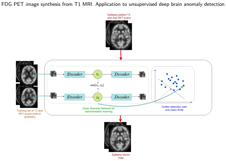
The machinery is a Cycle-GAN for T1-to-PET translation, augmented with a mean-squared-error loss between paired real and synthetic PET, and configured either as 2.5D slice triplets or 3D patches; the best version is the 3D-patch model, followed by Gaussian smoothing and histogram matching of the reconstructed volumes. The downstream UAD model is a siamese convolutional autoencoder whose latent vectors feed a per-voxel one-class SVM, producing ranked anomaly clusters. The paper's new quality metrics, global reconstruction MSE and Mahalanobis distance in the autoencoder latent space, check whether fake PET behaves as in-distribution for the detector before the detection experiment is run.
What would settle it
Take the same UAD pipeline and the same 17-patient test set, but train the detector on a larger real-PET normative database that is scanner-matched to the patients; if its sensitivity reaches or exceeds 74%, the synthetic advantage is not caused by better normative fidelity. Independently, recompute detection counts using only the top-1 or top-3 clusters instead of the top-10 rule described in Appendix C; if the synthetic-trained sensitivity drops below the real-trained 42%, the reported gain is an artifact of the cluster-counting protocol.
Extended reading notes
Core claim
The central claim, stated directly in the conclusion, is that synthetic FDG PET data generated from T1 MRI can efficiently replace true FDG PET images in normative databases for training unsupervised anomaly detection models. The paper supports this with three linked results: a Cycle-GAN with an added mean-squared-error loss, trained on paired T1 and PET data, produces synthetic PET with SSIM around 0.9 and PSNR around 23.8; the synthetic PET falls in-distribution relative to real PET under the proposed OOD metrics; and the UAD model trained on real T1 plus synthetic 3D-patch PET detects more subtle epileptogenic lesions than the same model trained on real PET. The authors caution that their explanation for the improvement, a denser synthetic latent distribution due to lower inter-individual variability, should be interpreted with caution.
Load-bearing premise
The load-bearing premise is that the synthetic-trained detector's higher sensitivity reflects a genuinely useful denser normative latent distribution, not an artifact of reduced inter-individual variability that happens to produce more or smaller candidate clusters on this particular 17-patient cohort.
Editorial extensions
If this is right
- A center with only T1 MRI of healthy subjects could build a PET-trained unsupervised anomaly detector without collecting a real FDG PET control database.
- The 3D-patch Cycle-GAN with MSE loss is the recommended synthesis configuration, giving the best visual metrics and the best downstream detection performance.
- The proposed OOD metrics offer a fast pre-screening check for whether a new patient's multimodal images are in-distribution for a trained UAD model.
- For localizing subtle epileptogenic zones, training on synthetic rather than real PET is reported to raise sensitivity from 42% to 74% in this 17-patient cohort.
Reading between the lines
- Because the authors themselves flag the denser-latent-distribution explanation as tentative, a direct test would be to measure the covariance of latent vectors for synthetic versus real control PET: a substantially narrower synthetic distribution would predict that the detection gain shrinks on more heterogeneous patient data.
- The top-ten cluster counting rule makes a hit easy, since a lesion counts as detected if it appears anywhere among the ten most suspicious clusters; recomputing the comparison with only the top-ranked cluster would show whether the synthetic-trained advantage reflects sharper localization or simply more candidate clusters.
- If the effect generalizes, the same replacement strategy could apply to other radiotracers or modalities where normative databases are scarce, such as amyloid or dopamine-transporter PET, provided a paired MRI-based synthesis model can be trained.
- A natural next experiment is mixing real and synthetic PET in the normative database; if detection improves monotonically with the amount of real data, the benefit is likely due to data scale rather than something unique to synthetic images.
Signed reviews
Editorial analysis
A structured set of objections, weighed in public.
Referee Report
Summary. The manuscript proposes to synthesize FDG PET images from T1-weighted MRI using GAN-based architectures, with the stated goal of using such synthetic PET data as normative training data for an unsupervised anomaly detection (UAD) model for epileptogenic zone screening. The authors compare several GAN configurations (2.5D vs 3D-patch, simple GAN vs Cycle-GAN, with and without an additional MSE loss), evaluate visual quality using PSNR/SSIM/LPIPS and task-oriented out-of-distribution metrics, and then train a siamese-autoencoder/OC-SVM UAD model on either 35 real paired T1/PET exams (UAD1) or on 35 T1 exams from a different control dataset paired with synthetic PET generated from those T1 scans (UAD2/UAD3). On 17 epilepsy patients with 19 subtle lesions, the model trained on 3D-patch synthetic PET achieves 74% sensitivity (14/19) versus 42% (8/19) for the model trained on real PET, with mean ranks of 2.1 and 3.9 respectively. The authors conclude that synthetic PET could efficiently replace true PET in multi-modality normative databases for training UAD models.
Significance. If the central claim were established, this would be a valuable contribution: it would relax a significant practical bottleneck for UAD training, namely the difficulty of acquiring normative PET databases, and it would extend prior work by evaluating synthetic data on a concrete downstream detection task rather than only with visual metrics. The paper has clear strengths: the code and normative image dataset are released, the visual-quality comparison includes formal statistical tests (Wilcoxon signed-rank), and the clinical evaluation targets a well-defined, difficult problem (MRI-negative epilepsy with subtle hypometabolism). The task-oriented OOD metrics are a reasonable attempt to bridge visual quality and utility, although their self-referential nature limits their probative value. However, the central claim—that synthetic PET can replace true PET—is supported by a comparison that is confounded by the training population, and the effect size is not statistically established on the small lesion cohort. Because these issues bear directly on the main conclusion, the current evidence supports a more cautious claim than the one stated in the Conclusion.
major comments (3)
- [Section 4.2 and Table 1] The comparison between UAD1 (trained on real PET) and UAD2/UAD3 (trained on synthetic PET) is confounded: UAD1 is trained on the 35 true T1/PET exams of DBC1, while UAD2 and UAD3 are trained on 35 T1 exams of DBC2 paired with synthetic PET generated from those T1 scans. The observed 74% vs 42% difference in Table 3 therefore conflates the effect of synthetic PET with differences in the normative T1 training population (DBC2 vs DBC1) and any differences in training-set composition. The paper calls this a 'fair comparison' in Section 4.2, but fairness would require true and synthetic PET for the same normative subjects, e.g., by generating synthetic PET from DBC1 T1 scans (the GANs are trained on DBC1) or by obtaining true PET for DBC2. This confound is load-bearing for the replacement claim and needs to be resolved with a within-dataset control.
- [Table 3 and Section 5.3] The key result, 14/19 vs 8/19 detections, is reported without confidence intervals or a significance test. A McNemar exact test on the discordant pairs from Table 3 yields p≈0.07, so even under the current (confounded) design the superiority of the synthetic-trained model is not statistically established. The authors should report an appropriate paired test or a bootstrap interval, and should frame the 74% vs 42% difference as an exploratory finding given the small lesion count.
- [Section 6 and Appendix C] The explanation for the improvement—that synthetic PET induces a 'denser latent distribution'—is acknowledged by the authors as speculative, but the evaluation protocol itself may contribute to the gap: clusters are thresholded per score map to yield at most 10 clusters (Appendix C), and a lesion counts as detected if it lies in any of the top-10 clusters. If the synthetic-trained models produce more or smaller clusters on this cohort, the sensitivity difference could be inflated. The paper should report the number of clusters per patient per model and evaluate sensitivity to the cluster threshold; without this, the 74% vs 42% difference cannot be confidently attributed to a genuinely more useful normative representation.
minor comments (5)
- [Section 3.1.3] The Gaussian smoothing FWHM (1.5 mm) and the histogram-matching reference image are selected on the validation set; a brief sensitivity analysis or justification would clarify how dependent the reported visual metrics are on these post-processing choices.
- [Section 5.3] In the comparison sentence, 'the real paired T1 MRI and FDG PET of D_B3Dpatch_C1' should read 'of DBC1'; the subscript appears to be a typo.
- [Figure 5] The scatter plots are dense and the three groups overlap; consider plotting marginal distributions or increasing transparency, and state the number of points per group in the caption.
- [Appendix C] The cluster-ranking criterion uses equal weights for cluster size and average score, and the OC-SVM RBF kernel is used without a reported hyperparameter search; a brief description of how these choices were made, or a sensitivity check, would improve reproducibility.
- [Introduction and Conclusion] The claim that GANs 'outperform transformer or diffusion models' rests on the cited review [6] and not on experiments in this paper; this limitation is acknowledged in the Discussion but could be stated more carefully in the Conclusion.
Circularity Check
No circularity found: the UAD comparison is an external evaluation on patient data; the training-set difference is a validity confound, not a circular reduction.
full rationale
The central claim—that synthetic FDG PET generated from T1 MRI can replace true PET in a normative database for training a UAD model—is tested on the 17 epilepsy-patient exams of DBCep (plus patient R), which are not used to train either the Cycle-GAN or the UAD models. UAD1 is trained on true T1+PET pairs from DBC1; UAD2 and UAD3 are trained on DBC2 T1 images paired with synthetic PET produced by a GAN trained on DBC1. The Table 3 sensitivities (8/19, 11/19, 14/19) are empirical outcomes on held-out patients, not quantities reconstructed from the training data by construction. The task-oriented OOD metrics in Section 3.3 and Figure 5 are computed with the same UAD architecture that performs detection and are therefore model-relative; however, they are not fitted to the detection result and are presented as a proxy, with the patient-level detection comparison providing an independent check. References [1] and [37] are self-citations, but they are used as method sources (the siamese-AE plus OC-SVM architecture and prior synthesis pipeline) rather than as theorems that force the paper's conclusion, so they are not load-bearing in a circular sense. The paper itself cautions that the 'denser latent distribution' explanation is speculative and that the cohort is small. The main validity concern is experimental rather than circular: UAD1 trains on DBC1 while UAD2 and UAD3 train on DBC2, so the 'synthetic vs. real' comparison is entangled with the control-population difference; this affects interpretation and generalizability but does not make the reported prediction equivalent to its inputs.
Assumptions & free parameters
free parameters (6)
- Gaussian smoothing FWHM for synthetic PET post-processing =
1.5 mm
- Histogram matching reference PET image =
Randomly chosen PET image from GAN training database
- UAD cluster threshold =
Set to yield at most n=10 detected clusters per patient
- Cluster ranking criterion weights =
Equal contribution of cluster size and score
- OC-SVM RBF kernel hyperparameters
- Training/validation/test split for final GAN retraining =
29 training, 3 validation, 3 test controls
assumptions (6)
- domain assumption FDG PET patterns of healthy subjects are sufficiently determined by T1-weighted MRI anatomy for the purpose of training an anomaly detector.
- domain assumption The GAN trained on DBC1 (35 paired exams) generalizes to T1 images of DBC2 and patient scans acquired on the same scanner with the same protocol.
- domain assumption Per-voxel OC-SVM support estimation in a 64-dimensional latent space is reliable with 35 training subjects.
- domain assumption The Mahalanobis distance and reconstruction error computed with the UAD model trained on synthetic data are valid proxies for whether a new sample is in-distribution for the detection task.
- domain assumption The Cycle-GAN with auxiliary MSE loss converges to a useful mapping with the described architectures and hyperparameters.
- standard math The empirical covariance matrix in the Mahalanobis distance is invertible and well-estimated from the training data.
Cite this review
Pith. "Pith review of GAN-based synthetic FDG PET images from T1 brain MRI can serve to improve performance of deep unsupervised anomaly detection models." pith.science (2026). https://pith.science/paper/CCFF2FXU
@misc{pith2026250507364,
  author       = {Pith},
  title        = {Pith review of: GAN-based synthetic FDG PET images from T1 brain MRI can serve to improve performance of deep unsupervised anomaly detection models},
  year         = {2026},
  howpublished = {\url{https://pith.science/paper/CCFF2FXU}},
  note         = {Machine review of arXiv:2505.07364}
}
read the original abstract
Background and Objective. Research in the cross-modal medical image translation domain has been very productive over the past few years in tackling the scarce availability of large curated multimodality datasets with the promising performance of GAN-based architectures. However, only a few of these studies assessed task-based related performance of these synthetic data, especially for the training of deep models. Method. We design and compare different GAN-based frameworks for generating synthetic brain [18F]fluorodeoxyglucose (FDG) PET images from T1 weighted MRI data. We first perform standard qualitative and quantitative visual quality evaluation. Then, we explore further impact of using these fake PET data in the training of a deep unsupervised anomaly detection (UAD) model designed to detect subtle epilepsy lesions in T1 MRI and FDG PET images. We introduce novel diagnostic task-oriented quality metrics of the synthetic FDG PET data tailored to our unsupervised detection task, then use these fake data to train a use case UAD model combining a deep representation learning based on siamese autoencoders with a OC-SVM density support estimation model. This model is trained on normal subjects only and allows the detection of any variation from the pattern of the normal population. We compare the detection performance of models trained on 35 paired real MR T1 of normal subjects paired either on 35 true PET images or on 35 synthetic PET images generated from the best performing generative models. Performance analysis is conducted on 17 exams of epilepsy patients undergoing surgery. Results. The best performing GAN-based models allow generating realistic fake PET images of control subject with SSIM and PSNR values around 0.9 and 23.8, respectively and in distribution (ID) with regard to the true control dataset. The best UAD model trained on these synthetic normative PET data allows reaching 74% sensitivity. Conclusion. Our results confirm that GAN-based models are the best suited for MR T1 to FDG PET translation, outperforming transformer or diffusion models. We also demonstrate the diagnostic value of these synthetic data for the training of UAD models and evaluation on clinical exams of epilepsy patients. Our code and the normative image dataset are available.
Figures


Figures from the paper (5 more)


Reference graph
Works this paper leans on
-
[1]
Alaverdyan, Z., Jung, J., Bouet, R., Lartizien, C., 2020. Regularized siamese neural network for unsupervised outlier detection on brain multiparametricmagneticresonanceimaging:applicationtoepilepsy lesion screening. Medical Image Analysis 60, 101618
work page 2020
-
[2]
Armanious, K., Jiang, C., Abdulatif, S., Küstner, T., Gatidis, S., Yang, B., 2019. Unsupervised medical image translation using cycle-medgan,in:201927thEuropeanSignalProcessingConference (EUSIPCO), IEEE. pp. 1–5
work page 2019
-
[3]
Autoencoders for unsupervised anomaly segmentation in brain MR images:Acomparativestudy
Baur, C., Denner, S., Wiestler, B., Navab, N., Albarqouni, S., 2021. Autoencoders for unsupervised anomaly segmentation in brain MR images:Acomparativestudy. MedicalImageAnalysis69. doi: https: //doi.org/10.1016/j.media.2020.101952
arXiv 2021
-
[4]
Burgos, N., Cardoso, M., Samper-González, J., Habert, M., Dur- rleman, S., Ourselin, S., Colliot, O., Initiative, A.D.N., Initiative, F.L.D.N.,2021.Anomalydetectionfortheindividualanalysisofbrain pet images. Journal of Medical Imaging 8. doi:10.1117/1.JMI.8.2. 024003
-
[5]
Resvit:Residualvisiontrans- formers for multimodal medical image synthesis
Dalmaz,O.,Yurt,M.,Çukur,T.,2022. Resvit:Residualvisiontrans- formers for multimodal medical image synthesis. IEEE Transactions on Medical Imaging 41, 2598–2614. doi:10.1109/TMI.2022.3167808
arXiv 2022
-
[6]
Deep learning based synthesis of mri, ct and pet: Review and analysis
Dayarathna, S., Islam, K.T., Uribe, S., Yang, G., Hayat, M., Chen, Z., 2024. Deep learning based synthesis of mri, ct and pet: Review and analysis. Medical Image Analysis 92, 103046. URL: https: //www.sciencedirect.com/science/article/pii/S1361841523003067, doi:https://doi.org/10.1016/j.media.2023.103046
arXiv 2024
-
[7]
Flaus, A., Jung, J., Ostrowky-Coste, K., Sylvain, R., Guénot, M., Bouvard, S., Janier, M., Yaakub, S.N., Lartizien, C., Costes, N., Hammers, A., 2023. Deep-learning predicted pet can be subtracted from the true clinical fluorodeoxyglucose pet co-registered to mri to identify the epileptogenic focus in focal epilepsy. Epilepsia Open 00, 1–12. doi:10.1002/e...
-
[8]
González, C., Gotkowski, K., Fuchs, M., Bucher, A., Dadras, A., Fischbach, R., Kaltenborn, I.J., Mukhopadhyay, A., 2022. Distance-based detection of out-of-distribution silent failures for covid-19 lung lesion segmentation. Medical Image Analysis 82, 102596. URL: https://www.sciencedirect.com/science/article/ pii/S1361841522002298, doi: https://doi.org/10...
Show all 38 references
-
[9]
Generative adversarial nets, in: Advances in neural information processing systems, pp
Goodfellow, I., Pouget-Abadie, J., Mirza, M., Xu, B., Warde-Farley, D., Ozair, S., Courville, A., Bengio, Y., 2014. Generative adversarial nets, in: Advances in neural information processing systems, pp. 2672–2680
2014
-
[10]
Three-dimensional maximum probability atlas of the human brain, with particular reference to the temporal lobe
Hammers, A., Allom, R., Koepp, M.J., Free, S.L., Myers, R., Lemieux, L., Mitchell, T.N., Brooks, D.J., Duncan, J.S., 2003. Three-dimensional maximum probability atlas of the human brain, with particular reference to the temporal lobe. Human Brain Mapping 19, 224–247. URL: http...
2003 doi
-
[11]
Tiling and stitching segmentation output for remote sensing: Basic challenges and recommendations
Huang, B., Reichman, D., Collins, L.M., Bradbury, K., Malof, J.M., 2018. Tiling and stitching segmentation output for remote sensing: Basic challenges and recommendations. arXiv preprint arXiv:1805.12219
2018 arXiv
-
[12]
One model to synthesize them all: Multi-contrast multi- scale transformer for missing data imputation
Liu, J., Pasumarthi, S., Duffy, B., Gong, E., Datta, K., Zaharchuk, G., 2023. One model to synthesize them all: Multi-contrast multi- scale transformer for missing data imputation. IEEE Transactions on Medical Imaging 42, 2577–2591. doi:10.1109/TMI.2023.3261707
2023
-
[13]
Least squares generative adversarial networks, in: Proceedings of the IEEE conference on computer vision (ICCV), pp
Mao, X., Li, Q., Xie, H., Lau, R.Y.K., Wang, Z., Smolley, S.P., 2017. Least squares generative adversarial networks, in: Proceedings of the IEEE conference on computer vision (ICCV), pp. 2813–2821
2017
-
[14]
On the pitfalls of using the residual as anomaly score, in: Medical Imaging with Deep Learning (MIDL), 2022 International Conference on
Meissen, F., Wiestler, B., Kaissis, G., Rueckert, D., 2022. On the pitfalls of using the residual as anomaly score, in: Medical Imaging with Deep Learning (MIDL), 2022 International Conference on. URL: https://openreview.net/forum?id=ZsoHLeupa1D
2022
-
[15]
Cermep- idb-mrxfdg: a database of 37 normal adult human brain [18f]fdg pet, t1 and flair mri, and ct images available for research
Mérida, I., Jung, J., Bouvard, S., Le Bars, D., Lancelot, S., Lavenne, F.,Bouillot,C.,Redouté,J.,Hammers,A.,Costes,N.,2021. Cermep- idb-mrxfdg: a database of 37 normal adult human brain [18f]fdg pet, t1 and flair mri, and ct images available for research. EJNMMI Research 11. d...
2021 doi
-
[16]
Muñoz-Ramírez, V., Pinon, N., Forbes, F., Lartizen, C., Dojat, M.,
-
[17]
Pan, Y., Chen, Y., Shen, D., Xia, Y., 2021. Collaborative image synthesisanddiseasediagnosisforclassificationofneurodegenerative disorders with incomplete multi-modal neuroimages, in: de Bruijne, M.,Cattin,P.C.,Cotin,S.,Padoy,N.,Speidel,S.,Zheng,Y.,Essert,C. (Eds.), Medical Im...
2021
-
[18]
Spatially- constrained fisher representation for brain disease identification with incompletemulti-modalneuroimages
Pan, Y., Liu, M., Lian, C., Xia, Y., Shen, D., 2020. Spatially- constrained fisher representation for brain disease identification with incompletemulti-modalneuroimages. IEEETransactionsonMedical Imaging 39, 2965–2975. doi:10.1109/TMI.2020.2983085
2020
-
[19]
Disease-image-specific learningfordiagnosis-orientedneuroimagesynthesiswithincomplete multi-modalitydata
Pan, Y., Liu, M., Xia, Y., Shen, D., 2022. Disease-image-specific learningfordiagnosis-orientedneuroimagesynthesiswithincomplete multi-modalitydata. IEEETransactionsonPatternAnalysisandMa- chine Intelligence 44, 6839–6853. doi:10.1109/TPAMI.2021.3091214
2022
-
[20]
Unsupervisedbrainimaging3danomalyde- tectionandsegmentationwithtransformers
Pinaya,W.H.,Tudosiu,P.D.,Gray,R.,Rees,G.,Nachev,P.,Ourselin, S.,Cardoso,M.J.,2022. Unsupervisedbrainimaging3danomalyde- tectionandsegmentationwithtransformers. MedicalImageAnalysis 79
2022
-
[21]
Pinon, N., Oudoumanessah, G., Trombetta, R., Dojat, M., Forbes, F., Lartizien, C., 2023a. Brain subtle anomaly detection based on auto-encoderslatentspaceanalysis:applicationtodenovoparkinson patients, in: ISBI 2023 - IEEE 20th International Symposium on Biomedical Imaging, IE...
2023
-
[22]
Pinon, N., Trombetta, R., Lartizien, C., 2023b. One-class svm on siamese neural network latent space for unsupervised anomaly detection on brain mri white matter hyperintensities, in: Medical ImagingwithDeepLearning(MIDL),2023InternationalConference on
-
[23]
Neural computation 13, 1443–1471
Schölkopf, B., Platt, J.C., Shawe-Taylor, J., Smola, A.J., Williamson, R.C.,2001.Estimatingthesupportofahigh-dimensionaldistribution. Neural computation 13, 1443–1471
2001
-
[24]
Sikka,A.,Peri,S.V.,Bathula,D.R.,2018.Mritofdg-pet:cross-modal synthesis using 3d u-net for multi-modal alzheimer’s classification, in: International Workshop on Simulation and Synthesis in Medical Imaging, Springer. pp. 80–89
2018
-
[25]
Mri to pet cross-modality translation using globally and locally aware gan (gla- gan) for multi-modal diagnosis of alzheimer’s disease
Sikka, A., Skand, Virk, J.S., Bathula, D.R., 2021. Mri to pet cross-modality translation using globally and locally aware gan (gla- gan) for multi-modal diagnosis of alzheimer’s disease. ArXiv abs/2108.02160. URL: https://api.semanticscholar.org/CorpusID: 236912958
2021 arXiv
-
[26]
Embracing imperfect datasets: A review of deep learning solutions for medical image segmentation
Tajbakhsh, N., Jeyaseelan, L., Li, Q., Chiang, J.N., Wu, Z., Ding, X., 2020. Embracing imperfect datasets: A review of deep learning solutions for medical image segmentation. Medical Image Analysis 63, 101693. URL: https://www.sciencedirect. com/science/article/pii/S1361841520...
2020
-
[27]
Image qualityassessment:fromerrorvisibilitytostructuralsimilarity
Wang, Z., Bovik, A.C., Sheikh, H.R., Simoncelli, E.P., 2004. Image qualityassessment:fromerrorvisibilitytostructuralsimilarity. IEEE transactions on image processing 13, 600–612
2004
-
[28]
Predictingpet-deriveddemyelinationfrommul- timodal mri using sketcher-refiner adversarial training for multiple sclerosis
Wei,W.,Poirion,E.,Bodini,B.,Durrleman,S.,Ayache,N.,Stankoff, B.,Colliot,O.,2019. Predictingpet-deriveddemyelinationfrommul- timodal mri using sketcher-refiner adversarial training for multiple sclerosis. Medical image analysis 58, 101546
2019
-
[29]
Pro- posal for a new classification of outcome with respect to epileptic seizures following epilepsy surgery
Wieser, H.G., Blume, W.T., Fish, D.R., Goldensohn, E.S., Hufnagel, A., King, D., Sperling, M.R., Lüders, H., Pedley, T.A., 2001. Pro- posal for a new classification of outcome with respect to epileptic seizures following epilepsy surgery. Epilepsia 42. URL: https: Zotova, Pino...
2001
-
[30]
Gener- ative adversarial networks for noise reduction in low-dose ct
Wolterink, J.M., Leiner, T., Viergever, M.A., Išgum, I., 2017. Gener- ative adversarial networks for noise reduction in low-dose ct. IEEE transactions on medical imaging 36, 2536–2545
2017
-
[31]
Generative adversarial networks: A primer for radiologists
Wolterink, J.M., Mukhopadhyay, A., Leiner, T., Vogl, T.J., Bucher, A.M., Išgum, I., 2021. Generative adversarial networks: A primer for radiologists. RadioGraphics 41, 840–857. URL: https://doi.org/10.1148/rg.2021200151, doi: 10.1148/rg.2021200151, arXiv:https://doi.org/10.114...
2021 doi
-
[32]
Synthesizing pet images from high-field and ultra-high-field mr images using joint diffusion attention model
Xie, T., Cao, C., Cui, Z., Guo, Y., Wu, C., Wang, X., Li, Q., Hu, Z., Sun, T., Sang, Z., Zhou, Y., Zhu, Y., Liang, D., Jin, Q., Zeng, H., Chen, G., Wang, H., 2024. Synthesizing pet images from high-field and ultra-high-field mr images using joint diffusion attention model. URL...
2024 arXiv
-
[33]
Yaakub, S.N., McGinnity, C.J., Clough, J.R., Kerfoot, E., Girard, N., Guedj, E., Hammers, A., 2019. Pseudo-normal pet synthesis with generative adversarial networks for localising hypometabolism inepilepsies,in:InternationalWorkshoponSimulationandSynthesis in Medical Imaging, ...
2019
-
[34]
Bpgan: Brain pet synthesis from mri using generative adversarial network for multi-modal alzheimer’s disease diagnosis
Zhang, J., He, X., Qing, L., Gao, F., Wang, B., 2022. Bpgan: Brain pet synthesis from mri using generative adversarial network for multi-modal alzheimer’s disease diagnosis. Computer Methods and Programs in Biomedicine 217, 106676. URL: https: //www.sciencedirect.com/science/a...
2022
-
[35]
The unreasonableeffectivenessofdeepfeaturesasaperceptualmetric,in: Proceedings of the IEEE conference on computer vision and pattern recognition, pp
Zhang,R.,Isola,P.,Efros,A.A.,Shechtman,E.,Wang,O.,2018. The unreasonableeffectivenessofdeepfeaturesasaperceptualmetric,in: Proceedings of the IEEE conference on computer vision and pattern recognition, pp. 586–595
2018
-
[36]
Unpaired image- to-image translation using cycle-consistent adversarial networks, in: Computer Vision (ICCV), 2017 IEEE International Conference on
Zhu, J.Y., Park, T., Isola, P., Efros, A.A., 2017. Unpaired image- to-image translation using cycle-consistent adversarial networks, in: Computer Vision (ICCV), 2017 IEEE International Conference on
2017
-
[37]
Zotova, D., Jung, J., Lartizien, C., 2021. Gan-based synthetic fdg pet images from t1 brain mri can serve to improve performance of deep unsupervisedanomalydetectionmodels,in:Svoboda,D.,Burgos,N., Wolterink,J.M.,Zhao,C.(Eds.),SimulationandSynthesisinMedical Imaging, Springer I...
2021
-
[2021]
Global Image-Based Unsupervised Anomaly Detec- tion in MR Brain Scans of Early Parkinsonian Patients, in: Machine Learning in Clinical Neuroimaging, Cham
Patch vs. Global Image-Based Unsupervised Anomaly Detec- tion in MR Brain Scans of Early Parkinsonian Patients, in: Machine Learning in Clinical Neuroimaging, Cham. pp. 34–43
Reviewed August 15, 2026 · model on record in the stance chip above.
Discussion (0). Continue with ORCID to comment.