REVIEW 3 major objections 5 minor 43 references
Generalizing imaging biomarker repeatability studies using Bayesian inference: Applications in detecting heterogeneous treatment response in whole-body diffusion-weighted MRI of metastatic prostate cancer
T0 review · 3 major / 5 minor · reviewed 2026-08-15 · deepseek-v4-flash
Pith's one-line read The paper proposes a Bayesian framework that generalizes imaging-biomarker repeatability studies to non-normal biomarkers, and reports an ~70% individual-tumor response rate in two mCRPC cohorts.
desk verdict Genuinely new simplex-biomarker repeatability model and a clean BA-equivalence proof, but the ~70% response-rate claim leans on unvalidated transfer of measurement error across scanner protocols. read the letter →
The pith
A machine-rendered reading of the paper's core claim, the machinery that carries it, and where it could break.
The reading
What carries the argument
The load-bearing object is the posterior predictive distribution under the null model M0, written as p(y|y0, yb, M0), approximated by Hamiltonian Monte Carlo samples and turned into a credible region by a kernel-distance criterion. Posterior odds PO10 come from a Bayesian mixture model over lesion labels (change versus no change), with a mixing weight lambda that estimates the population response rate. For habitat biomarkers the likelihood is a Dirichlet-Multinomial model on unnormalised voxel counts, with baseline proportions fixed at (0.1, 0.8, 0.1) from ADC percentiles; this is what lets the framework handle bounded, compositional biomarker data.
What would settle it
Acquire true test-retest data using the exact acquisition parameters of Study 1 and Study 2 (same b-values, in-plane resolution, and slice thickness), fit the same null model, and compare the resulting posterior predictive credible regions with those derived from Study 3. If the regions differ materially, or if lesions known by an independent clinical or biological marker to be stable are classified as responders at a rate above the nominal level, the transfer of measurement error from Study 3 is the breaking point.
Extended reading notes
Core claim
On the paper's own terms, the central discovery is that conventional repeatability analysis is a special case of Bayesian novelty detection: with normally distributed biomarkers and intraclass correlation tending to one, the posterior predictive null distribution reduces to the classical Bland-Altman result, so the Bayesian credible region and the Repeatability Coefficient coincide. Once this is in place, the same machinery extends to arbitrary biomarker likelihoods. For habitat biomarkers, which are counts of voxels falling into ADC-defined tissue classes, the paper models the null and alternative cases with Dirichlet-Multinomial distributions and estimates posterior odds for every lesion via Bayesian mixture modeling. In two mCRPC treatment studies, the posterior odds reveal inter- and intra-lesion heterogeneity and yield an approximately 70% response rate among individual tumors.
Load-bearing premise
The classifications and the ~70% response rate depend on using Study 3's double-baseline data (10 patients, 73 lesions) as the measurement-error model for Studies 1 and 2, even though the acquisition protocols differ; if the noise in the treatment studies is not the same, every posterior odds estimate is miscalibrated.
Editorial extensions
If this is right
- Repeatability studies no longer need to assume real-valued normal biomarkers; any biomarker with a defined likelihood, such as counts, proportions, or bounded values, can get a credible region and a per-subject response decision.
- Classical Bland-Altman repeatability coefficients and the Student-t form with Nb degrees of freedom emerge as limiting cases, so the framework is backward-compatible with existing precision results.
- Posterior odds computed from the mixture model give a per-lesion classifier and a population-level response-rate estimate; in the two mCRPC studies this rate is about 70%.
- Because posterior odds can exceed the 95% specificity ceiling of p-value-based testing, lesion classifications can in principle be more specific than conventional thresholding.
- Habitat-style simplex biomarkers, previously without a repeatability method, can be monitored in the same decision framework.
Reading between the lines
- An implication the paper leaves implicit is that the 70% figure is a posterior summary of the two cohorts, not a universal response rate; applying it to a new trial requires re-estimating the measurement-error and mixing parameters for that trial's protocol.
- Because the paper notes that including post-treatment data changed the habitat measurement-precision estimate in Study 1, a simulation study varying the baseline proportions and precision priors would show how sensitive the 70% and per-lesion labels are to model misspecification.
- A natural next step, which the paper lists as future work, is to add per-patient response-rate parameters lambda_i; one could then correlate those per-patient posterior distributions with survival endpoints such as progression-free or overall survival.
- The same anomaly-detection machinery could be applied to other constrained biomarkers, such as fat fraction bounded on [0,1] or count-based radiomic features, by substituting the appropriate likelihood and rerunning the repeatability study.
Signed reviews
Editorial analysis
A structured set of objections, weighed in public.
Referee Report
Summary. The paper proposes a Bayesian framework for imaging-biomarker repeatability that extends conventional Bland-Altman-style analysis to biomarkers that are not univariate normal, in particular to Dirichlet-Multinomial distributed 'habitat' proportions on the simplex. The authors derive an analytical link between Bayesian anomaly detection and the conventional repeatability coefficient, propose posterior odds (PO10) via Bayesian mixture modeling to classify individual lesions as responding or not, and apply the framework to whole-body diffusion-weighted MRI data from two mCRPC treatment cohorts (Studies 1 and 2) using a separate double-baseline cohort (Study 3) to estimate measurement error. They report an approximately 70% response rate among individual tumors across both studies, based on the posterior median of the mixing weight λ. The manuscript includes simulation studies, a prior sensitivity analysis, and Stan code in the supplementary material.
Significance. If the methodological and clinical claims are valid, the paper provides a genuinely useful generalization of repeatability assessment to non-normal, simplex-valued imaging biomarkers, with a principled Bayesian alternative to Bland-Altman for such data. The theoretical derivations in the supplement are internally consistent, and the simulation study demonstrates low bias and good coverage in most parameter regions, which supports the computational implementation (modulo the Stan-code issues noted below). The framework also gives a concrete, interpretable way to quantify inter-lesion response heterogeneity, which is clinically relevant to mCRPC. However, the key clinical quantity -- the 70% response rate -- rests on an untested assumption about the transferability of measurement error across cohorts, and the printed Stan code for the habitat model is not syntactically executable as written; these issues currently limit the reliability and reproducibility of the central claims.
major comments (3)
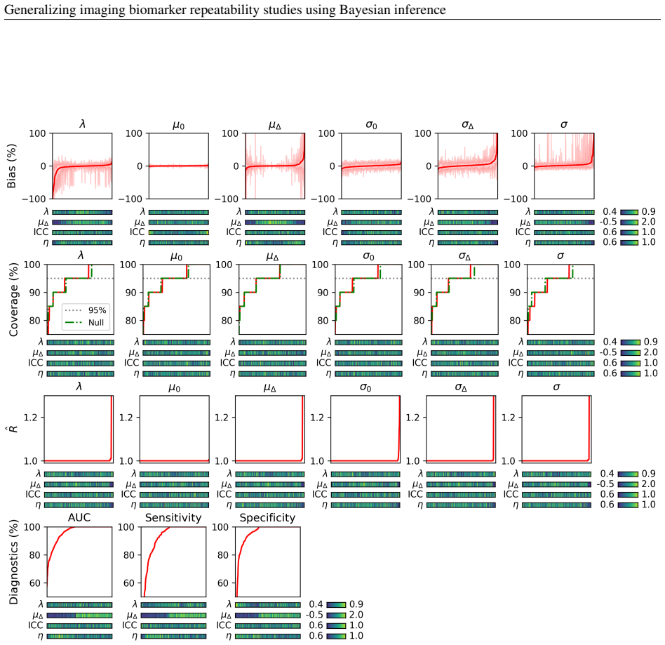
- [Sections 3.2 and 2.5; Figure 11] The transferability of the double-baseline measurement error from Study 3 to Studies 1 and 2 is a load-bearing premise for the per-lesion posterior odds and the reported ~70% response rate. The three studies differ in slice thickness (6 mm vs 5 mm), in-plane resolution, b-value sampling, and lesion volume (median 5.1 ml vs 12.5 and 22.0 ml), yet Section 2.5 only asserts that the double-baseline data 'should be representative' without providing evidence or a sensitivity analysis. Moreover, the paper's own prior-sensitivity analysis in Section 4.3 (Figure 11) shows that for habitat biomarkers in Study 1, the posterior of the measurement precision τ shifts when post-treatment data are included, which the authors attribute to 'some misalignment between the assumed model and the acquired post-treatment data.' The recommendation to rely on repeat-baseline-only estimates does not resolve the issue because those estimates come from a different cohort. I ask the authors to provide a direct assessment of how the posterior odds and λ estimates would change under plausible perturbations of τ (e.g., a sensitivity interval over τ), or to present an empirical demonstration that measurement repeatability is similar across the three acquisition protocols.
- [Section 7.4 (Supplementary Stan code)] The Stan code for the Dirichlet-Multinomial habitat model is syntactically malformed and, as printed, cannot be executed. The custom function `dir_mult` is declared with a 2D array argument (`array[,] int y`) and separate `vector mu` and `real prec` arguments, but in both the model block and the generated-quantities block it is called with `yp[n]`, which is a 1D array of length K, and with a single expression `mu0 * prec` (a vector) rather than separate `mu` and `prec` arguments. Additionally, the likelihood statement `yb ~ dirichlet_multinomial(mu0, prec);` does not reference the custom `dir_mult_lpmf`; if it relies on Stan's built-in `dirichlet_multinomial` distribution, the custom function is redundant and the inconsistency still prevents the code from running as written. Because the habitat results are a central contribution, the authors must correct the code and confirm that the reported results are reproducible from the provided model code.
- [Abstract and Section 4.2] The headline 'approximately 70% response rate among individual tumors across both studies' is stated without any uncertainty quantification. The posterior distribution of λ is shown in Figure 9, but the text reports no numerical estimate, credible interval, or posterior predictive check for this quantity. Given that the 70% figure is a posterior median based on the mixture model, the authors should report the posterior median and 95% credible interval for λ for each study and each biomarker type, and should moderate the claim if the posterior is wide or if the credible interval includes values far from 0.7.
minor comments (5)
- [Throughout] The manuscript contains numerous typographical and spelling errors, including 'Hamiltonean', 'disitributed', 'paramters', 'lections', 'seperated' (Figure 7 caption), 'respoonse', and 'depcted'. A careful proofread is needed before resubmission.
- [Section 4.1 / Figure 6] The caption of Figure 6 states 'median ADC in this instance' even though the figure describes the Dirichlet-Multinomial habitat model; this is misleading and should be corrected.
- [Section 4.2 / Figure 7] The color-coding description in the text refers to bins defined by posterior odds values, but the last bin is labeled 'BF10 > 10 (dark green)' while the rest are labeled with PO10; for consistency, all bins should be labeled as PO10.
- [Section 2.6] The assumption that the baseline habitat proportion is known and fixed at μ0 = (0.1, 0.8, 0.1) is ad hoc; please add a sentence acknowledging this as a modeling assumption and, if possible, report a sensitivity analysis or at least describe how deviations from this prior value would affect inference.
- [Table 5] In the simulation study, the measurement precision τ is fixed to 11.54 and the voxel-size parameters are fixed to values derived from the clinical studies; because the transferability of τ across cohorts is a central concern (see Major Comment 1), the simulation design should include a grid over τ or otherwise acknowledge this limitation.
Circularity Check
No significant circularity: theoretical derivations are self-contained, simulation studies are computational self-consistency checks, and the cited self-works are contextual rather than load-bearing.
full rationale
The paper's derivation chain is internally coherent. The equivalence between the Bayesian posterior predictive and Bland-Altman analysis in Section 2.3.1 is obtained from an explicit normal-normal model with stated priors, and the Bayes-factor expression in Section 2.4.1 is derived from the assumed generative distributions, not assumed from the result. The mixture-model posterior odds in Section 2.5 are estimated from the likelihoods of the fitted models; neither the per-lesion classifications nor the approximately 70% response rate is a parameter fitted to one subset and then reported as a prediction of a closely related quantity. The simulation studies generate data from the same models and assess parameter recovery; this is a computational self-consistency check, and the paper explicitly frames it as demonstrating that inference is achievable for well-defined models, not as external validation. Self-citations, such as Blackledge et al. (2017) for habitat mapping and Thrussell et al. (2022) for the IMS naming, provide context or terminology rather than carrying the central statistical argument, and no uniqueness theorem is imported to force the chosen model. The internal caveat in Section 4.3 about possible misalignment of the habitat measurement precision and the use of Study 3 double-baseline data for Studies 1 and 2 is a measurement-assumption or external-validity concern, not a circular step; it does not reduce any derived quantity to its own input. Accordingly, no circular step meeting the required evidentiary standard was found.
Assumptions & free parameters
free parameters (5)
- Prior width sigma_mu for median ADC model =
not stated in main text
- Prior width gamma_sigma for median ADC model =
not stated in main text
- Prior width gamma_tau for habitat model =
not stated in main text
- Baseline habitat proportions mu0 =
(0.1, 0.8, 0.1)
- Number of components in Dirichlet mixture density approximation =
5
assumptions (5)
- domain assumption Measurement error for real biomarkers is zero-mean normal and homoskedastic
- domain assumption Change in a real biomarker under M1 is independent of baseline value and normally distributed
- ad hoc to paper Habitat counts follow a Dirichlet-Multinomial distribution with a simplex baseline mean mu0 = (0.1, 0.8, 0.1)
- domain assumption The double-baseline cohort is representative of measurement error in the treatment cohorts
- domain assumption Lesions are statistically independent
Cite this review
Pith. "Pith review of Generalizing imaging biomarker repeatability studies using Bayesian inference: Applications in detecting heterogeneous treatment response in whole-body diffusion-weighted MRI of metastatic prostate cancer." pith.science (2026). https://pith.science/paper/2OYCR4A2
@misc{pith2026250509197,
  author       = {Pith},
  title        = {Pith review of: Generalizing imaging biomarker repeatability studies using Bayesian inference: Applications in detecting heterogeneous treatment response in whole-body diffusion-weighted MRI of metastatic prostate cancer},
  year         = {2026},
  howpublished = {\url{https://pith.science/paper/2OYCR4A2}},
  note         = {Machine review of arXiv:2505.09197}
}
read the original abstract
The assessment of imaging biomarkers is critical for advancing precision medicine and improving disease characterization. Despite the availability of methods to derive disease heterogeneity metrics in imaging studies, a robust framework for evaluating measurement uncertainty remains underdeveloped. To address this gap, we propose a novel Bayesian framework to assess the precision of disease heterogeneity measures in biomarker studies. Our approach extends traditional methods for evaluating biomarker precision by providing greater flexibility in statistical assumptions and enabling the analysis of biomarkers beyond univariate or multivariate normally-distributed variables. Using Hamiltonian Monte Carlo sampling, the framework supports both, for example, normally-distributed and Dirichlet-Multinomial distributed variables, enabling the derivation of posterior distributions for biomarker parameters under diverse model assumptions. Designed to be broadly applicable across various imaging modalities and biomarker types, the framework builds a foundation for generalizing reproducible and objective biomarker evaluation. To demonstrate utility, we apply the framework to whole-body diffusion-weighted MRI (WBDWI) to assess heterogeneous therapeutic responses in metastatic bone disease. Specifically, we analyze data from two patient studies investigating treatments for metastatic castrate-resistant prostate cancer (mCRPC). Our results reveal an approximately 70% response rate among individual tumors across both studies, objectively characterizing differential responses to systemic therapies and validating the clinical relevance of the proposed methodology. This Bayesian framework provides a powerful tool for advancing biomarker research across diverse imaging-based studies while offering valuable insights into specific clinical applications, such as mCRPC treatment response.
Figures


Figures from the paper (9 more)



Reference graph
Works this paper leans on
-
[1]
B(A+1) where the necessary conditionsA> −1 and Re(B)> 0 are guaranteed by the data, we arrive at p(d|db)∝ d2 + NbX j=1 d2 bj − Nb+1 2 = d2 +Nbd2 b − Nb+1 2 ∝ d2/d2 b Nb + 1 !− Nb+1 2 where d2 b = 1 Nb NbX i=1 d2 bj We notice from its functional form that this is a Student t distribution St(t;ν) = Γ ν+1 2 √νπΓ ν 2 t2 ν + 1 −ν+1 2 witht2 =d2/d2 b and...
work page 2010
-
[6]
doi:10.18632/oncotarget.17752. Armando Manduca, Philip J Bayly, Richard L Ehman, Arunark Kolipaka, Thomas J Royston, Ingolf Sack, Ralph Sinkus, and Bernard E Van Beers. Mr elastography: Principles, guidelines, and terminology. Magn Reson Med, 85 (5):2377–2390, May
-
[13]
Sarah R Amend and Kenneth J Pienta
doi:10.1038/nrc.2017.69. Sarah R Amend and Kenneth J Pienta. Ecology meets cancer biology: the cancer swamp promotes the lethal cancer phenotype. Oncotarget, 6(12):9669–78,
-
[14]
E Sala, E Mema, Y Himoto, H Veeraraghavan, J D Brenton, A Snyder, B Weigelt, and H A Vargas
doi:10.18632/oncotarget.3430. E Sala, E Mema, Y Himoto, H Veeraraghavan, J D Brenton, A Snyder, B Weigelt, and H A Vargas. Unravelling tumour heterogeneity using next-generation imaging: radiomics, radiogenomics, and habitat imaging. Clin Radiol, 72(1): 3–10, Jan
-
[15]
doi:10.1016/j.crad.2016.09.013. Mireia Crispin-Ortuzar, Marcel Gehrung, Stephan Ursprung, Andrew B Gill, Anne Y Warren, Lucian Beer, Ferdia A Gallagher, Thomas J Mitchell, Iosif A Mendichovszky, Andrew N Priest, Grant D Stewart, Evis Sala, and Florian Markowetz. Three-dimensional printed molds for image-guided surgical biopsies: An open source computation...
-
[17]
doi:10.1007/s00330-020-07560-8. Konstantinos Zormpas-Petridis, Evon Poon, Matthew Clarke, Neil P Jerome, Jessica K R Boult, Matthew D Blackledge, Fernando Carceller, Alexander Koers, Giuseppe Barone, Andrew D J Pearson, Lucas Moreno, John Anderson, Neil Sebire, Kieran McHugh, Dow-Mu Koh, Louis Chesler, Yinyin Yuan, Simon P Robinson, and Yann Jamin. Noninv...
-
[18]
doi:10.1158/0008-5472.CAN-20-0133. Camilla Panico, Giacomo Avesani, Konstantinos Zormpas-Petridis, Leonardo Rundo, Camilla Nero, and Evis Sala. Radiomics and radiogenomics of ovarian cancer: Implications for treatment monitoring and clinical management. Radiol Clin North Am , 61(4):749–760, Jul
-
[19]
doi:10.1016/j.rcl.2023.02.006. Nancy A Obuchowski. Interpreting change in quantitative imaging biomarkers. Acad Radiol, 25(3):372–379, Mar
Show all 43 references
-
[20]
doi:10.1016/j.acra.2017.09.023
ISSN 1878-4046 (Electronic); 1076-6332 (Print); 1076-6332 (Linking). doi:10.1016/j.acra.2017.09.023. Nancy A. Obuchowski and Andrew J. Buckler. Estimating the precision of quantitative imaging biomarkers without test-retest studies. Academic Radiology , 29(4):543–549,
2017 doi
-
[22]
doi:10.1148/radiol.2017161965. Jessica M Winfield, Aisha B Miah, Dirk Strauss, Khin Thway, David J Collins, Nandita M deSouza, Martin O Leach, Veronica A Morgan, Sharon L Giles, Eleanor Moskovic, Andrew Hayes, Myles Smith, Shane H Zaidi, Daniel Henderson, and Christina Messiou...
-
[25]
doi:10.1200/JCO.2015.64.2702
ISSN 15277755. doi:10.1200/JCO.2015.64.2702. Matthew D. Blackledge, David J. Collins, Nina Tunariu, Matthew R. Orton, Anwar R. Padhani, Martin O. Leach, and Dow Mu Koh. Assessment of treatment response by total tumor volume and global apparent diffusion coefficient using diffu...
2015 doi
-
[27]
doi:10.1016/j.eururo.2016.05.033
ISSN 18737560. doi:10.1016/j.eururo.2016.05.033. Anne M. Euser, Friedo W. Dekker, and Saskia le Cessie. A practical approach to bland-altman plots and variation coefficients for log transformed variables. Journal of Clinical Epidemiology, 61(10):978–982,
2016 doi
-
[29]
23 Generalizing imaging biomarker repeatability studies using Bayesian inference Andrew Gelman and Donald B Rubin
doi:10.1016/j.acra.2022.08.031. 23 Generalizing imaging biomarker repeatability studies using Bayesian inference Andrew Gelman and Donald B Rubin. Inference from iterative simulation using multiple sequences. Statistical science, 7(4):457–472,
2022 doi
-
[30]
Michael D Lee and Eric-Jan Wagenmakers
doi:10.1016/j.jmp.2017.09.005. Michael D Lee and Eric-Jan Wagenmakers. Bayesian cognitive modeling: A practical course . Cambridge university press,
2017 doi
-
[31]
doi:10.3389/fonc.2022.899180. Dow-Mu Koh, Matthew Blackledge, David J Collins, Anwar R Padhani, Toni Wallace, Benjamin Wilton, N Jane Taylor, J James Stirling, Rajesh Sinha, Pat Walicke, Martin O Leach, Ian Judson, and Paul Nathan. Reproducibility and changes in the apparent d...
2022
-
[32]
Matthew David Blackledge, DM Koh, David J Collins, Erica Scurr, Julie Hughes, M Leach, et al
doi:10.1007/s00330-009- 1469-4. Matthew David Blackledge, DM Koh, David J Collins, Erica Scurr, Julie Hughes, M Leach, et al. Assessing response heterogeneity following radium 223 administration using whole body diffusion weighted mri. In International Society for Magnetic Res...
-
[34]
Computer-aided detection of focal bone metastases from whole-body multi-modal mri
Jakub Ceranka, Frédéric Lecouvet, Johan De Mey, and Jef Vandemeulebroucke. Computer-aided detection of focal bone metastases from whole-body multi-modal mri. In Medical Imaging 2020: Computer-Aided Diagnosis , volume 11314, pages 174–180. SPIE,
2020
-
[35]
doi:10.1007/s00330-022-08536-6. Lianghui Zhu, Huijuan Shi, Huiting Wei, Chengjiang Wang, Shanshan Shi, Fenfen Zhang, Renao Yan, Yiqing Liu, Tingting He, Liyuan Wang, Junru Cheng, Hufei Duan, Hong Du, Fengjiao Meng, Wenli Zhao, Xia Gu, Linlang Guo, Yingpeng Ni, Yonghong He, Tia...
-
[36]
doi:10.1016/j.ebiom.2022.104426. Lauren Brady, Michelle Kriner, Ilsa Coleman, Colm Morrissey, Martine Roudier, Lawrence D True, Roman Gulati, Stephen R Plymate, Zoey Zhou, Brian Birditt, Rhonda Meredith, Gary Geiss, Margaret Hoang, Joseph Beechem, and Peter S Nelson. Inter- an...
2022
-
[37]
doi:10.1038/s41467-021-21615-4. 24 Generalizing imaging biomarker repeatability studies using Bayesian inference Raquel Perez-Lopez, Joaquin Mateo, Helen Mossop, Matthew D Blackledge, David J Collins, Mihaela Rata, Veronica A Morgan, Alison Macdonald, Shahneen Sandhu, David Lo...
-
[38]
doi:10.1148/radiol.2016160646. Raquel Perez-Lopez, David Lorente, Matthew D Blackledge, David J Collins, Joaquin Mateo, Diletta Bianchini, Aurelius Omlin, Andrea Zivi, Martin O Leach, Johann S de Bono, Dow-Mu Koh, and Nina Tunariu. V olume of bone metastasis assessed with whol...
-
[39]
Fei Tony Liu, Kai Ming Ting, and Zhi-Hua Zhou
doi:10.1148/radiol.2015150799. Fei Tony Liu, Kai Ming Ting, and Zhi-Hua Zhou. Isolation forest. In 2008 eighth ieee international conference on data mining, pages 413–422. IEEE,
2008 doi
-
[41]
Hania Paverd, Konstantinos Zormpas-Petridis, Hannah Clayton, Sarah Burge, and Mireia Crispin-Ortuzar
doi:10.1038/s41598-022-18986-z. Hania Paverd, Konstantinos Zormpas-Petridis, Hannah Clayton, Sarah Burge, and Mireia Crispin-Ortuzar. Radiology and multi-scale data integration for precision oncology. NPJ Precision Oncology, 8(1):158,
-
[1986]
doi:10.1016/S0140-6736(86)90837-8
ISSN 0140-6736. doi:10.1016/S0140-6736(86)90837-8. Howard I. Scher, Michael J. Morris, Walter M. Stadler, Celestia Higano, Ethan Basch, Karim Fizazi, Emmanuel S. Antonarakis, Tomasz M. Beer, Michael A. Carducci, Kim N. Chi, Paul G. Corn, Johann S. De Bono, Robert Dreicer, Dani...
-
[2001]
Stanislav Pidhorskyi, Ranya Almohsen, and Gianfranco Doretto
doi:10.1162/089976601750264965. Stanislav Pidhorskyi, Ranya Almohsen, and Gianfranco Doretto. Generative probabilistic novelty detection with adversarial autoencoders. Advances in neural information processing systems , 31,
-
[2004]
Matthew D Blackledge, David J Collins, Dow-Mu Koh, and Martin O Leach
doi:10.1007/s10278-004-1014-6. Matthew D Blackledge, David J Collins, Dow-Mu Koh, and Martin O Leach. Rapid development of image analysis research tools: bridging the gap between researcher and clinician with pyosirix. Computers in biology and medicine , 69:203–212,
-
[2006]
Dow-Mu Koh and David J Collins
doi:10.1102/1470-7330.2006.0021. Dow-Mu Koh and David J Collins. Diffusion-weighted mri in the body: applications and challenges in oncology. AJR Am J Roentgenol, 188(6):1622–35, Jun
2006
-
[2007]
Alexey Surov, Hans Jonas Meyer, and Andreas Wienke
doi:10.2214/AJR.06.1403. Alexey Surov, Hans Jonas Meyer, and Andreas Wienke. Correlation between apparent diffusion coefficient (adc) and cellularity is different in several tumors: a meta-analysis. Oncotarget, 8(35):59492–59499, Aug
-
[2009]
doi:10.1016/J.EJCA.2008.10.026
ISSN 1879-0852. doi:10.1016/J.EJCA.2008.10.026. URL https://pubmed.ncbi.nlm.nih. gov/19097774/. Elizabeth M Charles-Edwards and Nandita M deSouza. Diffusion-weighted magnetic resonance imaging and its application to cancer. Cancer Imaging, 6(1):135–43, Sep
2008
-
[2010]
25 Generalizing imaging biomarker repeatability studies using Bayesian inference Supplementary Material 7.1 Mathematical derivations Theorem 7.1
doi:10.1016/j.cogpsych.2009.12.001. 25 Generalizing imaging biomarker repeatability studies using Bayesian inference Supplementary Material 7.1 Mathematical derivations Theorem 7.1. If y∼N (x0,σ ) y0∼N (x0,σ ) x0∼N (µ0,σ
2009 doi
-
[2012]
Parvinder Sujlana, Jan Skrok, and Laura M Fayad
doi:10.1007/s00330-012-2446-x. Parvinder Sujlana, Jan Skrok, and Laura M Fayad. Review of dynamic contrast-enhanced mri: Technical aspects and applications in the musculoskeletal system. J Magn Reson Imaging , 47(4):875–890, Apr
-
[2014]
doi:10.1371/journal.pone.0091779
ISSN 19326203. doi:10.1371/journal.pone.0091779. Anwar R. Padhani, Frederic E. Lecouvet, Nina Tunariu, Dow Mu Koh, Frederik De Keyzer, David J. Collins, Evis Sala, Heinz Peter Schlemmer, Giuseppe Petralia, H. Alberto Vargas, Stefano Fanti, H. Bertrand Tombal, and Johann de Bon...
-
[2015]
doi:10.1016/j.ccell.2014.12.001. Carlo C Maley, Athena Aktipis, Trevor A Graham, Andrea Sottoriva, Amy M Boddy, Michalina Janiszewska, Ariosto S Silva, Marco Gerlinger, Yinyin Yuan, Kenneth J Pienta, Karen S Anderson, Robert Gatenby, Charles Swanton, David Posada, Chung-I Wu, ...
2014 doi
-
[2016]
doi:10.1148/radiol.2015151169. James P B O’Connor, Eric O Aboagye, Judith E Adams, Hugo J W L Aerts, Sally F Barrington, Ambros J Beer, Ronald Boellaard, Sarah E Bohndiek, Michael Brady, Gina Brown, David L Buckley, Thomas L Chenevert, Laurence P Clarke, Sandra Collette, Gary ...
-
[2017]
doi:10.1038/nrclinonc.2016.162. E. A. Eisenhauer, P. Therasse, J. Bogaerts, L. H. Schwartz, D. Sargent, R. Ford, J. Dancey, S. Arbuck, S. Gwyther, M. Mooney, L. Rubinstein, L. Shankar, L. Dodd, R. Kaplan, D. Lacombe, and J. Verweij. New response evaluation criteria in solid tu...
2016 doi
-
[2018]
doi:10.1002/jmri.25810
ISSN 1522-2586 (Electronic); 1053-1807 (Linking). doi:10.1002/jmri.25810. Emily Hoffmann, Max Masthoff, Wolfgang G Kunz, Max Seidensticker, Stefanie Bobe, Mirjam Gerwing, Wolfgang E Berdel, Christoph Schliemann, Cornelius Faber, and Moritz Wildgruber. Multiparametric mri for c...
-
[2019]
doi:10.3389/fonc.2019.00280. J. Martin Bland and Douglas G. Altman. Statistical methods for assessing agreement between two methods of clinical measurement. The Lancet, 327:307–310, 2
2019
-
[2020]
doi:10.1200/CCI.20.00026. Lucian Beer, Paula Martin-Gonzalez, Maria Delgado-Ortet, Marika Reinius, Leonardo Rundo, Ramona Woitek, Stephan Ursprung, Lorena Escudero, Hilal Sahin, Ionut-Gabriel Funingana, Joo-Ern Ang, Mercedes Jimenez-Linan, Tristan Lawton, Gaurav Phadke, Sally ...
-
[2021]
Ingolf Sack
doi:10.1002/mrm.28627. Ingolf Sack. Magnetic resonance elastography from fundamental soft-tissue mechanics to diagnostic imaging. Na- ture Reviews Physics, 5(1):25–42,
-
[2022]
doi:https://doi.org/10.1016/j.acra.2021.06.009
ISSN 1076-6332. doi:https://doi.org/10.1016/j.acra.2021.06.009. URL https://www.sciencedirect.com/science/article/ pii/S1076633221002798. Jessica M Winfield, Nina Tunariu, Mihaela Rata, Keiko Miyazaki, Neil P Jerome, Michael Germuska, Matthew D Blackledge, David J Collins, Joh...
2021 doi
-
[2023]
URL https://doi.org/10.1038/ s42254-022-00543-2
doi:10.1038/s42254-022-00543-2. URL https://doi.org/10.1038/ s42254-022-00543-2 . M O Leach, B Morgan, P S Tofts, D L Buckley, W Huang, M A Horsfield, T L Chenevert, D J Collins, A Jackson, D Lomas, B Whitcher, L Clarke, R Plummer, I Judson, R Jones, R Alonzi, T Brunner, D M K...
-
[2024]
Nicholas McGranahan and Charles Swanton
doi:10.1038/s41571-024-00891-1. Nicholas McGranahan and Charles Swanton. Biological and therapeutic impact of intratumor heterogeneity in cancer evolution. Cancer Cell, 27(1):15–26, Jan
-
[4356]
URL https://www.sciencedirect.com/science/ article/pii/S0895435607004131
doi:https://doi.org/10.1016/j.jclinepi.2007.11.003. URL https://www.sciencedirect.com/science/ article/pii/S0895435607004131. Nancy A Obuchowski, Erich Huang, Nandita M deSouza, David Raunig, Jana Delfino, Andrew Buckler, Charles Hatt, Xiaofeng Wang, Chaya Moskowitz, Alexander...
2007 doi
Reviewed August 15, 2026 · model on record in the stance chip above.
Discussion (0). Continue with ORCID to comment.