REVIEW 5 major objections 6 minor 41 references
SAMba-UNet: SAM2-Mamba UNet for Cardiac MRI in Medical Robotic Perception
T0 review · 5 major / 6 minor · reviewed 2026-08-07 · deepseek-v4-flash
Pith's one-line read Fusing SAM2 with Mamba in a UNet skeleton yields the best reported ACDC cardiac MRI segmentation, with a 0.9103 mDice.
desk verdict New architecture, uncontrolled comparison: the 0.71pp ACDC gap over LeViT-UNet-384 is not established as a real gain. read the letter →
The pith
A machine-rendered reading of the paper's core claim, the machinery that carries it, and where it could break.
The reading
What carries the argument
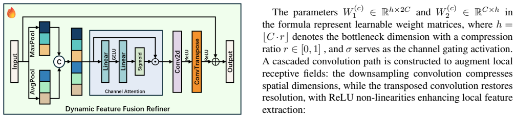
The load-bearing mechanism is the Heterogeneous Omni-Attention Convergence Module (HOACM), a fusion block that combines Omniscient Contextual Attention (OCA), which re-establishes global pixel-level positional semantics for SAM2's hierarchical features, with Bifurcated Selective Emphasis Attention (BSEA), which dynamically weights Mamba's global and locally salient features before cross-attention merges the two streams. The Dynamic Feature Fusion Refiner, a multi-scale pooling and channel–spatial calibration adapter, carries the domain-transfer work of pulling SAM2's natural-image representations toward MRI.
What would settle it
Re-run all ten baselines plus SAMba-UNet on a single, pre-registered ACDC protocol with identical preprocessing and seeds, and check whether the 0.71-point mDice lead over LeViT-UNet-384 survives; if it does not, the headline gain is protocol artifact.
Extended reading notes
Core claim
The paper's central claim is that SAM2's window-based positional detail and Mamba's long-range global context are complementary, and that a dedicated fusion module—not just a concatenation—lets a UNet exploit both at once. To that end it introduces a Dynamic Feature Fusion Refiner that adapts SAM2's natural-image features to MRI via multi-scale pooling and channel–spatial calibration, and a Heterogeneous Omni-Attention Convergence Module (HOACM) whose Omniscient Contextual Attention and Bifurcated Selective Emphasis Attention jointly reweight the two encoder streams. The reported results on ACDC—mDice 0.9103, mHD95 1.0859 mm—and the ablation tables are offered as evidence that every component contributes, with the right ventricle and myocardium showing the clearest gains.
Load-bearing premise
The reported 0.71-point margin assumes every baseline result in Table I comes from a comparable protocol—same ACDC split, preprocessing, and metric implementation—so the gap reflects architecture, not measurement differences.
Editorial extensions
If this is right
- If the reported margin is real, SAMba-UNet is the strongest known single model on the ACDC benchmark, with a 0.71-percentage-point mDice lead over the next-best entry.
- The gains concentrate on the right ventricle and myocardium—the structures with the most complex boundaries—so the approach is aimed at exactly the cases where manual annotation is hardest.
- Because the Mamba branch processes sequences in linear time, the added global-context capacity does not force quadratic attention cost, keeping the fused model practical for clinical image volumes.
- The frozen SAM2 encoder with a lightweight adapter means the method inherits foundation-model priors without full fine-tuning, a recipe that could transfer to other MRI targets with modest data.
- The authors position the model as a perception module for surgical robotic systems, where its boundary precision would support navigation and complication screening.
Reading between the lines
- The paper argues the segmentation maps are 'directly applicable' to robotic perception, but it reports no robot-in-the-loop or latency experiment; treating that as a transfer claim rather than a demonstrated result would be my reading.
- The headline comparison mixes re-trained baselines with numbers copied from original papers; if protocols differ, the 0.71-point gap could shrink or vanish, so an independent re-benchmark under one protocol would be the natural stress test.
- A testable next step is to freeze the trained encoder and probe whether the fusion module transfers to other cardiac MRI views or other modalities (e.g., CT), since the domain-shift argument is generic.
- The 'first to synergize SAM2, Mamba, and UNet' claim is about architectural novelty; whether that combination is necessary—versus a well-tuned single encoder—is only answered by the ablations, which show each module adds a small increment.
Editorial analysis
A structured set of objections, weighed in public.
Referee Report
Summary. The paper proposes SAMba-UNet, a U-shaped dual-encoder architecture that combines a frozen SAM2 Hiera encoder, a VMamba encoder, and a VMamba decoder, with two new modules: a Dynamic Feature Fusion Refiner (DFFR) and a Heterogeneous Omni-Attention Convergence Module (HOACM). The method is evaluated on the ACDC cardiac MRI segmentation benchmark, reporting an mDice of 0.9103 and HD95 of 1.0859 mm, and the authors claim it surpasses all compared baselines. The paper also states that the model is directly applicable as a perception module for intelligent medical and surgical robotic systems. The experiments include a comparison table, two ablation tables, and qualitative visualizations.
Significance. If the performance claim were established under a controlled protocol, the architecture would be a useful contribution: integrating a frozen SAM2 encoder with a linear-complexity Mamba branch for medical segmentation is a plausible and timely direction, and the use of a public benchmark (ACDC) is a strength. The paper also promises open-sourced code, which would support reproducibility. However, the central empirical claim is currently not established because the baseline comparison in Table I is not controlled, the boundary metrics that are highlighted in the abstract are not compared against any baseline, and several modules referenced in the ablations are undefined. The claimed robotic-perception applicability is not tested by the experiments. The contribution is therefore more of a proposal with promising but unverified results than a validated method at this stage.
major comments (5)
- [IV-D, Table I] The main performance claim rests on an uncontrolled baseline comparison. The text says that models with official implementations were 'retraining and testing on an A800 server (or using official weights directly)', while others use 'the officially reported results from their respective papers', but it never lists which baseline falls into which category, what ACDC train/validation split was used, what 2D preprocessing was applied (cropping, resampling, slice selection), or what metric implementation was used. ACDC has no standardized 2D split, and published Dice values on it vary by several points with preprocessing choices. No standard deviations, confidence intervals, or multiple-seed results are reported for any method. Consequently, the 0.71 percentage-point mDice advantage over LeViT-UNet-384 (Section IV-D) is within the range of protocol and seed variation and cannot be interpreted as an architectural gain. The authors need to retrain all baselines under the same split, preprocessing, and metric code, report per-class Dice for all, and provide statistical significance or at least multiple-seed variance.
- [Table I and Abstract] The abstract and conclusion emphasize boundary localization and report HD95 as a headline result, but Table I lists only mDice, RV, MYO, and LV for the baselines. HD95 and ASD, which are defined in Section IV-C, are reported for the proposed method only through the ablation tables. This makes it impossible to support the claim of 'notably improving boundary localization' over prior work. Baseline HD95/ASD values must be included for a fair boundary-performance comparison.
- [Table II, Table III, Section III-C] The ablations contain an undefined module: Table II includes a row 'w/o GCAA', but no module named GCAA is defined in Section III; the defined modules are OCA and BSEA, and a 'gated spatial attention (GSA)' appears inside OCA. As written, the reader cannot determine what is being ablated. In addition, the ablation differences are small (e.g., ALL 0.9103 vs. w/o BSEA 0.9064) and no variance or significance measures are reported, so it is unclear whether these differences are meaningful. The authors should define every ablated component, align the naming with the method section, and report multiple-seed results.
- [IV-B] The implementation details are not reproducible as stated. The paper reports Python 3.12.0 with PyTorch 1.10 and CUDA 12.1, but PyTorch 1.10 does not support Python 3.12, and the combination as written is not a valid released configuration. Additionally, the paper says 'the preprocessed ACDC dataset' was used but does not specify the preprocessing steps, image resolution, slice selection, or data split. This blocks reproduction. The authors must provide the exact software versions, preprocessing pipeline, split definitions, and either the released code or a precise configuration file.
- [Abstract, Fig. 1, Conclusion] The paper repeatedly claims that SAMba-UNet is 'directly applicable as a perception module within intelligent medical and surgical robotic systems' and includes a robotic workflow figure, but no experiment evaluates robotic perception, inference latency, real-time constraints, or integration with a robot controller. The ACDC segmentation benchmark alone does not test this claim. Either the robotic claim should be removed from the title, abstract, and conclusion, or supporting experiments (e.g., latency, deployment on a robotic platform) should be added.
minor comments (6)
- [Eq. (12)] The symbol M is reused for both the attention weight matrix in Eq. (10) and the spatial attention output in Eq. (12), which is confusing; please use distinct notation for these quantities.
- [Eq. (4)] In Eq. (4), X_cat has shape B×2C while X_sp is not given an explicit channel dimension in Eq. (3); the addition X_cat + X_sp appears dimensionally ambiguous and should be clarified.
- [III-A] The text 'we integrate design principles from FE-UNet and Medical SAM Adapter' references FE-UNet without a citation; a reference or a brief description is needed.
- [References] Reference [37] is cited to support the claim about SAM2's window-based absolute position encoding, but the cited paper is about mitigating position bias in large language models and does not appear relevant; please cite the appropriate source.
- [Figure 4 caption] The caption contains a typo: 'eterogeneous' should be 'Heterogeneous'.
- [Abstract and Introduction] The claim of being 'the first pioneering framework that successfully synergizes SAM2, Mamba, and UNet' should be tempered or supported by a more thorough recent-literature comparison, since several SAM+Mamba works already exist (e.g., SAM-Mamba [33]) and the novelty claim as stated is difficult to verify.
Circularity Check
No circularity: the paper's central claims are empirical benchmark results, not derivations that reduce to their own inputs.
full rationale
This is an empirical architecture paper, not a claimed derivation. The central quantitative claim (mDice 0.9103 and HD95 1.0859 mm on ACDC) is obtained by training the proposed SAMba-UNet and comparing it to independently published or retrained baselines; no parameter is fitted to the target metric, and the reported advantage over LeViT-UNet-384 is an external benchmark result rather than a construct. The ablations vary real architectural components and are evaluated on a held-out validation set, so the final score is not equivalent to the choice of modules by construction. The only self-citation is reference [23] (MaskSAM, by co-author Hao Tang et al.), used to support the general statement that SAM underperforms on medical images; this claim is corroborated by independent references [20]–[22] and is not load-bearing for any derived result. No equation defines an input in terms of the target output, no fitted parameter is renamed as a prediction, and no uniqueness theorem is imported from the authors' own prior work. Concerns about baseline comparability, missing error bars, or mismatch between described and ablated components are experimental-validity and reproducibility issues, not circularity. Therefore the derivation chain is self-contained with respect to its inputs.
Assumptions & free parameters
free parameters (3)
- Compression ratio r in DFFR channel bottleneck =
not reported
- Attention kernel size 7x7 in OCA/GSA =
7
- Training hyperparameters: initial LR 0.01, batch size 12, iterations 10k, weight decay 1e-4, momentum 0.9 =
as listed in Section IV-B
assumptions (4)
- domain assumption ACDC preprocessing and patient-wise split are assumed to match prior benchmark practice.
- domain assumption Baseline numbers taken from original papers are computed under the same evaluation protocol as the retrained ones.
- domain assumption Frozen SAM2 Hiera-Large features can be adapted to cardiac MRI with lightweight adapters.
- standard math Standard linear algebra, pooling, and attention operations compute as written.
Cite this review
Pith. "Pith review of SAMba-UNet: SAM2-Mamba UNet for Cardiac MRI in Medical Robotic Perception." pith.science (2026). https://pith.science/paper/VRP7QBWQ
@misc{pith2026250516304,
  author       = {Pith},
  title        = {Pith review of: SAMba-UNet: SAM2-Mamba UNet for Cardiac MRI in Medical Robotic Perception},
  year         = {2026},
  howpublished = {\url{https://pith.science/paper/VRP7QBWQ}},
  note         = {Machine review of arXiv:2505.16304}
}
read the original abstract
To address complex pathological feature extraction in automated cardiac MRI segmentation, we propose SAMba-UNet, a novel dual-encoder architecture that synergistically combines the vision foundation model SAM2, the linear-complexity state-space model Mamba, and the classical UNet to achieve cross-modal collaborative feature learning; to overcome domain shifts between natural images and medical scans, we introduce a Dynamic Feature Fusion Refiner that employs multi-scale pooling and channel-spatial dual-path calibration to strengthen small-lesion and fine-structure representation, and we design a Heterogeneous Omni-Attention Convergence Module (HOACM) that fuses SAM2's local positional semantics with Mamba's long-range dependency modeling via global contextual attention and branch-selective emphasis, yielding substantial gains in both global consistency and boundary precision-on the ACDC cardiac MRI benchmark, SAMba-UNet attains a Dice of 0.9103 and HD95 of 1.0859 mm, notably improving boundary localization for challenging structures like the right ventricle, and its robust, high-fidelity segmentation maps are directly applicable as a perception module within intelligent medical and surgical robotic systems to support preoperative planning, intraoperative navigation, and postoperative complication screening; the code will be open-sourced to facilitate clinical translation and further validation.
Figures


Figures from the paper (2 more)


Reference graph
Works this paper leans on
-
[1]
S. Sanyaolu, “Integration of machine learning in imaging analysis for clinical diagnosis of cardiovascular diseases,”Cardiology, vol. 2, p. 100006, 2025
work page 2025
-
[2]
U-net: Convolutional networks for biomedical image segmentation,
O. Ronneberger, P. Fischer, and T. Brox, “U-net: Convolutional networks for biomedical image segmentation,” inMedical image computing and computer-assisted intervention–MICCAI 2015: 18th international conference, Munich, Germany, October 5-9, 2015, pro- ceedings, part III 18. Springer, 2015, pp. 234–241
2015
-
[3]
Unet++: Redesigning skip connections to exploit multiscale features in image segmentation,
Z. Zhou, M. M. R. Siddiquee, N. Tajbakhsh, and J. Liang, “Unet++: Redesigning skip connections to exploit multiscale features in image segmentation,”IEEE TMI, vol. 39, no. 6, pp. 1856–1867, 2019
work page 2019
-
[4]
Densely connected convolutional networks,
G. Huang, Z. Liu, L. Van Der Maaten, and K. Q. Weinberger, “Densely connected convolutional networks,” inCVPR, 2017, pp. 4700–4708
2017
-
[5]
Deep residual learning for image recognition,
K. He, X. Zhang, S. Ren, and J. Sun, “Deep residual learning for image recognition,” inCVPR, 2016, pp. 770–778
2016
-
[6]
Cbam: Convolutional block attention module,
S. Woo, J. Park, J.-Y . Lee, and I. S. Kweon, “Cbam: Convolutional block attention module,” inProceedings of the European conference on computer vision (ECCV), 2018, pp. 3–19
2018
-
[7]
Mobilenets: Efficient convolutional neural networks for mobile vision applications,
A. G. Howard, M. Zhu, B. Chen, D. Kalenichenko, W. Wang, T. Weyand, M. Andreetto, and H. Adam, “Mobilenets: Efficient convolutional neural networks for mobile vision applications,”arXiv preprint arXiv:1704.04861, 2017
arXiv 2017
-
[8]
Multi-scale context aggregation by dilated convolutions,
F. Yu and V . Koltun, “Multi-scale context aggregation by dilated convolutions,”arXiv preprint arXiv:1511.07122, 2015
arXiv 2015
Show all 41 references
-
[9]
Acnn: a full resolution dcnn for medical image segmentation,
X.-Y . Zhou, J.-Q. Zheng, P. Li, and G.-Z. Yang, “Acnn: a full resolution dcnn for medical image segmentation,” in2020 IEEE International Conference on Robotics and Automation (ICRA). IEEE, 2020, pp. 8455–8461
2020
-
[10]
Transunet: Transformers make strong encoders for medical image segmentation,
J. Chen, Y . Lu, Q. Yu, X. Luo, E. Adeli, Y . Wang, L. Lu, A. L. Yuille, and Y . Zhou, “Transunet: Transformers make strong encoders for medical image segmentation,”arXiv preprint arXiv:2102.04306, 2021
2021 arXiv
-
[11]
Transfuse: Fusing transformers and cnns for medical image segmentation,
Y . Zhang, H. Liu, and Q. Hu, “Transfuse: Fusing transformers and cnns for medical image segmentation,” inMedical image computing and computer assisted intervention–MICCAI 2021: 24th international conference, Strasbourg, France, September 27–October 1, 2021, pro- ceedings, Par...
2021
-
[12]
Medical transformer: Gated axial-attention for medical image segmentation,
J. M. J. Valanarasu, P. Oza, I. Hacihaliloglu, and V . M. Patel, “Medical transformer: Gated axial-attention for medical image segmentation,” inMedical image computing and computer assisted intervention– MICCAI 2021: 24th international conference, Strasbourg, France, September...
2021
-
[13]
Swin unetr: Swin transformers for semantic segmentation of brain tumors in mri images,
A. Hatamizadeh, V . Nath, Y . Tang, D. Yang, H. R. Roth, and D. Xu, “Swin unetr: Swin transformers for semantic segmentation of brain tumors in mri images,” inInternational MICCAI brainlesion workshop. Springer, 2021, pp. 272–284
2021
-
[14]
An image is worth 16x16 words: Transformers for image recognition at scale,
A. Dosovitskiy, L. Beyer, A. Kolesnikov, D. Weissenborn, X. Zhai, T. Unterthiner, M. Dehghani, M. Minderer, G. Heigold, S. Gellyet al., “An image is worth 16x16 words: Transformers for image recognition at scale,”arXiv preprint arXiv:2010.11929, 2020
2010 arXiv
-
[15]
Segment anything,
A. Kirillov, E. Mintun, N. Ravi, H. Mao, C. Rolland, L. Gustafson, T. Xiao, S. Whitehead, A. C. Berg, W.-Y . Loet al., “Segment anything,” inProceedings of the IEEE/CVF international conference on computer vision, 2023, pp. 4015–4026
2023
-
[16]
Foundation models for generalist medical artificial intelligence,
M. Moor, O. Banerjee, Z. S. H. Abad, H. M. Krumholz, J. Leskovec, E. J. Topol, and P. Rajpurkar, “Foundation models for generalist medical artificial intelligence,”Nature, vol. 616, no. 7956, pp. 259– 265, 2023
2023
-
[17]
Foundation model for advancing healthcare: challenges, opportunities and future directions,
Y . He, F. Huang, X. Jiang, Y . Nie, M. Wang, J. Wang, and H. Chen, “Foundation model for advancing healthcare: challenges, opportunities and future directions,”IEEE Reviews in Biomedical Engineering, 2024
2024
-
[18]
A comprehensive survey of foundation models in medicine,
W. Khan, S. Leem, K. B. See, J. K. Wong, S. Zhang, and R. Fang, “A comprehensive survey of foundation models in medicine,”IEEE Reviews in Biomedical Engineering, 2025
2025
-
[19]
Sam 2: Segment anything in images and videos,
N. Ravi, V . Gabeur, Y .-T. Hu, R. Hu, C. Ryali, T. Ma, H. Khedr, R. R ¨adle, C. Rolland, L. Gustafsonet al., “Sam 2: Segment anything in images and videos,”arXiv preprint arXiv:2408.00714, 2024
2024 arXiv
-
[20]
Segment anything model for medical images?
Y . Huang, X. Yang, L. Liu, H. Zhou, A. Chang, X. Zhou, R. Chen, J. Yu, J. Chen, C. Chenet al., “Segment anything model for medical images?”Medical Image Analysis, vol. 92, p. 103061, 2024
2024
-
[21]
Accuracy of segment- anything model (sam) in medical image segmentation tasks,
S. He, R. Bao, J. Li, P. E. Grant, and Y . Ou, “Accuracy of segment- anything model (sam) in medical image segmentation tasks,”CoRR, 2023
2023
-
[22]
Sam. md: Zero-shot medical image segmentation capabilities of the segment anything model,
S. Roy, T. Wald, G. Koehler, M. R. Rokuss, N. Disch, J. Holzschuh, D. Zimmerer, and K. H. Maier-Hein, “Sam. md: Zero-shot medical image segmentation capabilities of the segment anything model,”arXiv preprint arXiv:2304.05396, 2023
2023 arXiv
-
[23]
Masksam: Towards auto-prompt sam with mask classification for volumetric medical image segmentation,
B. Xie, H. Tang, B. Duan, D. Cai, Y . Yan, and G. Agam, “Masksam: Towards auto-prompt sam with mask classification for volumetric medical image segmentation,”arXiv preprint arXiv:2403.14103, 2024
2024 arXiv
-
[24]
Vmamba: Visual state space model,
Y . Liu, Y . Tian, Y . Zhao, H. Yu, L. Xie, Y . Wang, Q. Ye, J. Jiao, and Y . Liu, “Vmamba: Visual state space model,”Advances in neural information processing systems, vol. 37, pp. 103 031–103 063, 2024
2024
-
[25]
Missformer: An effective transformer for 2d medical image segmentation,
X. Huang, Z. Deng, D. Li, X. Yuan, and Y . Fu, “Missformer: An effective transformer for 2d medical image segmentation,”IEEE TMI, vol. 42, no. 5, pp. 1484–1494, 2022
2022
-
[26]
Segmamba: Long- range sequential modeling mamba for 3d medical image segmenta- tion,
Z. Xing, T. Ye, Y . Yang, G. Liu, and L. Zhu, “Segmamba: Long- range sequential modeling mamba for 3d medical image segmenta- tion,” inInternational Conference on Medical Image Computing and Computer-Assisted Intervention. Springer, 2024, pp. 578–588
2024
-
[27]
Mambamil: Enhancing long se- quence modeling with sequence reordering in computational pathol- ogy,
S. Yang, Y . Wang, and H. Chen, “Mambamil: Enhancing long se- quence modeling with sequence reordering in computational pathol- ogy,” inInternational Conference on Medical Image Computing and Computer-Assisted Intervention. Springer, 2024, pp. 296–306
2024
-
[28]
Embedded prompt tuning: Towards enhanced calibration of pretrained models for medical images,
W. Zu, S. Xie, Q. Zhao, G. Li, and L. Ma, “Embedded prompt tuning: Towards enhanced calibration of pretrained models for medical images,”Medical Image Analysis, vol. 97, p. 103258, 2024
2024
-
[29]
Medical sam adapter: Adapting segment anything model for medical image segmentation,
J. Wu, Z. Wang, M. Hong, W. Ji, H. Fu, Y . Xu, M. Xu, and Y . Jin, “Medical sam adapter: Adapting segment anything model for medical image segmentation,”Medical image analysis, vol. 102, p. 103547, 2025
2025
-
[30]
Mamba: Linear-time sequence modeling with selective state spaces,
A. Gu and T. Dao, “Mamba: Linear-time sequence modeling with selective state spaces,”arXiv preprint arXiv:2312.00752, 2023
2023 arXiv
-
[31]
Mamba-unet: Unet-like pure visual mamba for medical image segmentation,
Z. Wang, J.-Q. Zheng, Y . Zhang, G. Cui, and L. Li, “Mamba-unet: Unet-like pure visual mamba for medical image segmentation,”arXiv preprint arXiv:2402.05079, 2024
2024 arXiv
-
[32]
U-mamba: Enhancing long-range dependency for biomedical image segmentation,
J. Ma, F. Li, and B. Wang, “U-mamba: Enhancing long-range dependency for biomedical image segmentation,”arXiv preprint arXiv:2401.04722, 2024
2024 arXiv
-
[33]
Sam-mamba: Mamba guided sam architecture for generalized zero-shot polyp segmentation,
T. K. Dutta, S. Majhi, D. R. Nayak, and D. Jha, “Sam-mamba: Mamba guided sam architecture for generalized zero-shot polyp segmentation,” in2025 IEEE/CVF Winter Conference on Applications of Computer Vision (WACV). IEEE, 2025, pp. 4655–4664
2025
-
[34]
Lfsamba: Marry sam with mamba for light field salient object detection,
Z. Liu, L. Wang, X. Fang, Z. Tu, and L. Wang, “Lfsamba: Marry sam with mamba for light field salient object detection,”IEEE Signal Processing Letters, 2024
2024
-
[35]
Masked autoencoders are scalable vision learners,
K. He, X. Chen, S. Xie, Y . Li, P. Doll ´ar, and R. Girshick, “Masked autoencoders are scalable vision learners,” inProceedings of the IEEE/CVF conference on computer vision and pattern recognition, 2022, pp. 16 000–16 009
2022
-
[36]
Hiera: A hierarchical vision transformer without the bells-and-whistles,
C. Ryali, Y .-T. Hu, D. Bolya, C. Wei, H. Fan, P.-Y . Huang, V . Ag- garwal, A. Chowdhury, O. Poursaeed, J. Hoffmanet al., “Hiera: A hierarchical vision transformer without the bells-and-whistles,” in International conference on machine learning. PMLR, 2023, pp. 29 441–29 454
2023
-
[37]
Mitigate position bias in large language models via scaling a single dimension,
Y . Yu, H. Jiang, X. Luo, Q. Wu, C.-Y . Lin, D. Li, Y . Yang, Y . Huang, and L. Qiu, “Mitigate position bias in large language models via scaling a single dimension,”arXiv preprint arXiv:2406.02536, 2024
2024 arXiv
-
[38]
Attention u-net: Learning where to look for the pancreas,
O. Oktay, J. Schlemper, L. L. Folgoc, M. Lee, M. Heinrich, K. Misawa, K. Mori, S. McDonagh, N. Y . Hammerla, B. Kainzet al., “Attention u-net: Learning where to look for the pancreas,”arXiv preprint arXiv:1804.03999, 2018
2018 arXiv
-
[39]
Swin-unet: Unet-like pure transformer for medical image segmentation,
H. Cao, Y . Wang, J. Chen, D. Jiang, X. Zhang, Q. Tian, and M. Wang, “Swin-unet: Unet-like pure transformer for medical image segmentation,” inEuropean conference on computer vision. Springer, 2022, pp. 205–218
2022
-
[40]
Unetr: Transformers for 3d medical image segmentation,
A. Hatamizadeh, Y . Tang, V . Nath, D. Yang, A. Myronenko, B. Land- man, H. R. Roth, and D. Xu, “Unetr: Transformers for 3d medical image segmentation,” inProceedings of the IEEE/CVF winter confer- ence on applications of computer vision, 2022, pp. 574–584
2022
-
[41]
Levit-unet: Make faster encoders with transformer for medical image segmentation,
G. Xu, X. Zhang, X. He, and X. Wu, “Levit-unet: Make faster encoders with transformer for medical image segmentation,” inChinese Confer- ence on Pattern Recognition and Computer Vision (PRCV). Springer, 2023, pp. 42–53
2023
Reviewed August 7, 2026 · model on record in the stance chip above.
Discussion (0). Sign in to comment.