REVIEW 4 major objections 5 minor 34 references
Promptable cancer segmentation using minimal expert-curated data
T0 review · 4 major / 5 minor · reviewed 2026-08-07 · deepseek-v4-flash
Pith's one-line read Prostate cancer segmentation from one click, trained on just 32 curated MRIs, beats SAM and MedSAM.
desk verdict A genuinely new method combination, but the write-up has a load-bearing architecture gap that must be fixed before the reported numbers can be trusted. read the letter →
The pith
A machine-rendered reading of the paper's core claim, the machinery that carries it, and where it could break.
The reading
What carries the argument
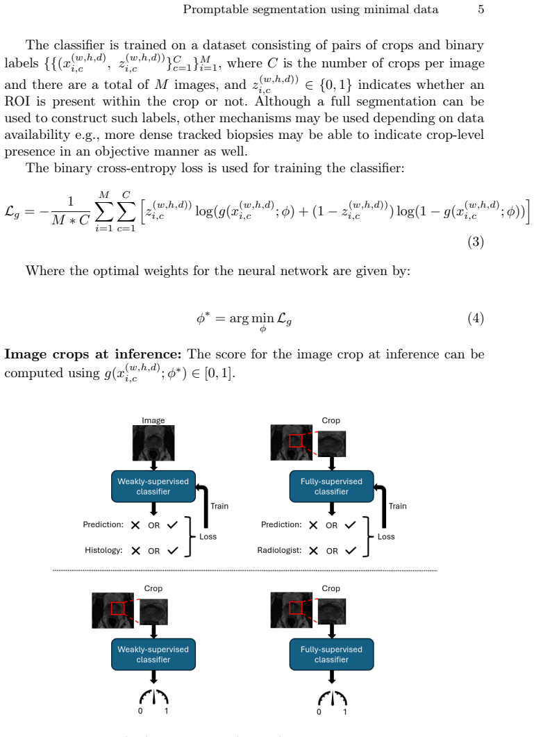
The load-bearing mechanism is a joint crop-scoring search. A weakly supervised classifier $f(x;\theta)$ is trained on whole images with binary ROI-presence labels and then applied to fixed-size crops; a fully supervised classifier $g(x;\phi)$ is trained directly on crops with crop-level presence labels. At inference a point prompt sets the first crop, and a spiral spline moves the crop center by $\delta_w = r\cos(\beta)$, $\delta_h = r\sin(\beta)$, with $r=t/s$ and $\beta=2\pi(t/\mu)$. Each crop receives the joint score $S=\alpha f(x;\theta^*)+(1-\alpha)g(x;\phi^*)$, is marked positive if $S>\tau$, and all positive crops are combined into the segmentation; repeated spirals with different parameters are merged by majority vote. This machinery converts two classification tasks into a dense segmentation without a pixel-level prediction head, which is why the training set can be so small.
What would settle it
Test the pipeline on elongated or multifocal lesions with prompts placed near lesion boundaries; if Dice drops far below centrally prompted runs, the spiral-search coverage assumption is falsified.
Extended reading notes
Core claim
The central claim is that weak binary labels plus a handful of segmentations are sufficient for promptable cancer segmentation: on a 200-image holdout, the method achieves a Dice score of 0.3085, above fine-tuned SAM (0.2361) and MedSAM (0.2673) and statistically indistinguishable from a fully supervised U-Net trained on 1,200 segmentations (0.3275, p = 0.07). The authors frame this as outperforming existing promptable methods while matching fully supervised methods with up to 100 times less annotated data. They further show that ablation over training set size gives convergence at 8 weak labels and 24 full labels, crop size 10x10x6, and that the spiral search runs in 4.2 seconds versus 439.6 seconds for an expert radiologist, at comparable Dice (0.2967 versus 0.2971).
Load-bearing premise
The load-bearing premise is that one click placed somewhere inside the cancer lets the fixed-size spiral sweep out the whole lesion, so irregular shapes or boundary clicks can be missed regardless of classifier accuracy.
Editorial extensions
If this is right
- Promptable cancer segmentation becomes feasible in settings where only a few dozen high-quality labels can be curated, since consensus-based and histology-based labels are affordable at this scale.
- At inference, a clinician needs only a single click to obtain a cancer map that matches fully supervised models trained on 200 to 1,200 segmentations.
- General-purpose promptable models such as SAM and MedSAM, even fine-tuned on 200 samples, remain below the proposed method's Dice, and fine-tuning with fewer samples performs worse still.
- The spiral search produces a segmentation in about four seconds, close to the agreement of an expert radiologist spending over seven minutes.
- The two-classifier design means the fully supervised classifier could be trained from crop-level labels (e.g., dense tracked biopsies) instead of full segmentations when segmentations are unavailable.
Reading between the lines
- If the recipe transfers, any weak signal correlated with a lesion's presence — lab values, symptoms, genomic flags — could drive spatial localization through crop scoring plus a geometric search, not just histology labels.
- The spiral's fixed size (10x10x6) and path are tuned to the roughly spherical prostate; on other organs the same framework would likely need an adaptive crop size or a contour-following search, which the paper's ablations suggest can be optimized from the same minimal data.
- The authors note over-prediction; a clinical reader could tune the threshold $\tau$ to favor over-segmentation for biopsy targeting or under-segmentation for treatment planning, a trade-off the paper leaves open.
- A direct test of generality would be to apply the same 24-plus-8 training recipe to a second cancer type and compare against fine-tuned SAM/MedSAM with equivalent total annotation budgets.
Editorial analysis
A structured set of objections, weighed in public.
Referee Report
Summary. The paper proposes a promptable cancer-segmentation framework for prostate MRI trained with 24 fully segmented volumes and 8 weakly labeled volumes. A weakly supervised classifier (trained on full images with binary histology labels) and a fully supervised classifier (trained on crops derived from the segmentations) are combined into a joint crop score, and a spiral search initiated by a single user-supplied point prompt accumulates positively scored crops into a final segmentation. On a 200-case holdout from a 500-case clinical dataset, the method reports Dice 0.3085, which exceeds the reported Dice of fine-tuned SAM and MedSAM and is not statistically different from a U-Net trained with much more data; ablations examine training-set size, crop size, search strategy, and hyperparameters.
Significance. If the central technical issues are resolved, the contribution is valuable: it demonstrates a route to promptable pathological-region segmentation with a small, curator-friendly training set, combining objective histology labels with a few high-quality segmentations. The paper deserves credit for evaluating on a real clinical dataset, for reporting prompt-location variability, and for ablating the amount of training data. However, the apparent architectural inconsistency in applying f to crops, the uncontrolled cross-dataset comparisons in Table 1, and the unsupported '100X' data-efficiency claim currently prevent the central claims from being accepted as stated.
major comments (4)
- [§2.1, §3.2] The weakly supervised classifier f is trained on full images in §2.1 and then applied to 10×10×6 crops at inference, but §3.2 specifies an architecture with three Conv+MaxPool3D(2,2,2) layers followed by flattening and dense layers, which fixes the input spatial size. No resizing, global/adaptive pooling, or fully convolutional modification is described, and a 10×10×6 volume cannot be processed by three successive (2,2,2) pools without padding or flooring complications. Unless the released code explicitly resizes crops or uses adaptive pooling, the joint score in Eq. (5) is undefined and the results in Table 1 are not reproducible from the manuscript. Please specify the exact input handling for both classifiers.
- [Table 1, §4.1] The rows for Yi et al. [33] and Yan et al. [32] are Dice values reported on test sets different from the authors' 200-image holdout, as the text acknowledges. Accordingly, the claim that the method 'performs comparably with fully-supervised methods' is supported only by the U-Net comparison, whose p-value (0.07) fails to reach significance and does not by itself establish performance equivalence. Please either evaluate [33] and [32] on the same holdout or restrict the comparative claim to the U-Net row and present the other rows as literature context.
- [Abstract, §3.3] The abstract and §3.3 state that the method uses 'up to 100X less' annotated data than the comparators. From Table 1 the largest ratio is at most 50× (1200 fully segmented images vs 24 fully segmented images for U-Net) and, counting the 8 weak labels as data, roughly 37.5×; the promptable comparators use only about 6× more data. Please correct the stated data-efficiency factor to the actual maximum supported by the table.
- [§4.2, Tables 1 and 4] The manuscript does not state the final values of α, T, μ, and n used to produce the headline Dice in Table 1, even though Table 4 reports ablations over these hyperparameters. Further, the 'Variance w.r.t. prompt' column in Table 1 is not defined (standard deviation? number of prompts? sampling protocol?), and the reported spread of ±0.2163 for the proposed method is large relative to the mean Dice of 0.3085. Please specify the final hyperparameter configuration and the prompt-sampling procedure.
minor comments (5)
- [Eq. (3)] Equation (3) contains a stray parenthesis ('z^{(w,h,d))}_{i,c}') and the BCE expression is missing a closing bracket; please correct.
- [§3.3, References] Reference [30] (ProMISe) is cited as 'SAM'; the original SAM is [12]. Please clarify which model was actually fine-tuned in the comparison.
- [Fig. 3] Figure 3's two horizontal axes are described only as 'bottom axis' and 'top axis' in the caption; please label both axes explicitly.
- [Table 4] In Table 4, when one hyperparameter is varied it is not stated whether the other hyperparameters are held at their optimal values; please state the fixed configuration for each ablation.
- [Table 2] Table 2 reports identical WSC accuracy (0.782) for 8, 12, and 16 weakly labeled samples; the convergence claim would be more convincing with multiple seeds or confidence intervals.
Circularity Check
No circular derivation: holdout comparison against external baselines supports the central claim; only minor, non-load-bearing self-citation.
full rationale
The paper's central claim—that the proposed two-classifier guided spiral search with a point prompt outperforms promptable baselines and matches fully-supervised U-Net on prostate cancer segmentation—is tested on a 200-image holdout set against independently trained SAM, MedSAM, U-Net, and prior method baselines, so the reported Dice values are not defined by the method's own training labels or by the fitting procedure. The weakly-supervised classifier f is trained on 8 binary histology labels and the fully-supervised classifier g on 24 full segmentations; these are external inputs, and the final segmentation is a thresholded combination of their crop scores. There is no equation in which a prediction reduces to its input by construction: Eq. 5 combines two independently learned probabilities, and the spiral search hyperparameters are tuned on the development split and then evaluated on the holdout. The authors self-cite their prior weakly-supervised segmentation work [24] when motivating full-image training with crop inference and low-sample convergence, but the same passages also cite independent works [19,29,34], and the convergence claim is substantiated by the paper's own ablation table, so the self-citation is not load-bearing. The discussion explicitly limits the spiral search to roughly spherical glands ('other strategies may need to be explored for other applications'), which is a generalizability limitation rather than circularity. The architecture description in Sec. 3.2 (fixed-size flatten/dense input) versus Sec. 2.1's 10x10x6 crops is a reproducibility inconsistency, not a circular reduction; if the released code does not resize or pool, Eq. 5 would be non-executable, but that is an engineering error and not an equivalence between the method's output and its input.
Assumptions & free parameters
free parameters (6)
- alpha (crop score balance) =
0.25 or 0.50 (best reported Dice)
- threshold tau =
0.05
- crop size (w,h,d) =
10x10x6
- spiral steps T =
not explicitly stated; best around 80 in Table 4
- spiral full-circle steps mu =
not explicitly stated; best 200 in Table 4
- spiral repeats n =
6 (best) or 8
assumptions (3)
- domain assumption Binary histology labels and consensus radiologist segmentations are accurate enough ground truth for training and evaluation.
- domain assumption The spiral search path and fixed crop geometry can cover the entire cancer lesion from a single point prompt.
- domain assumption Crop-level classification accuracy from classifiers trained on 8-24 samples transfers to the holdout population.
Cite this review
Pith. "Pith review of Promptable cancer segmentation using minimal expert-curated data." pith.science (2026). https://pith.science/paper/JCWZWBDA
@misc{pith2026250517915,
  author       = {Pith},
  title        = {Pith review of: Promptable cancer segmentation using minimal expert-curated data},
  year         = {2026},
  howpublished = {\url{https://pith.science/paper/JCWZWBDA}},
  note         = {Machine review of arXiv:2505.17915}
}
read the original abstract
Automated segmentation of cancer on medical images can aid targeted diagnostic and therapeutic procedures. However, its adoption is limited by the high cost of expert annotations required for training and inter-observer variability in datasets. While weakly-supervised methods mitigate some challenges, using binary histology labels for training as opposed to requiring full segmentation, they require large paired datasets of histology and images, which are difficult to curate. Similarly, promptable segmentation aims to allow segmentation with no re-training for new tasks at inference, however, existing models perform poorly on pathological regions, again necessitating large datasets for training. In this work we propose a novel approach for promptable segmentation requiring only 24 fully-segmented images, supplemented by 8 weakly-labelled images, for training. Curating this minimal data to a high standard is relatively feasible and thus issues with the cost and variability of obtaining labels can be mitigated. By leveraging two classifiers, one weakly-supervised and one fully-supervised, our method refines segmentation through a guided search process initiated by a single-point prompt. Our approach outperforms existing promptable segmentation methods, and performs comparably with fully-supervised methods, for the task of prostate cancer segmentation, while using substantially less annotated data (up to 100X less). This enables promptable segmentation with very minimal labelled data, such that the labels can be curated to a very high standard.
Figures


Figures from the paper (1 more)

Reference graph
Works this paper leans on
-
[33]
T2-Only Prostate Cancer Prediction by Meta-Learning from Bi-Parametric MR Imaging
Weixi Yi et al. “T2-Only Prostate Cancer Prediction by Meta-Learning from Bi-Parametric MR Imaging”. In: arXiv preprint arXiv:2411.07416 (2024)
work page Pith review arXiv 2024
-
[32]
Wen Yan et al. “The impact of using voxel-level segmentation metrics on evaluating multifocal prostate cancer localisation”. In: International Workshop on Applications of Medical AI. Springer. 2022, pp. 128–138
work page 2022
-
[1]
Early cancer detection using deep learning and medical imaging: A survey
Istiak Ahmad and Fahad Alqurashi. “Early cancer detection using deep learning and medical imaging: A survey”. In:Critical Reviews in Oncol- ogy/Hematology (2024), p. 104528
work page 2024
-
[2]
Hashim U Ahmed et al. “Diagnostic accuracy of multi-parametric MRI and TRUS biopsy in prostate cancer (PROMIS): a paired validating con- firmatory study”. In:The Lancet389.10071 (2017), pp. 815–822
work page 2017
-
[3]
A systematic review on supervised and unsu- pervised machine learning algorithms for data science
Mohamed Alloghani et al. “A systematic review on supervised and unsu- pervised machine learning algorithms for data science”. In:Supervised and unsupervised learning for data science(2020), pp. 3–21
work page 2020
-
[4]
Prostate-specific–antigen testing for early diagnosis of prostate cancer
Michael J Barry. “Prostate-specific–antigen testing for early diagnosis of prostate cancer”. In: New England Journal of Medicine 344.18 (2001), pp. 1373–1377
work page 2001
-
[5]
Mohammad Mahdi Behzadi et al. “Weakly-supervised deep learning model for prostate cancer diagnosis and gleason grading of histopathology im- ages”. In:Biomedical Signal Processing and Control95 (2024), p. 106351
work page 2024
-
[6]
The role of MRI/TRUS fusion biopsy in the diag- nosis of clinically significant prostate cancer
Andrea Benelli et al. “The role of MRI/TRUS fusion biopsy in the diag- nosis of clinically significant prostate cancer”. In:Therapeutic Advances in Urology 12 (2020), p. 1756287220916613
work page 2020
Show all 34 references
-
[7]
Development and evaluation of intraoperative ul- trasound segmentation with negative image frames and multiple observer labels
Liam F Chalcroft et al. “Development and evaluation of intraoperative ul- trasound segmentation with negative image frames and multiple observer labels”. In: Simplifying Medical Ultrasound: Second International Work- shop, ASMUS 2021, Held in Conjunction with MICCAI 2021, Stra...
2021
-
[8]
Image registration: Fundamentals and recent advances based on deep learning
Min Chen et al. “Image registration: Fundamentals and recent advances based on deep learning”. In:Machine Learning for Brain Disorders(2023), pp. 435–458
2023
-
[9]
Progress in the cryoablation and cryoimmunotherapy for tumor
Zenan Chen et al. “Progress in the cryoablation and cryoimmunotherapy for tumor”. In:Frontiers in immunology14 (2023), p. 1094009. 14 Karam et al
2023
-
[10]
Is segmentation uncertainty useful?
Steffen Czolbe et al. “Is segmentation uncertainty useful?” In:Information Processing in Medical Imaging: 27th International Conference, IPMI 2021, Virtual Event, June 28–June 30, 2021, Proceedings 27. Springer. 2021, pp. 715–726
2021
-
[11]
Cryoablation: mechanism of action and devices
Joseph P Erinjeri and Timothy WI Clark. “Cryoablation: mechanism of action and devices”. In:Journal of Vascular and Interventional Radiology 21.8 (2010), S187–S191
2010
-
[12]
Segmentanything
AlexanderKirillovetal.“Segmentanything”.In: Proceedings of the IEEE/CVF international conference on computer vision. 2023, pp. 4015–4026
2023
-
[13]
Imagenet classi- fication with deep convolutional neural networks
Alex Krizhevsky, Ilya Sutskever, and Geoffrey E Hinton. “Imagenet classi- fication with deep convolutional neural networks”. In:Advances in neural information processing systems25 (2012)
2012
-
[14]
Prism: A promptable and robust interactive segmenta- tion model with visual prompts
Hao Li et al. “Prism: A promptable and robust interactive segmenta- tion model with visual prompts”. In:International Conference on Medi- cal Image Computing and Computer-Assisted Intervention. Springer. 2024, pp. 389–399
2024
-
[15]
Weaklysupervisedhistopathologyimagesegmentationwith self-attention
KailuLietal.“Weaklysupervisedhistopathologyimagesegmentationwith self-attention”. In:Medical Image Analysis86 (2023), p. 102791
2023
-
[16]
Segment anything in medical images and videos: Benchmark and deployment
Jun Ma et al. “Segment anything in medical images and videos: Benchmark and deployment”. In:arXiv preprint arXiv:2408.03322(2024)
2024 arXiv
-
[17]
An introduction to convolutional neural networks
Keiron O’shea and Ryan Nash. “An introduction to convolutional neural networks”. In:arXiv preprint arXiv:1511.08458(2015)
2015 arXiv
-
[18]
Surgery, brachytherapy, and external-beam radiotherapy for early prostate cancer
Richard E Peschel and John W Colberg. “Surgery, brachytherapy, and external-beam radiotherapy for early prostate cancer”. In:The lancet on- cology 4.4 (2003), pp. 233–241
2003
-
[19]
Weakly supervised localisation of prostate cancer using reinforcement learning for bi-parametric MR images
Martynas Pocius et al. “Weakly supervised localisation of prostate cancer using reinforcement learning for bi-parametric MR images”. In:2024 IEEE International Symposium on Biomedical Imaging (ISBI).IEEE.2024,pp.1– 5
2024
-
[20]
Mixed supervision of histopathology improves prostate cancer classification from MRI
Abhejit Rajagopal et al. “Mixed supervision of histopathology improves prostate cancer classification from MRI”. In:IEEE Transactions on Med- ical Imaging(2024)
2024
-
[21]
Addressing AI algorithmic bias in health care
Raj M Ratwani, Karey Sutton, and Jessica E Galarraga. “Addressing AI algorithmic bias in health care”. In:JAMA 332.13 (2024), pp. 1051–1052
2024
-
[22]
U-net: Convolu- tional networks for biomedical image segmentation
Olaf Ronneberger, Philipp Fischer, and Thomas Brox. “U-net: Convolu- tional networks for biomedical image segmentation”. In: Medical image computing and computer-assisted intervention–MICCAI 2015: 18th inter- national conference, Munich, Germany, October 5-9, 2015, proceedings...
2015
-
[23]
Oncologic errors in diagnostic radiology: a 10-year analysis based on medical malpractice claims
Andrew B Rosenkrantz et al. “Oncologic errors in diagnostic radiology: a 10-year analysis based on medical malpractice claims”. In:Journal of the American College of Radiology18.9 (2021), pp. 1310–1316
2021
-
[24]
Competing for pixels: a self-play algorithm for weakly-supervised semantic segmentation
Shaheer U Saeed et al. “Competing for pixels: a self-play algorithm for weakly-supervised semantic segmentation”. In:IEEE Transactions on Pat- tern Analysis and Machine Intelligence(2024). Promptable segmentation using minimal data 15
2024
-
[25]
Image quality assessment by overlapping task- specific and task-agnostic measures: application to prostate multiparamet- ric MR images for cancer segmentation
Shaheer U Saeed et al. “Image quality assessment by overlapping task- specific and task-agnostic measures: application to prostate multiparamet- ric MR images for cancer segmentation”. In:Machine Learning in Biomed- ical Imaging (MELBA)(2022)
2022
-
[26]
The PI-CAI challenge: public training and develop- ment dataset
Anindo Saha et al. “The PI-CAI challenge: public training and develop- ment dataset”. In:(No Title)(2022)
2022
-
[27]
Cryoablation Treatment Guide
Boston Scientific. “Cryoablation Treatment Guide”. In:Advancing science for life (2021)
2021
-
[28]
Prostate cancer review: genetics, diagnosis, treatmentoptions, andalternativeapproaches
Mamello Sekhoacha et al. “Prostate cancer review: genetics, diagnosis, treatmentoptions, andalternativeapproaches”. In: Molecules 27.17(2022), p. 5730
2022
-
[29]
Grad-CAM: visual explanations from deep networks via gradient-based localization
Ramprasaath R Selvaraju et al. “Grad-CAM: visual explanations from deep networks via gradient-based localization”. In:International journal of computer vision128 (2020), pp. 336–359
2020
-
[30]
ProMISe: Promptable Medical Image Segmentation using SAM
Jinfeng Wang et al. “ProMISe: Promptable Medical Image Segmentation using SAM”. In:arXiv preprint arXiv:2403.04164(2024)
2024 arXiv
-
[31]
Scribbleprompt: fast and flexible interactive segmen- tation for any biomedical image
Hallee E Wong et al. “Scribbleprompt: fast and flexible interactive segmen- tation for any biomedical image”. In:European Conference on Computer Vision. Springer. 2024, pp. 207–229
2024
-
[34]
Grad-CAM-based explain- able artificial intelligence related to medical text processing
Hongjian Zhang and Katsuhiko Ogasawara. “Grad-CAM-based explain- able artificial intelligence related to medical text processing”. In:Bioengi- neering 10.9 (2023), p. 1070
2023
Reviewed August 7, 2026 · model on record in the stance chip above.
Discussion (0). Sign in to comment.