REVIEW 4 major objections 4 minor 48 references
Concentrate on Weakness: Mining Hard Prototypes for Few-Shot Medical Image Segmentation
T0 review · 4 major / 4 minor · reviewed 2026-08-07 · deepseek-v4-flash
Pith's one-line read This paper claims that focusing on weak features—the support pixels a global prototype would mislabel—and turning them into hard prototypes produces state-of-the-art few-shot medical image segmentation.
desk verdict A genuinely new hard-prototype mechanism, but the paper's own Table 7 shows the SOTA claim is an artifact of unmatched MS-COCO pretraining. read the letter →
The pith
A machine-rendered reading of the paper's core claim, the machinery that carries it, and where it could break.
The reading
What carries the argument
The load-bearing object is the hard prototype: a feature vector produced from support pixels whose predicted support mask disagrees with its ground-truth label. The Support Self-Prediction module creates the disagreement map, the Hard Prototype Generation module selects those pixels, resamples them, and projects them through an MLP to form multiple hard prototypes for foreground and background, and the Multiple Similarity Maps Fusion module combines cosine-similarity maps from all prototypes in a dual-path decoder. A boundary loss computed as one minus the F1 score of predicted and ground-truth boundary maps constrains the edges. Together these pieces convert the paper's insight—boundary errors are caused by neglected weak features—into a working segmentation pipeline.
What would settle it
Run every baseline in Tables 1 and 2 with the same MS-COCO-pretrained ResNet-101 backbone and training recipe as CoW; if CoW's mean Dice margins shrink to near zero while the hard-prototype modules are kept unchanged, the core claim that hard prototypes drive the improvement is refuted.
Extended reading notes
Core claim
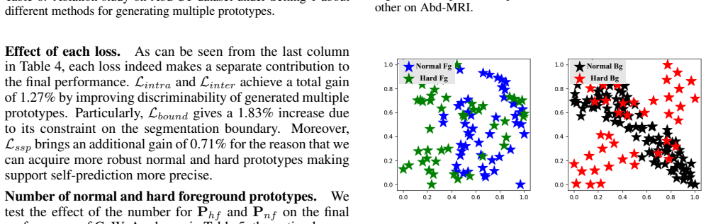
The central claim is that deliberately concentrating on weak features—those whose self-prediction disagrees with the true support label—lets a few-shot segmenter represent a class distribution more completely than random sampling or local averaging does. The paper states this as follows: normal prototypes capture the organ interior, hard prototypes capture boundary and easily confused regions, and the combination yields clearer segmentation boundaries. Evidence offered includes ablations in which each component adds a positive contribution, t-SNE plots showing hard prototypes fill gaps in the normal-prototype distribution, and quantitative tables where CoW outperforms the listed methods on all three datasets under both experimental settings.
Load-bearing premise
The paper's reported superiority over prior methods assumes the comparison is apples-to-apples, yet its own Table 7 shows that changing only which dataset the backbone was pretrained on, from ImageNet to MS-COCO, raises the mean Dice score on Abd-MRI by 7.09 points, so a large part of the measured gain could come from pretraining rather than from the hard-prototype mechanism.
Editorial extensions
If this is right
- On Abd-MRI under Setting 1, CoW reports a mean Dice score of 84.10, compared with the best listed prior result of 82.90 from GMRD.
- On Abd-CT under Setting 1, CoW reports 82.49 mean Dice, 3.97 points above GMRD, and on CMR it reports 80.00, above all listed methods.
- Ablations attribute each module a positive gain: SSP and HPG lift the baseline to 81.44, MSMF adds another 2.66 points, and the boundary loss alone contributes 1.83 points.
- A 50/50 split of hard to normal foreground prototypes performs best; relying on either type alone lowers mean Dice by 2.01 and 1.04 points respectively.
- Hard prototype generation also beats alternative prototype-construction methods such as k-means, prototype mixture models, and GMRD's random descriptors on Abd-CT.
Reading between the lines
- The paper's own Table 7 suggests a confound: switching only the backbone pretraining from ImageNet to MS-COCO raises Abd-MRI mean Dice by 7.09 points, a swing larger than some reported margins, so a controlled rerun of all baselines on the same pretrained backbone would test whether the hard-prototype mechanism, not the pretraining, carries the gain.
- Because boundary misclassification is a general weakness of prototype segmentation, the same self-prediction disagreement could define hard points in few-shot natural-image segmentation, not just medical images.
- The fixed hard-to-normal prototype ratio is a hyperparameter; a testable extension would allocate hard prototypes adaptively according to the length or complexity of the organ boundary in each episode.
- The SSP disagreement signal could be re-run at inference with the fused prototypes, generating a second round of harder instructions and potentially tighter boundaries.
Editorial analysis
A structured set of objections, weighed in public.
Referee Report
Summary. The paper proposes CoW, a few-shot medical image segmentation method built around four components: a Support Self-Prediction (SSP) module that identifies weak features by comparing a support self-prediction with the support ground truth; a Hard Prototypes Generation (HPG) module that samples hard and normal prototypes for foreground and background; a Multiple Similarity Maps Fusion (MSMF) module that fuses similarity maps from multiple prototypes; and a boundary loss for edge refinement. Experiments are reported on Abd-MRI, Abd-CT, and CMR under two settings, and the central claim in Section 4.3 is that CoW "significantly outperforms all listed methods in terms of the mean dice score on three different datasets."
Significance. If the empirical claim holds under a fair comparison, the paper makes a useful contribution: the idea of deriving hard prototypes from support self-prediction errors is well motivated, the method is not circular (the prototypes are built from support ground-truth error, not from query predictions), and the paper provides code, algorithm pseudocode, component-wise ablations, and an efficiency comparison in Table 9. The reported gains are, however, small relative to recent baselines, and the current comparison protocol leaves a major confound unresolved, so the significance of the claim depends on additional experiments that control the training protocol.
major comments (4)
- [Section 4.3, Tables 1-2, Table 7] The claimed state-of-the-art comparison is confounded by unmatched backbone pretraining. The paper states in Section 3.3 that it follows Q-Net in using a ResNet-101 backbone pretrained on MS-COCO, whereas the published baseline numbers in Tables 1 and 2 (e.g., GMRD, RPT) were obtained with ImageNet-pretrained backbones. Appendix Table 7 shows that this choice alone moves Abd-MRI mean DSC from 77.01 to 84.10 under Setting 1 and from 75.07 to 81.64 under Setting 2, swings of 7.09 and 6.57 points. These swings are larger than the claimed margins over the best prior method (1.20, 3.97, and 0.81 points in Tables 1-2). Moreover, with ImageNet pretraining, CoW scores 77.01 on Abd-MRI Setting 1, which is below the published GMRD value of 82.90 in Table 1. The statement in Section 4.3 is therefore not supported by the reported experiments; the authors should either rerun all baselines under MS-COCO pretraining or report CoW with ImageNet pretraining in the main tables and compare under matched protocols.
- [Tables 1-2 and Section 4.3] All numerical comparisons are single point estimates of the mean DSC, with no error bars, no standard deviations, and no significance tests. In episodic few-shot segmentation, fold and seed variability is substantial, and several reported margins over prior methods are less than one point (e.g., 0.81 on CMR). Without repeated runs or a statistical test, the claim of significant improvement cannot be evaluated.
- [Table 3] The ablation baseline is not an in-codebase reimplementation. The first row lists PA-Net numbers taken from published results (50.40, 32.19, 30.99, 40.58, 38.54), while rows 2 and 3 use CoW's own pipeline, including MS-COCO pretraining. The gains attributed to SSP and HPG therefore conflate architectural changes with backbone pretraining and implementation differences. The first row should be reimplemented within the same training protocol, or clearly identified as a literature number and excluded from incremental ablation claims.
- [Section 4.2 and Tables 5, 8] No held-out validation split is described for hyperparameter selection. The prototype counts Nhf, Nnf, Nhb, and Nnb and the loss weights lambda_0 and lambda_1 appear to be tuned on the same Abd-MRI test fold that is then used to report the final numbers (Tables 5 and 8). This creates a selection-on-test issue; the authors should use a validation split or explicitly report the selection procedure.
minor comments (4)
- [Equation (17)] The notation in the intra-class loss is unclear: the expression max over p_s^k in P_s* with P_q* in the cosine term is not formally defined, and the indices of the query prototypes are not specified.
- [Table 3 versus Table 1] The mean DSC for the PA-Net baseline is 38.54 in Table 3 but 38.53 in Table 1; please make the numbers consistent.
- [Figure 2] The labels "Cx2 Conv" and "expand Cx2 Conv" are not explained in the caption or the text; please define these operations.
- [Section 5] The phrase "Numerous experimental results also demonstrate" is vague; please point to the specific tables or figures that support the conclusion.
Circularity Check
No circularity: CoW's hard prototypes are derived from support labels and query-independent, and the reported gains are empirical comparisons, not reductions to fitted quantities.
full rationale
The paper reports an empirical few-shot segmentation method. The claimed derivation chain is: SSP uses masked average pooling on the support feature and the true support mask (Eqs. 1-6) to identify hard points; HPG builds hard prototypes from those support-derived points (Eqs. 7-11); MSMF fuses cosine similarity maps to predict the query mask (Eqs. 12-14). The query mask Mq is only used in the final segmentation and boundary losses (Eqs. 15, 19), never to construct the prototypes, so the central prediction is not an input to its own derivation. No parameter is fitted to a subset of data and then renamed as a prediction of that same data; the prototype counts and loss weights (Tables 5, 8; λ0, λ1 in Section 4.2) are hyperparameters tuned on the Abd-MRI test fold, which is an evaluation-leakage concern rather than a definitional circularity. The self-references (RPT, GMRD as prior work by co-author Haofeng Zhang) are used only as comparison baselines or inspiration and are not load-bearing for the method's derivation. The appendix's Table 7 confound between MS-COCO and ImageNet pretraining is a threat to the fairness of the SOTA comparison, but it does not make the derivation circular. Under the stated circularity criteria, there is no quoted equation or fitted parameter whose output is equivalent to its input by construction.
Assumptions & free parameters
free parameters (4)
- Foreground prototype counts N_hf and N_nf =
50 hard + 50 normal
- Background prototype counts N_hb and N_nb =
100 hard + 500 normal
- Loss weights lambda_0 and lambda_1 =
0.5 and 0.3
- Backbone pretraining dataset =
MS-COCO (chosen over ImageNet)
assumptions (6)
- domain assumption Episodic meta-learning with disjoint base and novel classes is the correct task formalization.
- domain assumption Masked average pooling yields a representative global support prototype.
- ad hoc to paper Support self-prediction errors identify weak features that are useful for query boundaries.
- domain assumption MS-COCO pretrained ResNet-101 features transfer to medical images.
- domain assumption Pseudo labels from 3D superpixel clustering are valid training targets.
- domain assumption Mean DSC over five folds, without variance, is a sufficient evaluation summary.
Cite this review
Pith. "Pith review of Concentrate on Weakness: Mining Hard Prototypes for Few-Shot Medical Image Segmentation." pith.science (2026). https://pith.science/paper/MTR7EQW6
@misc{pith2026250521897,
  author       = {Pith},
  title        = {Pith review of: Concentrate on Weakness: Mining Hard Prototypes for Few-Shot Medical Image Segmentation},
  year         = {2026},
  howpublished = {\url{https://pith.science/paper/MTR7EQW6}},
  note         = {Machine review of arXiv:2505.21897}
}
read the original abstract
Few-Shot Medical Image Segmentation (FSMIS) has been widely used to train a model that can perform segmentation from only a few annotated images. However, most existing prototype-based FSMIS methods generate multiple prototypes from the support image solely by random sampling or local averaging, which can cause particularly severe boundary blurring due to the tendency for normal features accounting for the majority of features of a specific category. Consequently, we propose to focus more attention to those weaker features that are crucial for clear segmentation boundary. Specifically, we design a Support Self-Prediction (SSP) module to identify such weak features by comparing true support mask with one predicted by global support prototype. Then, a Hard Prototypes Generation (HPG) module is employed to generate multiple hard prototypes based on these weak features. Subsequently, a Multiple Similarity Maps Fusion (MSMF) module is devised to generate final segmenting mask in a dual-path fashion to mitigate the imbalance between foreground and background in medical images. Furthermore, we introduce a boundary loss to further constraint the edge of segmentation. Extensive experiments on three publicly available medical image datasets demonstrate that our method achieves state-of-the-art performance. Code is available at https://github.com/jcjiang99/CoW.
Figures


Figures from the paper (6 more)


Reference graph
Works this paper leans on
-
[1]
Swin-unet: Unet-like pure transformer for medi- cal image segmentation
[Cao et al., 2022] Hu Cao, Yueyue Wang, Joy Chen, Dong- sheng Jiang, Xiaopeng Zhang, Qi Tian, and Manning Wang. Swin-unet: Unet-like pure transformer for medi- cal image segmentation. In ECCV, pages 205–218,
work page 2022
-
[6]
Few-shot semantic segmentation with prototype learning
[Dong and Xing, 2018] Nanqing Dong and Eric P Xing. Few-shot semantic segmentation with prototype learning. In BMVC, pages 1–11,
work page 2018
-
[7]
[El Naqa et al., 2007] Issam El Naqa, Deshan Yang, Aditya Apte, Divya Khullar, Sasa Mutic, Jie Zheng, Jeffrey D Bradley, Perry Grigsby, and Joseph O Deasy. Concur- rent multimodality image segmentation by active contours for radiotherapy treatment planning a. Medical physics , 34(12):4738–4749,
work page 2007
-
[11]
[Gu et al., 2020] Ran Gu, Guotai Wang, Tao Song, Rui Huang, Michael Aertsen, Jan Deprest, S´ebastien Ourselin, Tom Vercauteren, and Shaoting Zhang. Ca-net: Com- prehensive attention convolutional neural networks for ex- plainable medical image segmentation.IEEE Transactions on Medical Imaging, 40(2):699–711,
work page 2020
-
[12]
[Hansen et al., 2022] Stine Hansen, Srishti Gautam, Robert Jenssen, and Michael Kampffmeyer. Anomaly detection- inspired few-shot medical image segmentation through self-supervision with supervoxels. Medical Image Anal- ysis, 78:102385,
work page 2022
-
[13]
Algorithm as 136: A k-means clustering algorithm
[Hartigan and Wong, 1979] John A Hartigan and Manchek A Wong. Algorithm as 136: A k-means clustering algorithm. Journal of the Royal Statistical Society. Series C (Applied Statistics) , 28(1):100–108,
work page 1979
-
[16]
Chaos challenge-combined (ct-mr) healthy abdominal organ segmentation
[Kavur et al., 2021] A Emre Kavur, N Sinem Gezer, Mustafa Barıs ¸, Sinem Aslan, Pierre-Henri Conze, Vladimir Groza, Duc Duy Pham, Soumick Chatterjee, Philipp Ernst, Savas ¸ ¨Ozkan, et al. Chaos challenge-combined (ct-mr) healthy abdominal organ segmentation. Medical Image Analysis , 69:101950,
work page 2021
-
[17]
Miccai multi-atlas labeling beyond the cranial vault–workshop and challenge
[Landman et al., 2015] Bennett Landman, Zhoubing Xu, J Igelsias, Martin Styner, Thomas Langerak, and Arno Klein. Miccai multi-atlas labeling beyond the cranial vault–workshop and challenge. In MICCAI Workshop Challenge, volume 5, page 12,
work page 2015
Show all 48 references
-
[19]
Microsoft coco: Com- mon objects in context
[Lin et al., 2014] Tsung-Yi Lin, Michael Maire, Serge Be- longie, James Hays, Pietro Perona, Deva Ramanan, Piotr Doll´ar, and C Lawrence Zitnick. Microsoft coco: Com- mon objects in context. In ECCV, pages 740–755,
2014
-
[22]
Semi-supervised medical image seg- mentation through dual-task consistency
[Luo et al., 2021] Xiangde Luo, Jieneng Chen, Tao Song, and Guotai Wang. Semi-supervised medical image seg- mentation through dual-task consistency. In AAAI, pages 8801–8809,
2021
-
[23]
Self- supervision with superpixels: Training few-shot medical image segmentation without annotation
[Ouyang et al., 2020] Cheng Ouyang, Carlo Biffi, Chen Chen, Turkay Kart, Huaqi Qiu, and Daniel Rueckert. Self- supervision with superpixels: Training few-shot medical image segmentation without annotation. In ECCV, pages 762–780,
2020
-
[24]
Self- supervised learning for few-shot medical image segmenta- tion
[Ouyang et al., 2022] Cheng Ouyang, Carlo Biffi, Chen Chen, Turkay Kart, Huaqi Qiu, and Daniel Rueckert. Self- supervised learning for few-shot medical image segmenta- tion. IEEE Transactions on Medical Imaging, 41(7):1837– 1848,
2022
-
[25]
A robust volumetric transformer for accurate 3d tumor segmentation
[Peiris et al., 2022] Himashi Peiris, Munawar Hayat, Zhaolin Chen, Gary Egan, and Mehrtash Harandi. A robust volumetric transformer for accurate 3d tumor segmentation. In MICCAI, pages 162–172,
2022
-
[26]
U-net: Convolutional networks for biomedical image segmentation
[Ronneberger et al., 2015] Olaf Ronneberger, Philipp Fis- cher, and Thomas Brox. U-net: Convolutional networks for biomedical image segmentation. In MICCAI, pages 234–241,
2015
-
[27]
‘squeeze & excite’guided few-shot segmentation of volu- metric images
[Roy et al., 2020] Abhijit Guha Roy, Shayan Siddiqui, Se- bastian P ¨olsterl, Nassir Navab, and Christian Wachinger. ‘squeeze & excite’guided few-shot segmentation of volu- metric images. Medical image analysis, 59:101587,
2020
-
[28]
Imagenet large scale visual recogni- tion challenge
[Russakovsky et al., 2015] Olga Russakovsky, Jia Deng, Hao Su, Jonathan Krause, Sanjeev Satheesh, Sean Ma, Zhiheng Huang, Andrej Karpathy, Aditya Khosla, Michael Bernstein, et al. Imagenet large scale visual recogni- tion challenge. International journal of computer vision , 1...
2015
-
[29]
One-shot learning for semantic segmentation
[Shaban et al., 2017] Amirreza Shaban, Shray Bansal, Zhen Liu, Irfan Essa, and Byron Boots. One-shot learning for semantic segmentation. arXiv preprint arXiv:1709.03410,
2017 arXiv
-
[30]
Q-net: Query-informed few-shot medical image segmentation
[Shen et al., 2023] Qianqian Shen, Yanan Li, Jiyong Jin, and Bin Liu. Q-net: Query-informed few-shot medical image segmentation. In Proceedings of SAI Intelligent Systems Conference, pages 610–628,
2023
-
[31]
Marginal loss and exclusion loss for par- tially supervised multi-organ segmentation
[Shi et al., 2021] Gonglei Shi, Li Xiao, Yang Chen, and S Kevin Zhou. Marginal loss and exclusion loss for par- tially supervised multi-organ segmentation. Medical Im- age Analysis, 70:101979,
2021
-
[32]
Prototypical networks for few-shot learning
[Snell et al., 2017] Jake Snell, Kevin Swersky, and Richard Zemel. Prototypical networks for few-shot learning. In NeurIPS, volume 30,
2017
-
[33]
Learning to compare: Relation network for few-shot learn- ing
[Sung et al., 2018] Flood Sung, Yongxin Yang, Li Zhang, Tao Xiang, Philip HS Torr, and Timothy M Hospedales. Learning to compare: Relation network for few-shot learn- ing. In CVPR, pages 1199–1208,
2018
-
[34]
Few-shot medical image segmentation with high-fidelity prototypes
[Tang et al., 2024] Song Tang, Shaxu Yan, Xiaozhi Qi, Jianxin Gao, Mao Ye, Jianwei Zhang, and Xiatian Zhu. Few-shot medical image segmentation with high-fidelity prototypes. Medical Image Analysis, page 103412,
2024
-
[35]
Integrat- ing segmentation information into cnn for breast cancer diagnosis of mammographic masses
[Tsochatzidis et al., 2021] Lazaros Tsochatzidis, Panagiota Koutla, Lena Costaridou, and Ioannis Pratikakis. Integrat- ing segmentation information into cnn for breast cancer diagnosis of mammographic masses. Computer Methods and Programs in Biomedicine, 200:105913,
2021
-
[36]
Visualizing data using t-sne.Journal of machine learning research, 9(11),
[Van der Maaten and Hinton, 2008] Laurens Van der Maaten and Geoffrey Hinton. Visualizing data using t-sne.Journal of machine learning research, 9(11),
2008
-
[39]
Panet: Few-shot im- age semantic segmentation with prototype alignment
[Wang et al., 2019] Kaixin Wang, Jun Hao Liew, Yingtian Zou, Daquan Zhou, and Jiashi Feng. Panet: Few-shot im- age semantic segmentation with prototype alignment. In ICCV, pages 9197–9206,
2019
-
[40]
Few-shot medical image segmentation regularized with self-reference and contrastive learning
[Wang et al., 2022] Runze Wang, Qin Zhou, and Guoyan Zheng. Few-shot medical image segmentation regularized with self-reference and contrastive learning. In MICCAI, pages 514–523,
2022
-
[41]
Vessel-net: Retinal vessel segmentation under multi- path supervision
[Wu et al., 2019] Yicheng Wu, Yong Xia, Yang Song, Dong- hao Zhang, Dongnan Liu, Chaoyi Zhang, and Weidong Cai. Vessel-net: Retinal vessel segmentation under multi- path supervision. In MICCAI, pages 264–272,
2019
-
[42]
Dual contrastive learning with anatomical auxiliary supervision for few-shot medical image segmentation
[Wu et al., 2022] Huisi Wu, Fangyan Xiao, and Chongxin Liang. Dual contrastive learning with anatomical auxiliary supervision for few-shot medical image segmentation. In ECCV, pages 417–434,
2022
-
[43]
Prototype mixture models for few-shot semantic segmentation
[Yang et al., 2020] Boyu Yang, Chang Liu, Bohao Li, Jian- bin Jiao, and Qixiang Ye. Prototype mixture models for few-shot semantic segmentation. In MICCAI, pages 763– 778,
2020
-
[44]
Pet-guided delineation of radiation therapy treatment vol- umes: a survey of image segmentation techniques
[Zaidi and El Naqa, 2010] Habib Zaidi and Issam El Naqa. Pet-guided delineation of radiation therapy treatment vol- umes: a survey of image segmentation techniques. Euro- pean Journal of Nuclear Medicine and Molecular Imag- ing, 37:2165–2187,
2010
-
[46]
Few-shot medical image segmenta- tion via a region-enhanced prototypical transformer
[Zhu et al., 2023] Yazhou Zhu, Shidong Wang, Tong Xin, and Haofeng Zhang. Few-shot medical image segmenta- tion via a region-enhanced prototypical transformer. In MICCAI, pages 271–280,
2023
-
[47]
Multivariate mixture model for myocardial segmentation combining multi-source im- ages
[Zhuang, 2018] Xiahai Zhuang. Multivariate mixture model for myocardial segmentation combining multi-source im- ages. IEEE Transactions on Pattern Analysis and Machine Intelligence, 41(12):2933–2946,
2018
-
[48]
Algorithm of HPG
Appendix In this section, we present the complete experimental results omitted from the main text, along with additional validation, analysis, and further details on the implementation of Hard Prototype Generation (HPG) module. Algorithm of HPG. To more clearly depict the HPG ...
2014
-
[1979]
Deep learning techniques for medical image segmentation: achievements and challenges
[Hesamian et al., 2019] Mohammad Hesam Hesamian, Wenjing Jia, Xiangjian He, and Paul Kennedy. Deep learning techniques for medical image segmentation: achievements and challenges. Journal of Digital Imaging, 32:582–596,
2019
-
[2007]
Multi- organ segmentation over partially labeled datasets with multi-scale feature abstraction
[Fang and Yan, 2020] Xi Fang and Pingkun Yan. Multi- organ segmentation over partially labeled datasets with multi-scale feature abstraction. IEEE Transactions on Medical Imaging, 39(11):3619–3629,
2020
-
[2008]
Matching networks for one shot learning
[Vinyals et al., 2016] Oriol Vinyals, Charles Blundell, Tim- othy Lillicrap, Daan Wierstra, et al. Matching networks for one shot learning. In NeurIPS, pages 3637–364,
2016
-
[2010]
Few-shot 3d volumetric segmentation with multi-surrogate fusion
[Zheng et al., 2024] Meng Zheng, Benjamin Planche, Zhongpai Gao, Terrence Chen, Richard J Radke, and Ziyan Wu. Few-shot 3d volumetric segmentation with multi-surrogate fusion. In MICCAI, pages 286–296,
2024
-
[2014]
Few shot medical image segmentation with cross attention transformer
[Lin et al., 2023] Yi Lin, Yufan Chen, Kwang-Ting Cheng, and Hao Chen. Few shot medical image segmentation with cross attention transformer. In MICCAI, pages 233–243,
2023
-
[2015]
H- denseunet: hybrid densely connected unet for liver and tu- mor segmentation from ct volumes
[Li et al., 2018] Xiaomeng Li, Hao Chen, Xiaojuan Qi, Qi Dou, Chi-Wing Fu, and Pheng-Ann Heng. H- denseunet: hybrid densely connected unet for liver and tu- mor segmentation from ct volumes. IEEE Transactions on Medical Imaging, 37(12):2663–2674,
2018
-
[2016]
Deepigeos: a deep interactive geodesic framework for medical image segmentation
[Wang et al., 2018] Guotai Wang, Maria A Zuluaga, Wenqi Li, Rosalind Pratt, Premal A Patel, Michael Aertsen, Tom Doel, Anna L David, Jan Deprest, S ´ebastien Ourselin, et al. Deepigeos: a deep interactive geodesic framework for medical image segmentation. IEEE Transactions on ...
2018
-
[2017]
Few-shot learning with graph neural networks
[Garcia and Bruna, 2017] Victor Garcia and Joan Bruna. Few-shot learning with graph neural networks. arXiv preprint arXiv:1711.04043,
2017 arXiv
-
[2018]
A deep learning-based auto-segmentation system for organs-at- risk on whole-body computed tomography images for ra- diation therapy
[Chen et al., 2021] Xuming Chen, Shanlin Sun, Narisu Bai, Kun Han, Qianqian Liu, Shengyu Yao, Hao Tang, Chu- peng Zhang, Zhipeng Lu, Qian Huang, et al. A deep learning-based auto-segmentation system for organs-at- risk on whole-body computed tomography images for ra- diation t...
2021
-
[2019]
nnu-net: a self-configuring method for deep learning- based biomedical image segmentation
[Isensee et al., 2021] Fabian Isensee, Paul F Jaeger, Si- mon AA Kohl, Jens Petersen, and Klaus H Maier-Hein. nnu-net: a self-configuring method for deep learning- based biomedical image segmentation. Nature methods , 18(2):203–211,
2021
-
[2020]
Model-agnostic meta-learning for fast adaptation of deep networks
[Finn et al., 2017] Chelsea Finn, Pieter Abbeel, and Sergey Levine. Model-agnostic meta-learning for fast adaptation of deep networks. In ICML, pages 1126–1135,
2017
-
[2021]
Few- shot medical image segmentation via generating multiple representative descriptors
[Cheng et al., 2024] Ziming Cheng, Shidong Wang, Tong Xin, Tao Zhou, Haofeng Zhang, and Ling Shao. Few- shot medical image segmentation via generating multiple representative descriptors. IEEE Transactions on Medical Imaging,
2024
-
[2022]
Drinet for medical image segmentation
[Chen et al., 2018] Liang Chen, Paul Bentley, Kensaku Mori, Kazunari Misawa, Michitaka Fujiwara, and Daniel Rueckert. Drinet for medical image segmentation. IEEE Transactions on Medical Imaging , 37(11):2453–2462,
2018
-
[2023]
Fully convolutional networks for seman- tic segmentation
[Long et al., 2015] Jonathan Long, Evan Shelhamer, and Trevor Darrell. Fully convolutional networks for seman- tic segmentation. In CVPR, pages 3431–3440,
2015
-
[2024]
Hyperdense-net: a hyper-densely connected cnn for multi-modal image segmentation
[Dolz et al., 2018] Jose Dolz, Karthik Gopinath, Jing Yuan, Herve Lombaert, Christian Desrosiers, and Ismail Ben Ayed. Hyperdense-net: a hyper-densely connected cnn for multi-modal image segmentation. IEEE Transactions on Medical Imaging, 38(5):1116–1126,
2018
Reviewed August 7, 2026 · model on record in the stance chip above.
Discussion (0). Sign in to comment.