REVIEW 4 major objections 5 minor 45 references
Single Domain Generalization for Alzheimer's Detection from 3D MRIs with Pseudo-Morphological Augmentations and Contrastive Learning
T0 review · 4 major / 5 minor · reviewed 2026-08-07 · deepseek-v4-flash
Pith's one-line read A 3D U-Net trained only on NACC, with learnable pseudo-erosion and pseudo-dilation augmentations plus supervised contrastive learning, beats generic single-domain-generalization baselines on ADNI and AIBL.
desk verdict The combination of pseudo-morphological augmentations and contrastive learning is a plausible new SDG pipeline for AD detection, but the ablation that supposedly isolates the morphology modules is confounded, so the central mechanistic claim is currently unsupported. read the letter →
The pith
A machine-rendered reading of the paper's core claim, the machinery that carries it, and where it could break.
The reading
What carries the argument
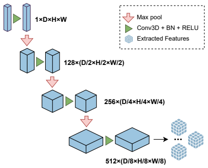
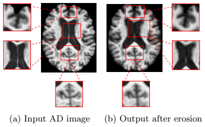
The load-bearing mechanism is the pair of learnable pseudo-morphological modules built from 3D convolutions followed by channel-wise max for dilation and the negation of a channel-wise min for erosion, with binary foreground masks so background zeros do not drive the operations. These approximate the classical morphological operators of mathematical morphology without a full lattice foundation; erosion is intended to simulate brain atrophy and dilation to simulate healthy expansion. Around them, a weighted supervised contrastive loss with class weights pulls same-class projections together and pushes other classes apart, while CutMix3D is applied to uniform MCI batches for extra intra-class diversity. Together the modules generate anatomically motivated class-specific augmentations during training, and the encoder learns representations that remain discriminative on unseen target distributions.
What would settle it
Compare the spatial pattern of changes induced by the pseudo-erosion module on healthy control MRIs against longitudinal atrophy maps of patients who later convert to Alzheimer's; if the simulated changes do not overlap known atrophy regions such as the hippocampus and medial temporal lobe significantly more than random intensity perturbations do, the anatomical mechanism is not supported.
Extended reading notes
Core claim
On the paper's own terms, the central discovery is that differentiable, class-aware morphological augmentation transfers to unseen imaging domains better than generic augmentation or feature-alignment baselines. Erosion and dilation are treated as disease models: applying learnable erosion to Alzheimer's samples intensifies atrophy, applying learnable dilation to healthy controls expands brain tissue, and affine augmentations plus 3D CutMix handle borderline mild cognitive impairment cases. These class-specific views are fed through a shared 3D U-Net encoder with a supervised contrastive loss, and the whole pipeline is trained end-to-end on NACC alone. On ADNI the method reaches 50.91% accuracy and macro-F1 0.424; on AIBL it reaches macro-F1 0.456, with gains over the strongest baseline on both balanced metrics. The paper interprets the results as evidence that morphological priors preserve disease-relevant structure while increasing intra-class diversity, which is exactly what an unseen-domain detector needs.
Load-bearing premise
The method's gains depend on the assumption that the learned erosion and dilation modules produce anatomically plausible class-specific changes, namely genuine atrophy and healthy brain expansion, rather than generic intensity distortions.
Editorial extensions
If this is right
- A model trained only on NACC can be deployed directly on ADNI and AIBL scans without any target-domain data or labels, since the single-domain-generalization protocol uses no target examples.
- Morphology-based augmentations improve balanced metrics such as macro-F1 more than generic MixUp, RSC, CCSDG, or EFDM under class imbalance.
- Removing the morphological modules drops macro-F1 by 6.5 points on ADNI and 11.8 points on AIBL in the ablations, indicating that the erosion and dilation views carry much of the generalization gain.
- Grad-CAM maps produced with the morphological modules concentrate on hippocampus and medial temporal lobe regions, indicating more disease-relevant localization.
- The added cost is modest in parameters (21.2M versus 19.6M for the baseline) but heavier in FLOPs and memory, and the paper treats this as an acceptable trade-off for the F1 gains.
Reading between the lines
- A natural next test, not run in the paper, is whether the same class-aware erosion and dilation recipe transfers to other neurodegenerative diseases with known atrophy patterns, such as frontotemporal dementia or Parkinson's disease.
- If the learned morphological operators are truly anatomy-based, their per-voxel effect on healthy scans should overlap known atrophy maps; quantifying that overlap would provide an independent check of the mechanism.
- The method's AIBL accuracy is lower than EFDM's, so a multi-objective evaluation that also optimizes accuracy could change the ranking; the paper's case rests on balanced macro-F1.
- With a larger multi-site training cohort, the contrastive and morphological components could be tested separately at scale, for instance by ablating only erosion or only dilation.
Editorial analysis
A structured set of objections, weighed in public.
Referee Report
Summary. This manuscript proposes a single-domain generalization (SDG) method for Alzheimer's disease detection from 3D MRIs. A 3D U-Net encoder is trained on NACC only; class-specific pseudo-dilation and pseudo-erosion modules generate augmented NC/AD samples, MCI samples receive affine and CutMix3D augmentation, and a supervised contrastive loss is optimized jointly with weighted cross-entropy. The method is evaluated on ADNI and AIBL and compared with MixUp, RSC, CCSDG, EFDM, and a no-SDG baseline; the authors report the highest macro-F1 on both target datasets and provide an ablation study, computational cost comparison, and Grad-CAM visualizations.
Significance. If the claims hold, the paper offers a well-motivated combination of morphological priors and supervised contrastive learning for single-domain generalization in neuroimaging, with macro-F1 as an appropriate primary metric under class imbalance. The use of three public datasets and a standard source-only evaluation protocol is a strength, and the paper attempts to ablate each component. However, the central empirical claim is currently supported by a mislabeled ablation row and by single-run numbers without error bars or significance tests, and the paper does not release code, so the significance of the contribution will only be clear after the experimental accounting is corrected.
major comments (4)
- [Table 4, Sec. 4.4] The first row of Table 4 ('No pseudo-morphology') is numerically identical to the Baseline row in Tables 2 and 3 (ADNI ACC 38.04, F1 0.359, SEN 0.359, SPE 0.679; AIBL ACC 38.50, F1 0.338, SEN 0.392, SPE 0.699). Since the baseline described in Sec. 4.2 has no SDG components at all (no CutMix3D, no contrastive loss), this row cannot represent removing only the pseudo-morphological modules while keeping the other components fixed. Therefore the statement in Sec. 4.4 that removing morphological modules causes F1 drops of 6.5 points on ADNI and 11.8 points on AIBL is unsupported. This is the central attribution of the paper, and the row must either be recomputed as a true single-component ablation or the text must be corrected.
- [Sec. 4.3, Tables 2 and 3] All results appear to be single-run numbers with no error bars, confidence intervals, or significance tests. The claimed improvement over the strongest baseline is small on ADNI (F1 0.424 vs. 0.407, a 1.7-point difference), and the claim that the method 'consistently achieved superior generalization performance' is also contradicted by Table 3, where EFDM reaches 69.94% accuracy on AIBL versus 62.27% for the proposed method. The authors should report means and standard deviations over at least three seeds for the main comparisons, and should qualify the superiority claim by specifying the metric (macro-F1, sensitivity, specificity) rather than accuracy.
- [Algorithm 1, Sec. 3.3] Algorithm 1 as printed contains unreachable code and is inconsistent with Sec. 3.3. When a minibatch has all labels equal, the pseudocode returns LCE after applying CutMix (for MCI) or immediately (for non-MCI), so the subsequent construction of the augmented set A, the computation of LSCL, and the combined loss Ltotal are never executed for such batches. When the batch has mixed labels, the homogeneous-batch branch is skipped. This makes it unclear when the supervised contrastive loss is actually applied, despite Eq. (6) assuming 2N original-plus-augmentation samples. The algorithm should be rewritten so that every code path and the exact loss composition are unambiguous.
- [Table 4, Sec. 4.4] The 'No supervised contrastive loss' ablation row reports AIBL accuracy of 73.28%, which is 11.01 points higher than the full method's 62.27%, while the macro-F1 drops from 0.456 to 0.305 and sensitivity from 0.452 to 0.343. The text only discusses the F1 and sensitivity drops and does not address this large accuracy swing. The authors should explain whether the full method intentionally trades accuracy for balanced performance, or whether this result indicates an unstable training run, and should provide a class-wise breakdown for this setting.
minor comments (5)
- [References] References [16] and [17] appear to describe the same paper: both are titled 'Learning with domain-knowledge for generalizable prediction of Alzheimer's disease from multi-site structural MRI' and both are listed as MICCAI 2023, Vancouver, pages 452–461. One of them should be removed or replaced with a distinct work.
- [Fig. 5] The caption of Figure 5 is malformed: the line 'xa xa xa (c) ˜xa' is not a complete caption and does not explain what is shown in each panel.
- [Eqs. (2) and (4)] Equations (2) and (4) are taken from the morphological module design in [23]; the text should state this explicitly in the contribution summary so the novelty is clearly the SDG application and the class-specific augmentation strategy, not the differentiable morphology operator itself.
- [Table 5] The 'Model size (MB)' of 494.56 MB is inconsistent with the reported 21.2M parameters (which would be about 85 MB as fp32). Please define whether this column measures GPU memory footprint during inference rather than parameter storage, and report both consistently.
- [Sec. 3.2.1] The foreground mask is written as 'M = 1 x̸=0'; this should use proper indicator notation, e.g., M = 1[x ≠ 0], and the mask application order should be stated precisely for both the dilation and erosion modules.
Circularity Check
No significant circularity: the paper's claims are empirical comparisons, and the only self-citation is not load-bearing.
full rationale
The paper does not claim a first-principles derivation whose output is built into its inputs. Its two novel-looking components are (i) differentiable pseudo-dilation/pseudo-erosion modules explicitly adopted from external prior work [23], used as class-conditioned training-time augmentations, and (ii) a standard supervised contrastive loss [36] combined with weighted cross-entropy. Equations (2) and (4) are presented as borrowed module definitions, not as evidence for generalization, so there is no self-definitional step. No fitted parameter is relabeled as a prediction: accuracy, F1, sensitivity, and specificity are measured on held-out ADNI and AIBL after training on NACC, so the central experimental claims are independent of the method's own outputs. The only self-citation, [33], appears in Related Work as a description of a prior distance-transform mixup strategy and is not used to justify the proposed architecture, augmentations, or loss; it is therefore not load-bearing. One non-circularity concern should be flagged for correctness rather than for the circularity score: in Table 4, the 'No pseudo-morphology' row is numerically identical to the 'Baseline [35]' row in Tables 2 and 3 (ADNI ACC 38.04 / F1 0.359 / SEN 0.359 / SPE 0.679; AIBL ACC 38.50 / F1 0.338 / SEN 0.392 / SPE 0.699), and Sec. 4.4 interprets this row as removing only the morphological modules while retaining CutMix and the contrastive loss. If those components were retained, the ablation row should differ from the no-SDG baseline. This is an internal-validity or table-consistency problem in the ablation attribution, not a case where a claimed prediction reduces by construction to its inputs, so it does not change the circularity finding.
Assumptions & free parameters
free parameters (3)
- temperature tau
- contrastive loss weight lambda
- pseudo-morphology kernel size k =
{3, 5}
assumptions (4)
- domain assumption Grayscale erosion and dilation mimic AD atrophy and healthy brain expansion
- ad hoc to paper The foreground mask M = 1_{x != 0} confines augmentations to anatomically relevant tissue
- domain assumption NACC is a representative single source domain for SDG evaluation
- domain assumption The pretrained Models Genesis 3D U-Net is an appropriate backbone
Cite this review
Pith. "Pith review of Single Domain Generalization for Alzheimer's Detection from 3D MRIs with Pseudo-Morphological Augmentations and Contrastive Learning." pith.science (2026). https://pith.science/paper/W6J4GDV2
@misc{pith2026250522465,
  author       = {Pith},
  title        = {Pith review of: Single Domain Generalization for Alzheimer's Detection from 3D MRIs with Pseudo-Morphological Augmentations and Contrastive Learning},
  year         = {2026},
  howpublished = {\url{https://pith.science/paper/W6J4GDV2}},
  note         = {Machine review of arXiv:2505.22465}
}
read the original abstract
Although Alzheimer's disease detection via MRIs has advanced significantly thanks to contemporary deep learning models, challenges such as class imbalance, protocol variations, and limited dataset diversity often hinder their generalization capacity. To address this issue, this article focuses on the single domain generalization setting, where given the data of one domain, a model is designed and developed with maximal performance w.r.t. an unseen domain of distinct distribution. Since brain morphology is known to play a crucial role in Alzheimer's diagnosis, we propose the use of learnable pseudo-morphological modules aimed at producing shape-aware, anatomically meaningful class-specific augmentations in combination with a supervised contrastive learning module to extract robust class-specific representations. Experiments conducted across three datasets show improved performance and generalization capacity, especially under class imbalance and imaging protocol variations. The source code will be made available upon acceptance at https://github.com/zobia111/SDG-Alzheimer.
Figures


Figures from the paper (3 more)


Reference graph
Works this paper leans on
-
[16]
Y. Zhou, Y. Li, F. Zhou, Y. Liu, and L. Tu. Learning with domain-knowledge for generalizable prediction of Alzheimer’s disease from multi-site structural MRI. In Proceedings of the International Conference on Medical Image Computing and Computer-Assisted Intervention , pages 452–461, Vancouver, Canada, 2023
work page 2023
-
[17]
Yanjie Zhou, Youhao Li, Feng Zhou, Yong Liu, and Liyun Tu. Learning with domain-knowledge for generalizable prediction of Alzheimer’s disease from multi-site structural MRI. In Proceedings of the International Conference on Medical Image Computing and Computer-Assisted Intervention , pages 452–461, Vancouver, Canada, 2023
work page 2023
-
[1]
Yuanyuan Chen, Yongsheng Pan, Yong Xia, and Yixuan Yuan. Disentangle first, then distill: a unified framework for missing modality imputation and Alzheimer’s disease diagnosis. IEEE Transactions on Medical Imaging, 42(12):3566–3578, 2023
work page 2023
-
[2]
Generalizing to unseen domains: A survey on domain generalization
Jindong Wang, Cuiling Lan, Chang Liu, Yidong Ouyang, Tao Qin, Wang Lu, Yiqiang Chen, Wenjun Zeng, and S Yu Philip. Generalizing to unseen domains: A survey on domain generalization. IEEE transactions on knowledge and data engineering , 35(8):8052–8072, 2022
work page 2022
-
[3]
Kangfu Han, Gang Li, Zhiwen Fang, and Feng Yang. Multi-template meta-information regularized network for Alzheimer’s disease diagnosis using structural MRI. IEEE Transactions on Medical Imaging , 43(5):1664–1676, 2023
work page 2023
-
[4]
Durga Supriya Hl, Swetha Mary Thomas, et al. A multimodal approach integrating convolutional and recurrent neural networks for Alzheimer’s disease temporal progression prediction. In Proceedings of the IEEE/CVF Conference on Computer Vision and Pattern Recognition , pages 5207–5215, 2024
work page 2024
-
[5]
M3t: three-dimensional medical image classifier using multi-plane and multi-slice transformer
Jinseong Jang and Dosik Hwang. M3t: three-dimensional medical image classifier using multi-plane and multi-slice transformer. In Proceedings of the IEEE/CVF Conference on Computer Vision and Pattern Recognition, pages 20718–20729, 2022
work page 2022
-
[6]
Multi-attention- based global 3d resnet for Alzheimer’s disease diagnosis
Yaozu Li, Yueheng Zhang, Jinfeng Wu, Xiaoshuang Zhang, Lili Han, and Xinchun Cui. Multi-attention- based global 3d resnet for Alzheimer’s disease diagnosis. In 2024 International Joint Conference on Neural Networks, pages 1–8, 2024
work page 2024
Show all 45 references
-
[7]
3d global fourier network for Alzheimer’s disease diagnosis using structural MRI
Shengjie Zhang, Xiang Chen, Bohan Ren, Haibo Yang, Ziqi Yu, Xiao-Yong Zhang, and Yuan Zhou. 3d global fourier network for Alzheimer’s disease diagnosis using structural MRI. In Proceedings of the International Conference on Medical Image Computing and Computer-Assisted Interve...
2022
-
[8]
An effective deep learning-based automatic prediction and classification of Alzheimer’s disease using EGELU-SZN technique
B Sathyabhama and M Kannan. An effective deep learning-based automatic prediction and classification of Alzheimer’s disease using EGELU-SZN technique. Neural Computing and Applications , pages 1–18, 2025
2025
-
[9]
Vitad: Leveraging modified vision transformer for Alzheimer’s disease multi-stage classification from brain MRI scans
Md Ashif Mahmud Joy, Shamima Nasrin, Ayesha Siddiqua, and Dewan Md Farid. Vitad: Leveraging modified vision transformer for Alzheimer’s disease multi-stage classification from brain MRI scans. Brain Research, 1847:149302, 2025
2025
-
[10]
Multimodal surface-based transformer model for early diagnosis of Alzheimer’s disease
Quan Anh Duong, Sy Dat Tran, and Jin Kyu Gahm. Multimodal surface-based transformer model for early diagnosis of Alzheimer’s disease. Scientific Reports, 15(1):5787, 2025
2025
-
[11]
Training vit with limited data for Alzheimer’s disease classification: An empirical study
Kassymzhomart Kunanbayev, Vyacheslav Shen, and Dae-Shik Kim. Training vit with limited data for Alzheimer’s disease classification: An empirical study. In Proceedings of the International Conference on Medical Image Computing and Computer-Assisted Intervention , pages 334–343,...
2024
-
[12]
Dense convolution-based attention network for Alzheimer’s disease classification
Yingtong Gan, Quan Lan, ChenXi Huang, Weichao Su, and Zhiyuan Huang. Dense convolution-based attention network for Alzheimer’s disease classification. Scientific Reports, 15(1):5693, 2025
2025
-
[13]
Anatomy-aware gating network for explainable Alzheimer’s disease diagnosis
Hongchao Jiang and Chunyan Miao. Anatomy-aware gating network for explainable Alzheimer’s disease diagnosis. In Proceedings of the International Conference on Medical Image Computing and Computer- Assisted Intervention, pages 90–100, Marrakesh, Marocco, 2024. 11
2024
-
[14]
Lteif, S
D. Lteif, S. Sreerama, S. A. Bargal, B. A. Plummer, R. Au, and V. B. Kolachalama. Disease-driven domain generalization for neuroimaging-based assessment of Alzheimer’s disease. Human Brain Mapping , 45(8):e26707, Jun. 2024
2024
-
[15]
Prototype-guided multi-scale domain adaptation for Alzheimer’s disease detection
Hongshun Cai, Qiongmin Zhang, and Ying Long. Prototype-guided multi-scale domain adaptation for Alzheimer’s disease detection. Computers in Biology and Medicine , 154:106570, 2023
2023
-
[18]
Mathematical morphology
Jean Serra. Mathematical morphology. In Encyclopedia of mathematical geosciences , pages 820–835. Springer, 2023
2023
-
[19]
Histopathological image analysis: A review
Metin N Gurcan, Laura E Boucheron, Ali Can, Anant Madabhushi, Nasir M Rajpoot, and Bulent Yener. Histopathological image analysis: A review. IEEE reviews in biomedical engineering , 2:147–171, 2009
2009
-
[20]
Deformable medical image registration: A survey
Aristeidis Sotiras, Christos Davatzikos, and Nikos Paragios. Deformable medical image registration: A survey. IEEE Transactions on Medical Imaging , 32(7):1153–1190, 2013
2013
-
[21]
Medical image fusion: A survey of the state of the art
Alex Pappachen James and Belur V Dasarathy. Medical image fusion: A survey of the state of the art. Information fusion , 19:4–19, 2014
2014
-
[22]
Multivariate mathematical morphology applied to colour image analysis
Erchan Aptoula and S´ ebastien Lef` evre. Multivariate mathematical morphology applied to colour image analysis. Multivariate image processing: methods and applications , pages 303–337, 2009
2009
-
[23]
Learning deep morphological networks with neural architecture search
Yufei Hu, Nacim Belkhir, Jesus Angulo, Angela Yao, and Gianni Franchi. Learning deep morphological networks with neural architecture search. Pattern Recognition, 131:108893, 2022
2022
-
[24]
Multi-scale morphology-aided deep medical image segmentation
Susmita Ghosh and Swagatam Das. Multi-scale morphology-aided deep medical image segmentation. Engineering Applications of Artificial Intelligence , 137:109047, 2024
2024
-
[25]
Differentiable soft morphological filters for medical image segmentation
Lisa Guzzi, Maria A Zuluaga, Fabien Lareyre, Gilles Di Lorenzo, S´ ebastien Goffart, Andrea Chierici, Juliette Raffort, and Herv´ e Delingette. Differentiable soft morphological filters for medical image segmentation. In Proceedings of the International Conference on Medical I...
2024
-
[26]
Hybrid morphological-convolutional neural networks for computer-aided diagnosis
Martha Rebeca Canales-Fiscal and Jos´ e Gerardo Tamez-Pe˜ na. Hybrid morphological-convolutional neural networks for computer-aided diagnosis. Frontiers in Artificial Intelligence , 6:1253183, 2023
2023
-
[27]
Topological properties of individual gray matter morphological networks in identifying the preclinical stages of Alzheimer’s disease: a preliminary study
Hongyuan Ding, Zhihao Wang, Yin Tang, Tong Wang, Ming Qi, Weiqiang Dou, Long Qian, Yaxin Gao, Qian Zhong, Xi Yang, et al. Topological properties of individual gray matter morphological networks in identifying the preclinical stages of Alzheimer’s disease: a preliminary study. ...
2023
-
[28]
Interpretation of brain morphology in association to Alzheimer’s disease dementia classification using graph convolutional networks on triangulated meshes
Emanuel A Azcona, Pierre Besson, Yunan Wu, Arjun Punjabi, Adam Martersteck, Amil Dravid, Todd B Parrish, S Kathleen Bandt, and Aggelos K Katsaggelos. Interpretation of brain morphology in association to Alzheimer’s disease dementia classification using graph convolutional netw...
2020
-
[29]
Adapt: Alzheimer diagnosis through adaptive profiling transformers
Yifeng Wang, Ke Chen, and Haohan Wang. Adapt: Alzheimer diagnosis through adaptive profiling transformers. arXiv preprint arXiv:2401.06349 , 2024. 12
2024 arXiv
-
[30]
Embracing the disharmony in medical imaging: A simple and effective framework for domain adaptation
Rongguang Wang, Pratik Chaudhari, and Christos Davatzikos. Embracing the disharmony in medical imaging: A simple and effective framework for domain adaptation. Medical image analysis , 76:102309, 2022
2022
-
[31]
Multi-site MRI harmonization via attention-guided deep domain adaptation for brain disorder identification
Hao Guan, Yunbi Liu, Erkun Yang, Pew-Thian Yap, Dinggang Shen, and Mingxia Liu. Multi-site MRI harmonization via attention-guided deep domain adaptation for brain disorder identification. Medical image analysis , 71:102076, 2021
2021
-
[32]
Towards better interpretable and generalizable ad detection using collective artificial intelligence
Huy-Dung Nguyen, Micha¨ el Cl´ ement, Boris Mansencal, and Pierrick Coup´ e. Towards better interpretable and generalizable ad detection using collective artificial intelligence. Computerized Medical Imaging and Graphics, 104:102171, 2023
2023
-
[33]
Distance transform guided mixup for Alzheimer’s detection
Zobia Batool, Huseyin Ozkan, and Erchan Aptoula. Distance transform guided mixup for Alzheimer’s detection. In IEEE Conference on Signal Processing and Communications Applications, Istanbul, T¨ urkiye,
-
[34]
Cutmix: Regularization strategy to train strong classifiers with localizable features
Sangdoo Yun, Dongyoon Han, Seong Joon Oh, Sanghyuk Chun, Junsuk Choe, and Youngjoon Yoo. Cutmix: Regularization strategy to train strong classifiers with localizable features. In Proceedings of the IEEE/CVF International Conference on Computer Vision , pages 6023–6032, 2019
2019
-
[35]
Z. Zhou, V. Sodha, M. M. R. Siddiquee, R. Feng, N. Tajbakhsh, M. B. Gotway, and J. Liang. Models Genesis: Generic autodidactic models for 3D medical image analysis. In Proceedings of the International Conference on Medical Image Computing and Computer-Assisted Intervention, pa...
2019
-
[36]
Supervised contrastive learning
Prannay Khosla, Piotr Teterwak, Chen Wang, Aaron Sarna, Yonglong Tian, Phillip Isola, Aaron Maschinot, Ce Liu, and Dilip Krishnan. Supervised contrastive learning. Advances in neural information processing systems, 33:18661–18673, 2020
2020
-
[37]
D. L. Beekly, E. M. Ramos, G. van Belle, W. Deitrich, A. D. Clark, M. E. Jacka, and W. A. Kukull. The national Alzheimer’s coordinating center (NACC) database: an Alzheimer disease database. Alzheimer Disease & Associated Disorders , 18(4):270–277, 2004
2004
-
[38]
R. C. Petersen, P. S. Aisen, L. A. Beckett, M. C. Donohue, A. C. Gamst, D. J. Harvey, C. R. Jack, W. J. Jagust, L. M. Shaw, A. W. Toga, and J. Q. Trojanowski. Alzheimer’s disease neuroimaging initiative (ADNI) clinical characterization. Neurology, 74(3):201–209, 2010
2010
-
[39]
K. A. Ellis, A. I. Bush, D. Darby, D. De Fazio, J. Foster, P. Hudson, N. T. Lautenschlager, N. Lenzo, R. N. Martins, R. Maruff, P. Masters, and the AIBL Research Group. The Australian imaging, biomarkers and lifestyle (AIBL) study of aging: methodology and baseline characteris...
2009
-
[40]
Multimodal deep learning for Alzheimer’s disease dementia assessment
Shangran Qiu, Matthew I Miller, Prajakta S Joshi, Joyce C Lee, Chonghua Xue, Yunruo Ni, Yuwei Wang, Ileana De Anda-Duran, Phillip H Hwang, Justin A Cramer, et al. Multimodal deep learning for Alzheimer’s disease dementia assessment. Nature communications, 13(1):3404, 2022
2022
-
[41]
Learning to learn single domain generalization
Fengchun Qiao, Long Zhao, and Xi Peng. Learning to learn single domain generalization. In Proceedings of the IEEE/CVF Conference on Computer Vision and Pattern Recognition , pages 12556–12565, 2020
2020
-
[42]
H. Zhang. Mixup: Beyond empirical risk minimization. arXiv preprint, 2017
2017
-
[43]
Huang, H
Z. Huang, H. Wang, E. P. Xing, and D. Huang. Self-challenging improves cross-domain generalization. In Proceedings of the European Conference on Computer Vision , pages 124—-140, Glasgow, UK, 2020
2020
-
[44]
Exact feature distribution matching for arbitrary style transfer and domain generalization
Yabin Zhang, Minghan Li, Ruihuang Li, Kui Jia, and Lei Zhang. Exact feature distribution matching for arbitrary style transfer and domain generalization. In Proceedings of the IEEE/CVF Conference on Computer Vision and Pattern Recognition , pages 8035–8045, 2022
2022
-
[45]
Devil is in channels: Contrastive single domain generalization for medical image segmentation
Shishuai Hu, Zehui Liao, and Yong Xia. Devil is in channels: Contrastive single domain generalization for medical image segmentation. In Proceedings of the International Conference on Medical Image Computing and Computer-Assisted Intervention , pages 14–23, Vancouver, Canada, 2023. 13
2023
Reviewed August 7, 2026 · model on record in the stance chip above.
Discussion (0). Sign in to comment.