REVIEW 3 major objections 6 minor 57 references
Multipath cycleGAN for harmonization of paired and unpaired low-dose lung computed tomography reconstruction kernels
T0 review · 3 major / 6 minor · reviewed 2026-08-07 · deepseek-v4-flash
Pith's one-line read A shared-latent multipath cycleGAN harmonizes CT reconstruction kernels across vendors, reducing emphysema quantification bias.
desk verdict A genuine two-stage extension of the authors' own multipath cycleGAN with a careful held-out evaluation, but the abstract overclaims: Table 3 still shows significant kernel effects for Philips C, Philips D, and GE LUNG after harmonization. read the letter →
The pith
A machine-rendered reading of the paper's core claim, the machinery that carries it, and where it could break.
The reading
What carries the argument
The load-bearing object is the multipath cycleGAN: for each domain, a ResNet encoder maps images into a shared latent space of size N×256×128×128, and a domain-specific decoder reads that space to produce the target kernel style, with a PatchGAN discriminator per domain deciding real-versus-synthetic. A cycle-consistency loss and an identity loss computed on downsampled images, so the loss is insensitive to kernel texture, keep content and radio-opacity stable. Two-stage training freezes the stage-one encoders and decoders when new kernels are added, so the new domains adapt to the existing shared representation rather than learning separate pairwise models.
What would settle it
Segment skeletal muscle, subcutaneous adipose tissue, and lung vessels manually, or with a kernel-agnostic tool, on the same subjects' original and harmonized GE BONE and GE STANDARD images; if the overlap is substantially lower than the reported TotalSegmentator Dice, or if chest-wall muscle appears relabelled as lung in the harmonized images, then the anatomical-consistency claim is refuted. A second check would be to recompute the general linear model on harmonized Philips D images, where the paper already finds p<0.05, and show whether the residual bias exceeds a clinically meaningful emphysema threshold such as one absolute percentage point.
Extended reading notes
Core claim
The central claim is that paired and unpaired kernel harmonization with a shared latent space multipath cycleGAN mitigates errors in emphysema quantification and maintains anatomical consistency after harmonization. The network represents every kernel as its own encoder-decoder pair, but all pairs meet in one shared, high-dimensional latent space, so a single model trained in two stages (four kernels first, then three more with frozen stage-one weights) covers all 42 kernel combinations. On 240 withheld scans per kernel, harmonizing hard kernels to a soft reference reduced Bland-Altman bias and RMSE for paired kernels, and harmonizing all kernels to the Siemens B30f reference brought most emphysema medians close to the reference; the general linear model showed several kernels losing statistical significance after harmonization. Anatomical consistency was assessed by Dice overlap between TotalSegmentator segmentations before and after harmonization, with high Dice for skeletal muscle and subcutaneous adipose tissue and reasonable overlap for lung vessels.
Load-bearing premise
The anatomical-consistency claim rests on the assumption that Dice overlap between TotalSegmentator segmentations of the original and harmonized images measures true anatomical preservation, yet the authors note that for GE BONE and GE STANDARD, skeletal muscle and subcutaneous fat are sometimes modified as lung tissue, so the metric itself can miss hallucinations.
Editorial extensions
If this is right
- A single shared-latent model can serve many kernel pairs, so multi-centre studies do not need to train and maintain one cycleGAN per vendor pair.
- Harmonizing all kernels to one reference soft kernel reduces kernel-related confounding in emphysema, making longitudinal and cross-vendor comparisons more trustworthy.
- The two-stage training scheme suggests new vendor kernels can be added by freezing old weights and training only the new encoders, decoders, and discriminators.
- Residual kernel effects remain for Philips D, Philips C, and GE LUNG, so harmonization to a single reference does not yet fully erase vendor-specific bias.
Reading between the lines
- Because the identity and cycle losses are defined on texture-invariant downsampled images, the same architecture could plausibly be extended to other kernel-sensitive quantitative biomarkers such as coronary calcium scoring or body composition, provided the anatomical-consistency limitations are addressed first.
- The reported residual bias on Philips C and D suggests that a shared latent space alone may not suffice for all vendors; a testable fix would be to add a per-domain adversarial or feature-alignment term in the latent space.
- The anatomical-consistency claim is only as strong as TotalSegmentator's reliability on hard-kernel images; an external validation with manual or multi-tool segmentations on GE BONE and GE STANDARD harmonized images would directly test it.
Editorial analysis
A structured set of objections, weighed in public.
Referee Report
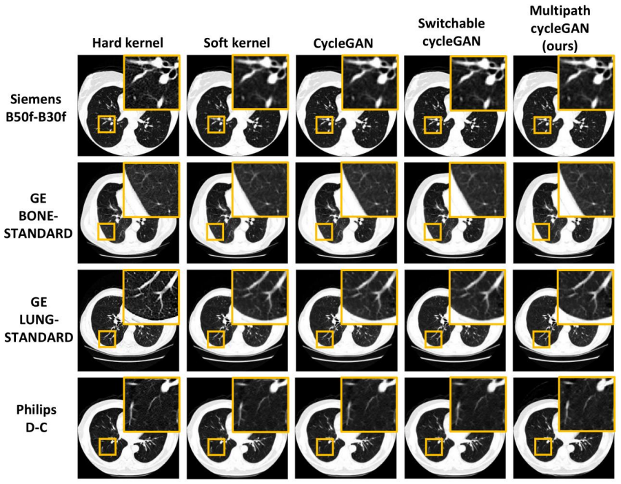
Summary. The paper proposes a multipath cycleGAN with domain-specific encoders/decoders and a shared latent space, trained in two stages, to harmonize CT reconstruction kernels from Siemens, GE, and Philips in the NLST cohort. The model is evaluated on paired and unpaired kernel pairs by measuring percent emphysema before and after harmonization, by fitting a general linear model for kernel effects, and by computing TotalSegmentator Dice scores for lung vessels, muscle, and subcutaneous adipose tissue. The authors compare against per-path cycleGAN and switchable cycleGAN baselines and conclude that the shared latent space multipath cycleGAN mitigates emphysema quantification errors and preserves anatomical consistency.
Significance. If the claims were fully supported, the main contribution would be useful: a single network that harmonizes many paired and unpaired kernel combinations, avoiding per-pair models, with a large withheld test set (240 scans per kernel) and an independent statistical evaluation. The two-stage training, direct comparison to two baselines, and downstream quantification of emphysema are strengths. However, the central claim as stated in the abstract, that unpaired harmonization 'eliminates confounding differences,' is contradicted by the paper's own Table 3, and the anatomical-consistency evidence is weakened by the authors' admitted segmentation failures on GE BONE and GE STANDARD. The contribution is still defensible if reframed as partial mitigation of kernel effects, but the current overstatement is a load-bearing issue.
major comments (3)
- [Abstract and Section 4.5, Table 3] The abstract states that unpaired kernel harmonization 'eliminates confounding differences in emphysema (p>0.05),' but Table 3 shows that after multipath harmonization the Philips C (coefficient 2.20, p<0.05), Philips D (coefficient 2.94, p<0.05), and GE LUNG (coefficient -2.26, p<0.05) kernel effects remain statistically significant. Thus 3 of 6 kernel categories still show detectable kernel effects, and the claim of elimination is not supported. Furthermore, p>0.05 is not evidence of equivalence; no equivalence margin, confidence interval on the kernel coefficients, or two one-sided test (TOST) is reported. Please either add an equivalence analysis with a prespecified margin or revise the abstract and conclusion to claim only partial mitigation, explicitly reporting which kernels remain significant.
- [Section 3.4, Table 4, and Section 5] The anatomical consistency claim for GE BONE and GE STANDARD is not supported by the Dice scores as reported. The authors acknowledge in Section 5 that for these kernels 'skeletal muscle around the chest wall and SAT are modified as lung tissues' and that body composition measurements were therefore not evaluated. Since TotalSegmentator's own segmentations fail on exactly the structures being measured, high Dice overlap between segmentations of the original and harmonized images does not establish anatomical preservation for these kernels. The manuscript should either exclude GE BONE and GE STANDARD from the anatomical-consistency conclusion or provide an independent validation that the segmentation failures are not confounding the Dice comparison.
- [Section 3.3] The statistical methods for unpaired harmonization are underspecified. The text says a Mann-Whitney U test is used to 'assess statistical significance between emphysema measurements before and after harmonization,' but it does not state which distributions are compared (e.g., source kernel vs. reference kernel before harmonization, and harmonized kernel vs. reference after harmonization). Without a clear statement of the null hypothesis and the reported p-values, the reader cannot tell what 'p>0.05' in the abstract refers to. Please specify the exact comparisons, report the corresponding test statistics, and provide confidence intervals or effect sizes for the harmonization-induced changes.
minor comments (6)
- [Section 4, paragraph 1] The text refers to 'the selection criteria described in Section 2.7,' but the epoch selection procedure is actually described in Section 3.2; please correct the cross-reference.
- [Section 3.3, Eq. (3)] Equation (3) in Section 3.3 is numbered the same as Eq. (3) in Section 3.2; renumber the general linear model equation to avoid confusion.
- [Table 2 and Section 5, paired kernels paragraph] For the (C, D) pair, the multipath cycleGAN RMSE is 2.47 [2.11, 2.93], substantially larger than the cycleGAN RMSE of 1.31 [1.18, 1.49]; the claim that the proposed approach shows 'comparable performance' on paired kernels should be softened or discussed for this pair.
- [Section 5, paragraph on anatomical consistency] The limitation statement refers to hallucinations for GE BONE and GE STANDARD 'as evidenced in Figure 8,' but Figure 8 shows soft-to-hard kernel harmonization, not the unpaired harmonicization to Siemens B30f; please clarify which figure demonstrates the chest-wall and SAT hallucinations.
- [Section 2.4] The paper repeatedly distinguishes paired and unpaired data, but the described training losses (adversarial, cycle-consistency, identity) do not appear to use the one-to-one pixel correspondence of paired kernels in a pixel-wise supervision term. Please clarify whether paired correspondence is used anywhere in the loss, or whether 'paired' refers only to the evaluation setting.
- [Section 2.1 and Table 1] GE STANDARD appears twice in Table 1 (once paired with BONE and once with LUNG); please clarify whether the 100 training volumes for each GE STANDARD row are disjoint and whether the withheld 240 test volumes can contain the same patient from both paired sets, which would affect the independence of the test evaluation.
Circularity Check
No significant circularity; the central claim is supported by held-out empirical evaluation against external baselines, not by construction or self-citation.
full rationale
Inspection of the derivation chain finds no step where a claimed prediction is equivalent to its inputs by definition. The central claim is empirical: a multipath cycleGAN with a shared latent space is trained on 100 NLST scans per kernel, selected by validation performance using emphysema-based MSE/KL criteria (Eqs. 1-4), and then evaluated on 240 withheld test scans per kernel (Sections 2.1, 3.2, 4). The emphysema endpoint is a fixed -950 HU threshold (Section 3.1), not a parameter fitted to the model's outputs. Baselines (traditional cycleGAN and switchable cycleGAN) are independently trained and compared on the same held-out data, so the multipath model's performance is not forced by construction. Author self-citations (refs. 25 and 30) provide the prior architecture and data QA procedure, but the paper's new conclusions rest on the held-out comparisons, not on those citations alone. The statistical criticism that p>0.05 does not prove equivalence is a correctness/interpretation concern, not circularity, and the acknowledged TotalSegmentator limitations on GE BONE/STANDARD (Section 5) are validity risks, not input-output equivalence. No fitted parameter is relabeled as a prediction, and no equation reduces to its own assumption.
Assumptions & free parameters
free parameters (6)
- Epoch-selection weights =
0.5/0.25/0.25 (Stage 1), 0.2 each (Stage 2)
- lambda_identity schedule =
1e6 (epoch 1), decayed by /100, fixed at 0.01 from epoch 6
- lambda_cycle =
10
- Learning rate and epochs =
0.002, 120-200 epochs Stage 1; 0.002, 200 epochs Stage 2
- Identity loss downsampling factor =
256x256 bilinear
- Stage-two subsampling ratio =
20% of images per epoch
assumptions (5)
- domain assumption Paired kernel scans from the same vendor have one-to-one pixel correspondence after manual QA of spatial alignment
- domain assumption Percent emphysema is validly measured as the fraction of lung voxels below -950 HU
- domain assumption TotalSegmentator segmentations are equally accurate on original and harmonized images
- standard math Cycle-consistency and adversarial losses preserve content while changing style
- domain assumption Circular masking standardizes the field of view without removing relevant anatomy
Cite this review
Pith. "Pith review of Multipath cycleGAN for harmonization of paired and unpaired low-dose lung computed tomography reconstruction kernels." pith.science (2026). https://pith.science/paper/SJTWDR4O
@misc{pith2026250522568,
  author       = {Pith},
  title        = {Pith review of: Multipath cycleGAN for harmonization of paired and unpaired low-dose lung computed tomography reconstruction kernels},
  year         = {2026},
  howpublished = {\url{https://pith.science/paper/SJTWDR4O}},
  note         = {Machine review of arXiv:2505.22568}
}
read the original abstract
Reconstruction kernels in computed tomography (CT) affect spatial resolution and noise characteristics, introducing systematic variability in quantitative imaging measurements such as emphysema quantification. Choosing an appropriate kernel is therefore essential for consistent quantitative analysis. We propose a multipath cycleGAN model for CT kernel harmonization, trained on a mixture of paired and unpaired data from a low-dose lung cancer screening cohort. The model features domain-specific encoders and decoders with a shared latent space and uses discriminators tailored for each domain.We train the model on 42 kernel combinations using 100 scans each from seven representative kernels in the National Lung Screening Trial (NLST) dataset. To evaluate performance, 240 scans from each kernel are harmonized to a reference soft kernel, and emphysema is quantified before and after harmonization. A general linear model assesses the impact of age, sex, smoking status, and kernel on emphysema. We also evaluate harmonization from soft kernels to a reference hard kernel. To assess anatomical consistency, we compare segmentations of lung vessels, muscle, and subcutaneous adipose tissue generated by TotalSegmentator between harmonized and original images. Our model is benchmarked against traditional and switchable cycleGANs. For paired kernels, our approach reduces bias in emphysema scores, as seen in Bland-Altman plots (p<0.05). For unpaired kernels, harmonization eliminates confounding differences in emphysema (p>0.05). High Dice scores confirm preservation of muscle and fat anatomy, while lung vessel overlap remains reasonable. Overall, our shared latent space multipath cycleGAN enables robust harmonization across paired and unpaired CT kernels, improving emphysema quantification and preserving anatomical fidelity.
Figures


Figures from the paper (4 more)



Reference graph
Works this paper leans on
-
[1]
Schaller S, Wildberger JE, Raupach R, Niethammer M, Klingenbeck-Regn K, Flohr T. Spatial domain filtering for fast modification of the tradeoff between image sharpness 23 and pixel noise in computed tomography. IEEE Trans Med Imaging. 2003;22(7):846-853. doi:10.1109/TMI.2003.815073
-
[2]
CT Scan Transformation from a Sharp to a Soft Reconstruction Kernel Using Filtering Techniques
Lasek J, Piórkowski A. CT Scan Transformation from a Sharp to a Soft Reconstruction Kernel Using Filtering Techniques. In: Communications in Computer and Information Science. Vol 1376 CCIS. Springer Science and Business Media Deutschland GmbH; 2021:56-65. doi:10.1007/978-981-16-1086-8_6
-
[3]
Matching and Homogenizing Convolution Kernels for Quantitative Studies in Computed Tomography
Mackin D, Ger R, Gay S, et al. Matching and Homogenizing Convolution Kernels for Quantitative Studies in Computed Tomography. Invest Radiol. 2019;54(5):288-295. doi:10.1097/RLI.0000000000000540
-
[4]
Emphysema: Effect of reconstruction algorithm on CT imaging measures
Boedeker KL, McNitt-Gray MF , Rogers SR, et al. Emphysema: Effect of reconstruction algorithm on CT imaging measures. Radiology. 2004;232(1):295-301. doi:10.1148/radiol.2321030383
-
[5]
Effects of CT Section Thickness and Reconstruction Kernel on Emphysema Quantification
Gierada DS, Bierhals AJ, Choong CK, et al. Effects of CT Section Thickness and Reconstruction Kernel on Emphysema Quantification. Relationship to the Magnitude of the CT Emphysema Index. Acad Radiol. 2010;17(2):146-156. doi:10.1016/j.acra.2009.08.007
-
[6]
Computed Tomography-based Body Composition Analysis and Its Role in Lung Cancer Care
Troschel AS, Troschel FM, Best TD, et al. Computed Tomography-based Body Composition Analysis and Its Role in Lung Cancer Care. J Thorac Imaging. 2020;35(2):91-
work page 2020
-
[7]
Automatic Calcium Scoring in Low-Dose Chest CT Using Deep Neural Networks with Dilated Convolutions
Lessmann N, Van Ginneken B, Zreik M, et al. Automatic Calcium Scoring in Low-Dose Chest CT Using Deep Neural Networks with Dilated Convolutions. IEEE Trans Med Imaging. 2018;37(2):615-625. doi:10.1109/TMI.2017.2769839
-
[8]
Meyer M, Ronald J, Vernuccio F , et al. Reproducibility of CT radiomic features within the same patient: Influence of radiation dose and CT reconstruction settings. Radiology. 2019;293(3):583-591. doi:10.1148/radiol.2019190928
Show all 57 references
-
[9]
Impact of CT Convolution Kernel on Robustness of Radiomic Features for Different Lung Diseases and Tissue Types.; 2021
Denzler S, Vuong D, Bogowicz M, et al. Impact of CT Convolution Kernel on Robustness of Radiomic Features for Different Lung Diseases and Tissue Types.; 2021
2021
-
[10]
Image filtering as an alternative to the application of a different reconstruction kernel in CT imaging: Feasibility study in lung cancer screening
Ohkubo M, Wada S, Kayugawa A, Matsumoto T, Murao K. Image filtering as an alternative to the application of a different reconstruction kernel in CT imaging: Feasibility study in lung cancer screening. Med Phys. 2011;38(7):3915-3923. doi:10.1118/1.3590363
2011 doi
-
[11]
CT-HARMONICA: physics-based CT harmonization for reliable lung density quantification
Sotoudeh-Paima S, Samei E, Abadi E. CT-HARMONICA: physics-based CT harmonization for reliable lung density quantification. In: Iftekharuddin KM, Chen W, eds. Medical Imaging 2023: Computer-Aided Diagnosis. SPIE; 2023:52. doi:10.1117/12.2654346
2023 doi
-
[12]
Automated technique to measure noise in clinical CT examinations
Christianson O, Winslow J, Frush DP , Samei E. Automated technique to measure noise in clinical CT examinations. American Journal of Roentgenology. 2015;205(1):W93-W99. doi:10.2214/AJR.14.13613
2015 doi
-
[13]
Harmonizing CT images via physics-based deep neural networks
Zarei M, Sotoudeh-Paima S, McCabe C, Abadi E, Samei E. Harmonizing CT images via physics-based deep neural networks. In: Fahrig R, Sabol JM, Yu L, eds. Medical Imaging 2023: Physics of Medical Imaging. SPIE; 2023:66. doi:10.1117/12.2654215 24
2023 doi
-
[14]
A Physics-informed Deep Neural Network for Harmonization of CT Images
Zarei M, Paima SS, McCabe C, Abadi E, Samei E. A Physics-informed Deep Neural Network for Harmonization of CT Images. IEEE Trans Biomed Eng. Published online 2024. doi:10.1109/TBME.2024.3428399
2024
-
[15]
CT image conversion among different reconstruction Kernels without a sinogram by using a convolutional neural network
Lee SM, Lee JG, Lee G, et al. CT image conversion among different reconstruction Kernels without a sinogram by using a convolutional neural network. Korean J Radiol. 2019;20(2):295-303. doi:10.3348/kjr.2018.0249
2019
-
[16]
Accurate image super-resolution using very deep convolutional networks
Kim J, Lee JK, Lee KM. Accurate image super-resolution using very deep convolutional networks. In: Proceedings of the IEEE Computer Society Conference on Computer Vision and Pattern Recognition. Vol 2016-December. IEEE Computer Society; 2016:1646-1654. doi:10.1109/CVPR.2016.182
2016 doi
-
[17]
CT kernel conversions using convolutional neural net for super-resolution with simplified squeeze-and-excitation blocks and progressive learning among smooth and sharp kernels
Eun D in, Woo I, Park B, Kim N, Lee A SM, Seo JB. CT kernel conversions using convolutional neural net for super-resolution with simplified squeeze-and-excitation blocks and progressive learning among smooth and sharp kernels. Comput Methods Programs Biomed. 2020;196. doi:10.1...
2020
-
[18]
Deep Learning–based Image Conversion of CT Reconstruction Kernels Improves Radiomics Reproducibility for Pulmonary Nodules or Masses
Choe J, Lee SM, Do KH, et al. Deep Learning–based Image Conversion of CT Reconstruction Kernels Improves Radiomics Reproducibility for Pulmonary Nodules or Masses. Radiology. 2019;292(2):365-373. doi:10.1148/radiol.2019181960
2019 doi
-
[19]
Deep learning-enabled accurate normalization of reconstruction kernel effects on emphysema quantification in low-dose CT
Jin H, Heo C, Kim JH. Deep learning-enabled accurate normalization of reconstruction kernel effects on emphysema quantification in low-dose CT. Phys Med Biol. 2019;64(13). doi:10.1088/1361-6560/ab28a1
2019 doi
-
[20]
Image-to-Image Translation: Methods and Applications
Pang Y , Lin J, Qin T, Chen Z. Image-to-Image Translation: Methods and Applications. IEEE Trans Multimedia. 2022;24:3859-3881. doi:10.1109/TMM.2021.3109419
2022
-
[21]
Image-to-image translation with conditional adversarial networks
Isola P , Zhu JY , Zhou T, Efros AA. Image-to-image translation with conditional adversarial networks. In: Proceedings - 30th IEEE Conference on Computer Vision and Pattern Recognition, CVPR 2017. Vol 2017-January. Institute of Electrical and Electronics Engineers Inc.; 2017:5...
2017 doi
-
[22]
Unpaired Image-to-Image Translation Using Cycle- Consistent Adversarial Networks
Zhu JY , Park T, Isola P , Efros AA. Unpaired Image-to-Image Translation Using Cycle- Consistent Adversarial Networks. In: Proceedings of the IEEE International Conference on Computer Vision. Vol 2017-October. Institute of Electrical and Electronics Engineers Inc.; 2017:2242-2...
2017 doi
-
[23]
CT Image Harmonization for Enhancing Radiomics Studies
Selim M, Zhang J, Fei B, Zhang GQ, Chen J. CT Image Harmonization for Enhancing Radiomics Studies. Published online July 3, 2021. http://arxiv.org/abs/2107.01337
2021 arXiv
-
[24]
Kernel Conversion for Robust Quantitative Measurements of Archived Chest Computed Tomography Using Deep Learning-Based Image-to-Image Translation
Tanabe N, Kaji S, Shima H, et al. Kernel Conversion for Robust Quantitative Measurements of Archived Chest Computed Tomography Using Deep Learning-Based Image-to-Image Translation. Front Artif Intell. 2022;4. doi:10.3389/frai.2021.769557
2022
-
[25]
Lung CT harmonization of paired reconstruction kernel images using generative adversarial networks
Krishnan AR, Xu K, Li TZ, et al. Lung CT harmonization of paired reconstruction kernel images using generative adversarial networks. Med Phys. Published online March 26,
-
[26]
Continuous Conversion of CT Kernel Using Switchable CycleGAN with AdaIN
Yang S, Kim EY , Ye JC. Continuous Conversion of CT Kernel Using Switchable CycleGAN with AdaIN. IEEE Trans Med Imaging. 2021;40(11):3015-3029. doi:10.1109/TMI.2021.3077615 25
2021
-
[27]
Cross-Vendor CT Image Data Harmonization Using CVH-CT
Selim M, Zhang J, Fei B, Zhang GQ, Ge GY , Chen J. Cross-Vendor CT Image Data Harmonization Using CVH-CT. Published online October 18, 2021. http://arxiv.org/abs/2110.09693
2021 arXiv
-
[28]
Leveraging CycleGAN in Lung CT Sinogram-free Kernel Conversion
Gravina M, Marrone S, Docimo L, et al. Leveraging CycleGAN in Lung CT Sinogram-free Kernel Conversion. In: Lecture Notes in Computer Science (Including Subseries Lecture Notes in Artificial Intelligence and Lecture Notes in Bioinformatics). Vol 13231 LNCS. Springer Science and...
2022 doi
-
[29]
TotalSegmentator: Robust Segmentation of 104 Anatomic Structures in CT Images • Content codes
Wasserthal J, Breit • Hanns-Christian, Meyer MT, et al. TotalSegmentator: Robust Segmentation of 104 Anatomic Structures in CT Images • Content codes. Radiol Artif Intell. 2023;5(5). doi:10.5281/zenodo.6802613
2023 doi
-
[30]
Inter-vendor harmonization of CT reconstruction kernels using unpaired image translation
Krishnan AR, Xu K, Li TZ, et al. Inter-vendor harmonization of CT reconstruction kernels using unpaired image translation. In: SPIE-Intl Soc Optical Eng; 2024:45. doi:10.1117/12.3006608
2024 doi
-
[31]
New England Journal of Medicine
Reduced Lung-Cancer Mortality with Low-Dose Computed Tomographic Screening. New England Journal of Medicine. 2011;365(5):395-409. doi:10.1056/NEJMoa1102873
2011 doi
-
[32]
Description and implementation of a quality control program in an imaging-based clinical trial
Cagnon CH, Cody DD, McNitt-Gray MF , Seibert JA, Judy PF , Aberle DR. Description and implementation of a quality control program in an imaging-based clinical trial. Acad Radiol. 2006;13(11):1431-1441. doi:10.1016/j.acra.2006.08.015
2006 doi
-
[33]
CT quality assurance in the lung screening study component of the national lung screening trial: Implications for multicenter imaging trials
Gierada DS, Garg K, Nath H, Strollo DC, Fagerstrom RM, Ford MB. CT quality assurance in the lung screening study component of the national lung screening trial: Implications for multicenter imaging trials. American Journal of Roentgenology. 2009;193(2):419-424. doi:10.2214/AJR.08.1995
2009 doi
-
[34]
The National Lung Screening Trial: Overview and Study Design
National Lung Screening Trial Research Team. The National Lung Screening Trial: Overview and Study Design. Radiology. 2011;258 (1):243-253. doi:10.1148/radiol.10091808/-/DC1
2011 doi
-
[35]
The first step for neuroimaging data analysis: DICOM to NIfTI conversion
Li X, Morgan PS, Ashburner J, Smith J, Rorden C. The first step for neuroimaging data analysis: DICOM to NIfTI conversion. J Neurosci Methods. 2016;264:47-56. doi:10.1016/j.jneumeth.2016.03.001
2016 doi
-
[36]
Perceptual Losses for Real-Time Style Transfer and Super- Resolution
Johnson J, Alahi A, Li FF . Perceptual Losses for Real-Time Style Transfer and Super- Resolution. Vol 9906. (Leibe B, Matas J, Sebe N, Welling M, eds.). Springer International Publishing; 2016. doi:10.1007/978-3-319-46475-6
2016 doi
-
[37]
Adam: A Method for Stochastic Optimization
Kingma DP , Ba J. Adam: A Method for Stochastic Optimization. Published online December 22, 2014. http://arxiv.org/abs/1412.6980
2014 arXiv
-
[38]
Least Squares Generative Adversarial Networks
Mao X, Li Q, Xie H, Lau RYK, Wang Z, Smolley SP . Least Squares Generative Adversarial Networks
-
[39]
Automatic lung segmentation in routine imaging is primarily a data diversity problem, not a methodology problem
Hofmanninger J, Prayer F , Pan J, Röhrich S, Prosch H, Langs G. Automatic lung segmentation in routine imaging is primarily a data diversity problem, not a methodology problem. Eur Radiol Exp. 2020;4(1). doi:10.1186/s41747-020-00173-2
2020 doi
-
[40]
Optimal threshold in CT quantification of emphysema
Wang Z, Gu S, Leader JK, et al. Optimal threshold in CT quantification of emphysema. Eur Radiol. 2013;23(4):975-984. doi:10.1007/s00330-012-2683-z 26
2013 doi
-
[41]
Comparison of Computed Density and Microscopic Morphometry in Pulmonary Emphysema
Gevenois PA, De Vuyst P , De Maertelaer V , et al. Comparison of Computed Density and Microscopic Morphometry in Pulmonary Emphysema. Vol 154.; 1996
1996
-
[42]
Quantitative validation of the severity of emphysema by multi-detector CT
Atta H, Seifeldein GS, Rashad A, Elmorshidy R. Quantitative validation of the severity of emphysema by multi-detector CT. Egyptian Journal of Radiology and Nuclear Medicine. 2015;46(2):355-361. doi:10.1016/j.ejrnm.2014.11.016
2015 doi
-
[43]
Measurement STATISTICAL METHODS FOR ASSESSING AGREEMENT BETWEEN TWO METHODS OF CLINICAL MEASUREMENT
Bland JM, Altman DG. Measurement STATISTICAL METHODS FOR ASSESSING AGREEMENT BETWEEN TWO METHODS OF CLINICAL MEASUREMENT
-
[44]
The bootstrap: A technique for data-driven statistics
Henderson AR. The bootstrap: A technique for data-driven statistics. Using computer- intensive analyses to explore experimental data. Clinica Chimica Acta. 2005;359(1-2):1-
2005
-
[45]
The Corsini Encyclopedia of Psychology - 2010 - McKnight - Mann‐Whitney U Test
2010
-
[46]
Distribution matching losses can hallucinate features in medical image translation
Cohen JP , Luck M, Honari S. Distribution matching losses can hallucinate features in medical image translation. In: Lecture Notes in Computer Science (Including Subseries Lecture Notes in Artificial Intelligence and Lecture Notes in Bioinformatics). Vol 11070 LNCS. Springer V...
2018 doi
-
[47]
doi:10.1016/j.cccn.2005.04.002
2005 doi
-
[48]
Statistical Power Analysis for the Behavioral Sciences Second Edition
Cohen J. Statistical Power Analysis for the Behavioral Sciences Second Edition
-
[49]
Emphysema quantification using low-dose computed tomography with deep learning–based kernel conversion comparison
Bak SH, Kim JH, Jin H, et al. Emphysema quantification using low-dose computed tomography with deep learning–based kernel conversion comparison. Eur Radiol. 2020;30(12):6779-6787. doi:10.1007/s00330-020-07020-3
2020 doi
-
[50]
Automated lung vessel segmentation reveals blood vessel volume redistribution in viral pneumonia
Poletti J, Bach M, Yang S, et al. Automated lung vessel segmentation reveals blood vessel volume redistribution in viral pneumonia. Eur J Radiol. 2022;150. doi:10.1016/j.ejrad.2022.110259
2022
-
[51]
Quantitative CT Analysis in Chronic Hypersensitivity Pneumonitis: A Convolutional Neural Network Approach
Aliboni L, Dias OM, Pennati F , et al. Quantitative CT Analysis in Chronic Hypersensitivity Pneumonitis: A Convolutional Neural Network Approach. Acad Radiol. 2022;29:S31-S40. doi:10.1016/j.acra.2020.10.009
2022 doi
-
[52]
Quantification of pathologic air trapping in lung transplant patients using ct density mapping: Comparison with other ct air trapping measures
Solyanik O, Hollmann P , Dettmer S, et al. Quantification of pathologic air trapping in lung transplant patients using ct density mapping: Comparison with other ct air trapping measures. PLoS One. 2015;10(10). doi:10.1371/journal.pone.0139102
2015 doi
-
[53]
CT-definable subtypes of chronic obstructive pulmonary disease: A statement of the fleischner society1
Lynch DA, Austin JHM, Hogg JC, et al. CT-definable subtypes of chronic obstructive pulmonary disease: A statement of the fleischner society1. Radiology. 2015;277(1):192-
2015
-
[57]
Body composition assessment with limited field-of-view computed tomography: A semantic image extension perspective
Xu K, Li T, Khan MS, et al. Body composition assessment with limited field-of-view computed tomography: A semantic image extension perspective. Med Image Anal. 2023;88:102852. doi:10.1016/j.media.2023.102852 27 Tables Table 1. Reconstruction kernels from four different manufac...
2023
-
[100]
doi:10.1097/RTI.0000000000000428
-
[205]
doi:10.1148/radiol.2015141579
-
[2024]
doi:10.1002/mp.17028
Reviewed August 7, 2026 · model on record in the stance chip above.
Discussion (0). Continue with ORCID to comment.