REVIEW 4 major objections 5 minor 45 references
pyMEAL: A Multi-Encoder Augmentation-Aware-Learning Toolbox for Robust Medical Image Translation
T0 review · 4 major / 5 minor · reviewed 2026-08-07 · deepseek-v4-flash
Pith's one-line read The paper claims that treating each augmentation as a distinct anatomical view, processed by its own encoder path and reweighted by a learned controller, makes CT-to-T1 MRI translation robust to geometric and intensity perturbations.
desk verdict A sensible multi-encoder augmentation-aware architecture for CT-to-T1, but the headline gains are confounded by a 273M-vs-4-14M parameter disparity and no external baselines. read the letter →
The pith
A machine-rendered reading of the paper's core claim, the machinery that carries it, and where it could break.
The reading
What carries the argument
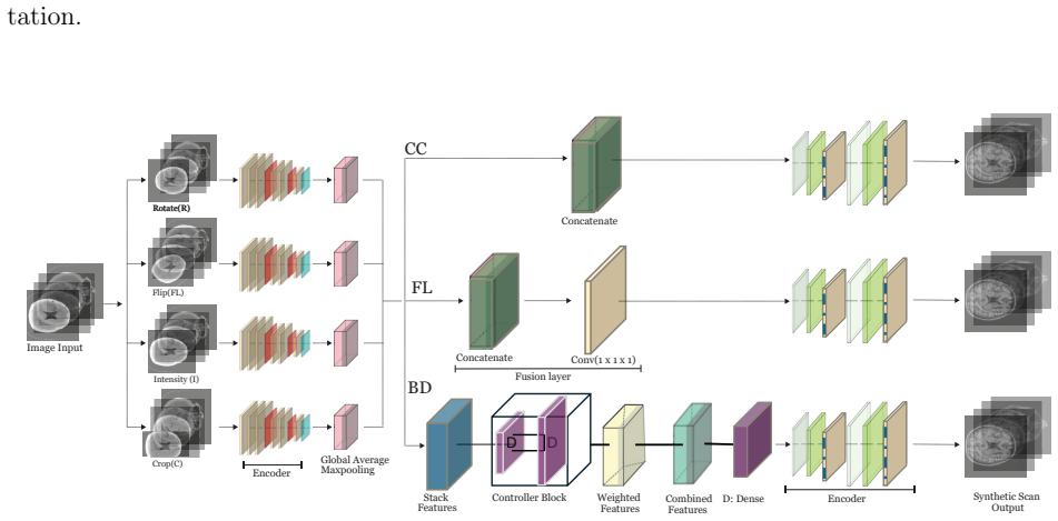
The load-bearing component is the controller block that computes per-stream attention weights $\alpha_k = \mathrm{softmax}(w^\top \mathrm{ReLU}(W \cdot \mathrm{GAP}(h_k)))$ and fuses shared-encoder features as $F_{\mathrm{BD}} = \sum_{k=1}^4 \alpha_k f_\theta(A_k(X))$, where $A_k$ are differentiable flip, rotation, crop, and intensity modules embedded in the computational graph. The shared encoder $f_\theta$ processes all four augmented views, and the decoder $\Gamma_\phi$ reconstructs the translated volume from the weighted combination. This mechanism lets the network suppress uninformative transformations and amplify informative ones during training, which the paper identifies as the source of robustness.
What would settle it
Run a controlled comparison in which every model receives the same four augmented views (for instance, as a four-channel input or through a shared stem) and the total trainable parameter count is matched to MEAL-BD's, then evaluate PSNR and SSIM on the same unseen test set under rotation, crop, flip, and intensity perturbations. If a single-stream baseline matches or beats MEAL-BD, the claim that controller-weighted fusion is responsible for the robustness fails.
Extended reading notes
Core claim
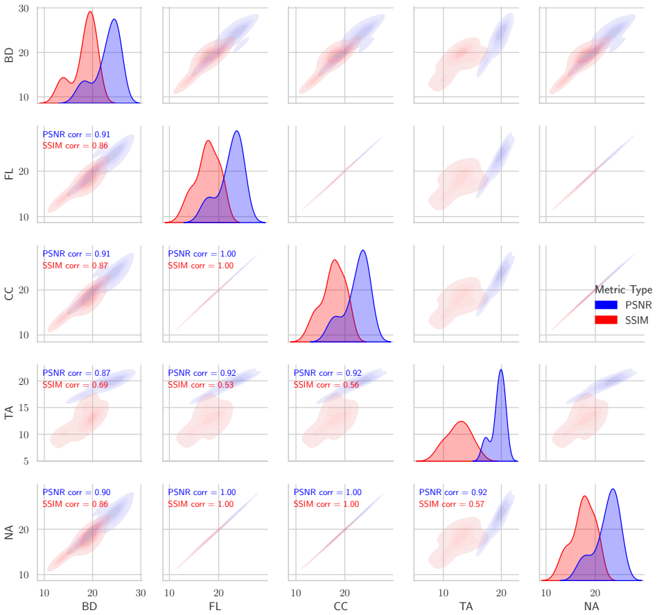
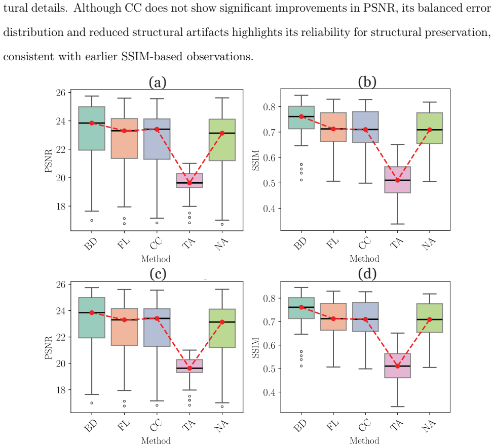
MEAL-BD, the variant with an adaptive controller block, consistently outperforms the other configurations (no augmentation, traditional augmentation, concatenation, and fusion-layer fusion) in CT-to-T1-weighted MRI translation. On unseen test data it reports the highest mean PSNR of 23.03 dB and SSIM of 0.733, and on the predefined test set 24.12 dB and 0.745; under rotation, crop, and flip augmentations it stays above roughly 23 dB and 0.72, while intensity perturbation yields lower but still leading scores. The authors attribute the gain to dynamic attention weighting of augmentation-specific features rather than to simple concatenation or averaging of those features.
Load-bearing premise
The load-bearing premise is that the comparison is fair: all methods get informationally equivalent inputs, and MEAL-BD's edge comes from the controller fusion rather than from receiving four augmented views while single-stream baselines receive one, or from having roughly 20 to 60 times more trainable parameters.
Editorial extensions
If this is right
- If MEAL-BD's advantage is real, augmentation-aware controller weighting becomes a design option for other 3D medical translation tasks, not just CT-to-MRI.
- The reported stability under rotation, crop, and flip suggests the architecture tolerates patient positioning and field-of-view differences without retraining.
- MEAL-BD's lead on unseen data implies the learned weighting transfers to scans the model never saw during training.
- The framing of augmentation as complementary views could be carried to tasks like segmentation and registration, as the paper itself proposes.
Reading between the lines
- The paper does not specify whether single-stream baselines received one or four augmented views at test time; if they received one while BD received four, part of the gain could be input diversity rather than the controller.
- The parameter counts in Section 2.5 (BD: 273M; CC/FL: 10–14M; NA/TA: 4.4M) contradict Section 2.1.4's claim of a 75% parameter reduction, so the efficiency advantage is not established.
- A natural testable extension is to replace the fixed set of four augmentations with a learned augmentation policy, where the differentiable modules $A_k$ are optimized by the same gradient signal.
- The paper mentions Dice on white and gray matter segmentation as an evaluation in Section 2.4 but reports no Dice results; reporting those numbers would test whether PSNR and SSIM gains translate to anatomical accuracy.
Editorial analysis
A structured set of objections, weighed in public.
Referee Report
Summary. The paper proposes pyMEAL, a multi-encoder augmentation-aware learning framework for 3D medical image translation, evaluated on CT-to-T1-weighted MRI synthesis using OASIS-3 data. The framework routes four augmentation variants through parallel encoder streams and compares three fusion strategies: concatenation (CC), a fusion layer (FL), and a dynamic controller block (BD). The central claim is that MEAL-BD, by dynamically weighting augmentation-specific features, consistently outperforms the other configurations and baseline no-augmentation/traditional-augmentation models in PSNR and SSIM on both predefined and unseen test data.
Significance. The core idea—treating augmentations as complementary views and learning to fuse them with an attention-style controller—is timely and potentially useful for robustness in medical image translation. The paper is also commendable for releasing pip-installable code and for applying a detailed statistical protocol (normality tests, Kruskal-Wallis, Dunn with Bonferroni). However, the empirical evidence as presented is confounded. The reported trainable-parameter counts contradict the claimed parameter reduction, the evaluation lacks external state-of-the-art baselines, and the test-time input protocol for single-stream baselines is unspecified. If these issues were resolved with controlled, parameter-matched comparisons and full pairwise reporting, the contribution could be solid; in its current form, the central claim is not established.
major comments (4)
- [Section 2.1.4 and Section 2.5] The claim in Section 2.1.4 that 'This architecture reduces parameters by 75% compared to other methods' is directly contradicted by the parameter counts in Section 2.5: BD has 273,265,538 trainable parameters, whereas CC has 13,725,953, FL has 10,760,577, and NA/TA have 4,428,545. BD is therefore 6 to 62 times larger than the alternatives, not 75% smaller. Because model capacity is a well-known confound for reconstruction quality, the reported PSNR/SSIM advantages of BD cannot be attributed to the controller or fusion mechanism without parameter-matched baselines or ablations that isolate the architectural contribution.
- [Section 3.2.1 and Table 2] The claim that MEAL-BD 'consistently outperformed competing approaches' is not supported by the reported pairwise tests. Under the intensity augmentation condition in Table 2, Dunn's tests are reported for BD vs FL and BD vs TA, but not for BD vs CC or BD vs NA, even though the mean scores show BD, NA, and CC close together (21.210, 20.590, 20.054). Similarly, in the no-augmentation results in Table 1, only BD vs TA is reported, not BD vs CC, FL, or NA. The central claim of consistent superiority requires these key pairwise comparisons to be reported; their absence raises the possibility that the differences are not significant.
- [Section 3 evaluation and Section 1] All comparative results are obtained against the paper's own internal configurations (NA, TA, CC, FL). The Introduction states that 'MEAL outperforms state-of-the-art single-stream and multi-modal baselines,' but no existing CT-to-MRI translation method (e.g., CycleGAN, Pix2Pix, or published U-Net variants) is evaluated. Without at least one external baseline, the robustness and superiority claims are not contextualized against the literature, and the reader cannot judge whether the gains are specific to this dataset and setup or representative of genuine improvement.
- [Section 3.2 and Section 2.2] The test-time evaluation protocol under augmentation-specific conditions is not specified. In Section 3.2.1, results are presented 'under various augmentation scenarios (rotation, crop, flip, and intensity),' but it is not stated whether all models receive the same augmented input at test time, or whether the multi-encoder models (BD, CC, FL) receive four augmented input views while the single-stream models (NA, TA) receive only one. If the input information differs across methods, the comparison is not fair, and the claimed advantage of BD could partly stem from receiving multiple views rather than from the fusion architecture. The exact input protocol for each method at test time must be described.
minor comments (5)
- [Section 2.3, Eq. (12)] The text states 'L1 the mean square error,' but L1 conventionally denotes mean absolute error. Either the notation or the definition should be corrected to avoid ambiguity.
- [Section 2.1.2, Eq. (2)] Equation (2) uses the symbol M for channel-wise concatenation, while the text and symbols list define L as the concatenation operator. Please use a single consistent notation.
- [Section 3.5 and Section 2.3] There are minor typos, including 'hightligts' in Section 3.5 and 'The, we define' in Section 2.3. A careful proofreading pass is needed.
- [Figure 2] The caption refers to 'Multi-Stream with a Build Controller method,' while the text and acronym list define BD as 'Builder Block.' The terminology should be made consistent.
- [Table 2] Several p-values are reported as 0.0000. These should be reported as p < 0.0001 or as exact values with a clear convention.
Circularity Check
No significant circularity: the central claim is an empirical benchmark with held-out test evaluation, not a derivation that assumes its conclusion.
full rationale
The paper's central claim is that the MEAL-BD architecture outperforms competing methods in CT-to-T1 MRI translation. This is an empirical result supported by held-out test evaluations, paired statistical comparisons, and qualitative error maps, not a derivation from a premise that already contains the conclusion. The fusion weights alpha_k are learned from training data, and the same loss L = L1 + 0.8(1-SSIM) is optimized by all model variants, so the reported BD advantage is not a definitional consequence of the training objective. No load-bearing step reduces by construction to its inputs: there is no fitted parameter renamed as a prediction, no self-citation chain invoked to forbid alternatives, no imported uniqueness theorem, and no ansatz smuggled in via citation. The main weaknesses are external-validity and fairness concerns: BD has 273,265,538 trainable parameters versus 4,428,545 to 13,725,953 for the baselines, which confounds the architectural attribution of the gains; Section 2.1.4's claim that the shared-encoder design 'reduces parameters by 75% compared to other methods' is contradicted by the parameter table in Section 2.5; and the test-time input protocol for the single-stream baselines is not fully specified. These are correctness and experimental-design issues rather than circularity, so under the strict circularity rubric the appropriate finding is no significant circularity with score 0.
Assumptions & free parameters
free parameters (5)
- Number of augmentation streams =
4
- Augmentation hyperparameters =
flip p=0.5; rotation k in {0,1,2,3}; intensity delta in [-0.1,0.1]; contrast alpha in [0.9,1.1]; crop offsets from Eq. 9
- SSIM loss weight =
0.8
- Controller projection dimension d =
unspecified
- Network width and depth schedule =
64-128-256 channels, two downsampling stages
assumptions (4)
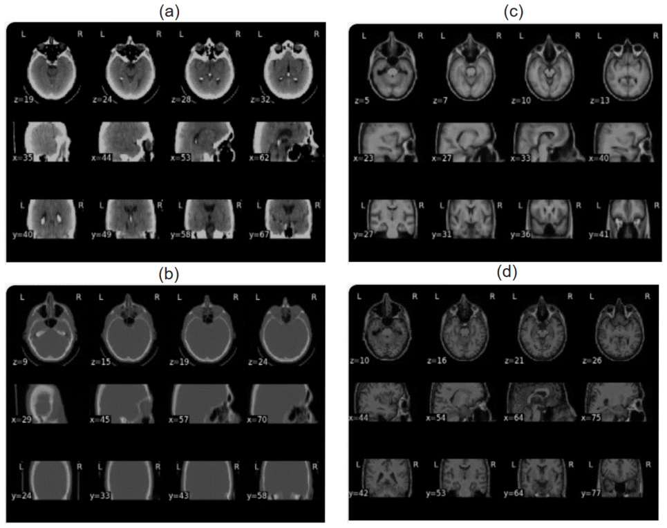
- domain assumption Paired OASIS-3 CT and T1 MRI volumes are accurately co-registered with ANTs so that voxel-wise losses are meaningful.
- domain assumption Synthetic augmentations (flips, rotations, crops, intensity shifts) are representative of clinically relevant acquisition variability.
- domain assumption The paired-data supervised learning setup with L1 plus SSIM loss is an adequate proxy for clinically useful CT-to-MRI translation.
- domain assumption The 204/51 train/validation split is at the subject level and does not leak the same subject across train and test sets.
Cite this review
Pith. "Pith review of pyMEAL: A Multi-Encoder Augmentation-Aware-Learning Toolbox for Robust Medical Image Translation." pith.science (2026). https://pith.science/paper/OPB3SIRQ
@misc{pith2026250524421,
  author       = {Pith},
  title        = {Pith review of: pyMEAL: A Multi-Encoder Augmentation-Aware-Learning Toolbox for Robust Medical Image Translation},
  year         = {2026},
  howpublished = {\url{https://pith.science/paper/OPB3SIRQ}},
  note         = {Machine review of arXiv:2505.24421}
}
read the original abstract
Medical imaging plays a vital role in clinical diagnosis, yet AI-driven imaging methods remain challenged by patient variability, image artifacts, and limited robustness across acquisition conditions. Although deep learning has advanced medical image analysis, 3D image translation remains hindered by limited training data and variability arising from scanner differences, imaging protocols, and patient motion. Conventional data augmentation typically relies on a single transformation pipeline, overlooking augmentation-specific characteristics and limiting representation learning. To address these challenges, we propose Multi-Encoder Augmentation-Aware Learning (MEAL), which processes multiple augmentation variants through dedicated encoder pathways. Three feature integration strategies are investigated: encoder concatenation (MEAL-CC), fusion layer (MEAL-FL), and an adaptive controller block (MEAL-BD). By dynamically weighting augmentation-specific features before decoding, MEAL-BD preserves complementary representations and improves robustness to clinically relevant variability. We evaluate MEAL using CT-to-T1-weighted MRI translation, a clinically relevant task when MRI is unavailable, contraindicated, or delayed. Across predefined and unseen test datasets, MEAL-BD consistently outperformed competing approaches under both geometric perturbations and standard imaging conditions, achieving higher peak signal-to-noise ratio (PSNR) and structural similarity index measure (SSIM). By prioritizing structural fidelity over perceptual realism, MEAL supports clinical interpretation and downstream image analysis rather than replacing diagnostic MRI, demonstrating that augmentation-aware representation learning improves the robustness and clinical applicability of medical image translation.
Figures

Figures from the paper (6 more)


Reference graph
Works this paper leans on
-
[1]
S. Hussain, I. Mubeen, N. Ullah, S. S. U. D. Shah, B. A. Khan, M. Zahoor, R. Ullah, F. A. Khan, M. A. Sultan, Modern diagnostic imaging technique applications and risk factors in the medical field: a review, BioMed research international 2022 (1) (2022) 5164970
work page 2022
-
[2]
E. Fountzilas, T. Pearce, M. A. Baysal, A. Chakraborty, A. M. Tsimberidou, Convergence of evolving artificial intelligence and machine learning techniques in precision oncology, npj Digital Medicine 8 (1) (2025) 75
work page 2025
-
[3]
H. N. Wagner Jr, P. S. Conti, Advances in medical imaging for cancer diagnosis and treatment, Cancer 67 (S4) (1991) 1121–1128
work page 1991
-
[4]
R. Attariwala, W. Picker, Whole body mri: improved lesion detection and characterization with diffusion weighted techniques, Journal of Magnetic Resonance Imaging 38 (2) (2013) 253–268
work page 2013
-
[5]
M. Cossio, Augmenting medical imaging: a comprehensive catalogue of 65 techniques for enhanced data analysis, arXiv preprint arXiv:2303.01178 (2023)
arXiv 2023
-
[6]
E. Ostertagova, O. Ostertag, J. Kováč, Methodology and application of the kruskal-wallis test, Applied mechanics and materials 611 (2014) 115–120
work page 2014
-
[7]
A. Alshardan, N. Alruwais, H. Alqahtani, A. Alshuhail, W. S. Almukadi, A. Sayed, Lever- aging transfer learning-driven convolutional neural network-based semantic segmentation model for medical image analysis using mri images, Scientific Reports 14 (1) (2024) 30549
work page 2024
- [8]
Show all 45 references
-
[9]
Alzubaidi, J
L. Alzubaidi, J. Zhang, A. J. Humaidi, A. Al-Dujaili, Y. Duan, O. Al-Shamma, J. San- tamaría, M. A. Fadhel, M. Al-Amidie, L. Farhan, Review of deep learning: concepts, cnn architectures, challenges, applications, future directions, Journal of big Data 8 (2021) 1–74. 32
2021
-
[10]
D. C. Elton, Z. Boukouvalas, M. D. Fuge, P. W. Chung, Deep learning for molecular design—a review of the state of the art, Molecular Systems Design & Engineering 4 (4) (2019) 828–849
2019
-
[11]
Huang, A
S.-C. Huang, A. Pareek, M. Jensen, M. P. Lungren, S. Yeung, A. S. Chaudhari, Self- supervised learning for medical image classification: a systematic review and implementa- tion guidelines, NPJ Digital Medicine 6 (1) (2023) 74
2023
-
[12]
Mumuni, F
A. Mumuni, F. Mumuni, Data augmentation: A comprehensive survey of modern ap- proaches, Array 16 (2022) 100258
2022
-
[13]
Alomar, H
K. Alomar, H. I. Aysel, X. Cai, Data augmentation in classification and segmentation: A survey and new strategies, Journal of Imaging 9 (2) (2023) 46
2023
-
[14]
Z. Yang, R. O. Sinnott, J. Bailey, Q. Ke, A survey of automated data augmentation algorithms for deep learning-based image classification tasks, Knowledge and Information Systems 65 (7) (2023) 2805–2861
2023
-
[15]
Garcea, A
F. Garcea, A. Serra, F. Lamberti, L. Morra, Data augmentation for medical imaging: A systematic literature review, Computers in Biology and Medicine 152 (2023) 106391
2023
-
[16]
Bosquet, D
B. Bosquet, D. Cores, L. Seidenari, V. M. Brea, M. Mucientes, A. Del Bimbo, A full data augmentation pipeline for small object detection based on generative adversarial networks, Pattern Recognition 133 (2023) 108998
2023
-
[17]
S. Kaji, S. Kida, Overview of image-to-image translation by use of deep neural networks: denoising, super-resolution, modality conversion, and reconstruction in medical imaging, Radiological physics and technology 12 (3) (2019) 235–248
2019
-
[18]
Shorten, T
C. Shorten, T. M. Khoshgoftaar, A survey on image data augmentation for deep learning, Journal of big data 6 (1) (2019) 1–48
2019
-
[19]
Kebaili, J
A. Kebaili, J. Lapuyade-Lahorgue, S. Ruan, Deep learning approaches for data augmen- tation in medical imaging: a review, Journal of imaging 9 (4) (2023) 81. 33
2023
-
[20]
Chlap, H
P. Chlap, H. Min, N. Vandenberg, J. Dowling, L. Holloway, A. Haworth, A review of medical image data augmentation techniques for deep learning applications, Journal of medical imaging and radiation oncology 65 (5) (2021) 545–563
2021
-
[21]
Abdollahi, N
B. Abdollahi, N. Tomita, S. Hassanpour, Data augmentation in training deep learning models for medical image analysis, Deep learners and deep learner descriptors for medical applications (2020) 167–180
2020
-
[22]
Goceri, Medical image data augmentation: techniques, comparisons and interpretations, Artificial Intelligence Review 56 (11) (2023) 12561–12605
E. Goceri, Medical image data augmentation: techniques, comparisons and interpretations, Artificial Intelligence Review 56 (11) (2023) 12561–12605
2023
-
[23]
Przewięźlikowski, M
M. Przewięźlikowski, M. Pyla, B. Zieliński, B. Twardowski, J. Tabor, M. Śmieja, Augmentation-aware self-supervised learning with conditioned projector, Knowledge- Based Systems 305 (2024) 112572
2024
-
[24]
M. Kim, J. Choi, S. Lee, J. Jung, U. Kang, Augward: Augmentation-aware representation learning for accurate graph classification, arXiv preprint arXiv:2503.21105 (2025)
2025 arXiv
-
[25]
Trzcinski, B
T. Trzcinski, B. Twardowski, B. Zieliński, K. Adamczewski, B. Wójcik, Zero-waste machine learning, in: ECAI 2024, IOS Press, 2024, pp. 43–49
2024
-
[26]
Sandfort, K
V. Sandfort, K. Yan, P. J. Pickhardt, R. M. Summers, Data augmentation using genera- tive adversarial networks (cyclegan) to improve generalizability in ct segmentation tasks, Scientific reports 9 (1) (2019) 16884
2019
-
[27]
M. A. Rasool, A. Abdusalomov, A. Kutlimuratov, M. A. Ahamed, S. Mirzakhalilov, A. Shavkatovich Buriboev, H. S. Jeon, Pixmed-enhancer: An efficient approach for medical image augmentation, Bioengineering 12 (3) (2025) 235
2025
-
[28]
Q. Yang, N. Li, Z. Zhao, X. Fan, E. I.-C. Chang, Y. Xu, Mri cross-modality image-to-image translation, Scientific reports 10 (1) (2020) 3753
2020
-
[29]
K.Choo, Y.Jun, M.Yun, S.J.Hwang, Slice-consistent3dvolumetricbrainct-to-mritrans- lation with 2d brownian bridge diffusion model, in: International Conference on Medical Image Computing and Computer-Assisted Intervention, Springer, 2024, pp. 657–667. 34
2024
-
[30]
H. Chen, Y. Zhang, J. Pang, Z. Wu, M. Jia, Q. Dong, W. Xu, The differentiation of soft tissue infiltration and surrounding edema in an animal model of malignant bone tu- mor: evaluation by dual-energy ct, Technology in cancer research & treatment 18 (2019) 1533033819846842
2019
-
[31]
P. W. Bearcroft, Imaging modalities in the evaluation of soft tissue complaints, Best Prac- tice & Research Clinical Rheumatology 21 (2) (2007) 245–259
2007
-
[32]
P. J. LaMontagne, T. L. Benzinger, J. C. Morris, S. Keefe, R. Hornbeck, C. Xiong, E. Grant, J. Hassenstab, K. Moulder, A. G. Vlassenko, et al., Oasis-3: longitudinal neu- roimaging, clinical, and cognitive dataset for normal aging and alzheimer disease, medrxiv (2019) 2019–12
2019
-
[33]
R. T. Shinohara, E. M. Sweeney, J. Goldsmith, N. Shiee, F. J. Mateen, P. A. Calabresi, S. Jarso, D. L. Pham, D. S. Reich, C. M. Crainiceanu, et al., Statistical normalization techniques for magnetic resonance imaging, NeuroImage: Clinical 6 (2014) 9–19
2014
-
[34]
J. A. Case, B. L. Hsu, S. J. Cullom, Fundamentals of computed tomography and computed tomography angiography, Nuclear Cardiology: Technical Applications (2008) 249
2008
-
[35]
Abadi, P
M. Abadi, P. Barham, J. Chen, Z. Chen, A. Davis, J. Dean, M. Devin, S. Ghemawat, G. Irving, M. Isard, et al.,{TensorFlow}: a system for{Large-Scale}machine learning, in: 12th USENIX symposium on operating systems design and implementation (OSDI 16), 2016, pp. 265–283
2016
-
[36]
N. J. Tustison, M. A. Yassa, B. Rizvi, P. A. Cook, A. J. Holbrook, M. T. Sathishku- mar, M. G. Tustison, J. C. Gee, J. R. Stone, B. B. Avants, Antsx neuroimaging-derived structural phenotypes of uk biobank, Scientific Reports 14 (1) (2024) 8848
2024
-
[37]
N.J.Tustison, P.A.Cook, A.J.Holbrook, H.J.Johnson, J.Muschelli, G.A.Devenyi, J.T. Duda, S. R. Das, N. C. Cullen, D. L. Gillen, et al., The antsx ecosystem for quantitative biological and medical imaging, Scientific reports 11 (1) (2021) 9068
2021
-
[38]
Montalt-Tordera, J
J. Montalt-Tordera, J. Steeden, V. Muthurangu, Tensorflow mri: a library for modern computational mri on heterogenous systems, in: Proceedings of the 31st Annual Meeting of ISMRM, London, UK, 2022, p. 2769. 35
2022
-
[39]
Raina, N
V. Raina, N. Molchanova, M. Graziani, A. Malinin, H. Muller, M. B. Cuadra, M. Gales, Tackling bias in the dice similarity coefficient: introducing ndsc for white matter lesion segmentation, in: 2023IEEE20thInternationalSymposiumonBiomedicalImaging(ISBI), IEEE, 2023, pp. 1–5
2023
-
[40]
N. M. Razali, Y. B. Wah, et al., Power comparisons of shapiro-wilk, kolmogorov-smirnov, lilliefors and anderson-darling tests, Journal of statistical modeling and analytics 2 (1) (2011) 21–33
2011
-
[41]
Rosner, R
B. Rosner, R. J. Glynn, M.-L. T. Lee, The wilcoxon signed rank test for paired comparisons of clustered data, Biometrics 62 (1) (2006) 185–192
2006
-
[42]
G. D. Ruxton, G. Beauchamp, Time for some a priori thinking about post hoc testing, Behavioral ecology 19 (3) (2008) 690–693
2008
-
[43]
Seabold, J
S. Seabold, J. Perktold, Statsmodels: econometric and statistical modeling with python., SciPy 7 (1) (2010) 92–96
2010
-
[44]
Virtanen, R
P. Virtanen, R. Gommers, T. E. Oliphant, M. Haberland, T. Reddy, D. Cournapeau, E.Burovski, P.Peterson, W.Weckesser, J.Bright, etal., Scipy1.0: fundamentalalgorithms for scientific computing in python, Nature methods 17 (3) (2020) 261–272
2020
-
[45]
M. A. Terpilowski, scikit-posthocs: Pairwise multiple comparison tests in python, Journal of Open Source Software 4 (36) (2019) 1169. 36
2019
Reviewed August 7, 2026 · model on record in the stance chip above.
Discussion (0). Sign in to comment.