REVIEW 3 major objections 6 minor 27 references
Efficient 3D Brain Tumor Segmentation with Axial-Coronal-Sagittal Embedding
T0 review · 3 major / 6 minor · reviewed 2026-08-07 · deepseek-v4-flash
Pith's one-line read A 50-epoch, single-fold nnU-Net with ImageNet-initialized ACS convolutions matches a 5-fold ensemble on brain tumor segmentation.
desk verdict Plausible efficiency win, but the 'outperform ensemble' claim rests on a mislabeled baseline and a sub-SD gap. read the letter →
The pith
A machine-rendered reading of the paper's core claim, the machinery that carries it, and where it could break.
The reading
What carries the argument
Axial-Coronal-Sagittal (ACS) convolutions, which factor a 3D convolution into three 2D convolutions applied along the three anatomical planes, serve as the bridge for transferring 2D ImageNet weights into the 3D nnU-Net encoder. Two transfer strategies are used: ACS-ResNet18 initializes only layers whose input/output channels match ResNet18, while ACS-All slices and reshapes connected ResNet18 layers (e.g., the first three 64x64 layers) to cover nnU-Net's 4x32, 32x32, and 32x64 stages. A third variant, JCS, freezes a separately trained HGG/LGG classification encoder and fuses its features into the segmentation encoder through six JCS adapters, each a concatenation followed by a Squeeze-and-Excitation block and a 3x3 convolution.
What would settle it
Compute the per-fold Dice of nnU-Net's 5-fold split on BraTS2018 and average over folds, or rerun the CV model on exactly the first fold for 50 epochs; if the first fold's mean Dice is notably above the 5-fold average, the 'outperform ensemble' claim collapses. Alternatively, train ACS-ResNet18-All+BN+DA with weights sliced from a different channel subset or with random initialization; if performance drops only slightly, the ImageNet transfer is not the operative ingredient.
Extended reading notes
Core claim
On BraTS2018 validation, ACS-ResNet18-All+BN+DA reports mean Dice 0.85665 and HD95 4.87734, outperforming the quoted 5-fold ensemble of the BraTS 2017 contribution (0.85600 Dice, 5.25667 HD95) despite training for 50 epochs on a single fold. With empirical post-processing, ACS-ResNet18+BN+DA+Post reaches 0.86231 Dice. On BraTS2020, ACS-ResNet18+BN+DA+Post attains 0.85546 Dice and 11.44526 HD95, close to the winning CV ensemble's 0.85580 Dice and better HD95. The authors interpret these results as evidence that 2D ImageNet-pretrained weights, transferred through ACS convolutions, can replace random initialization and lengthy multi-model ensembling.
Load-bearing premise
The quoted 5-fold ensemble scores were obtained under a different training protocol, and the paper assumes those published numbers are directly comparable to its own single-fold, 50-epoch results without rerunning the baselines under the same fold, augmentation, and post-processing.
Editorial extensions
If this is right
- A single model trained for 50 epochs on one fold can substitute for a five-model cross-validation ensemble, cutting training cost by roughly two orders of magnitude in epoch-fold product.
- ImageNet's 2D representations survive a hand-designed slicing into 4-channel and 320-channel 3D convolutions, so pre-training on natural images is a usable initialization for 3D medical segmentation.
- Batch normalization plus BraTS-specific augmentation and a voxel-count post-processing threshold can recover HD95 performance without extra epochs.
- The 18.6M-parameter ACS encoder is about one-fifth the size of the 88.6M-parameter nnU-Net baseline, which helps deployment on memory-limited hardware.
- The JCS adapter mechanism offers a template for injecting classification knowledge into segmentation, and the paper reports it lifts the challenging enhancing-tumor Dice to 0.8016.
Reading between the lines
- The decisive comparison to a 5-fold ensemble rests on quoted published numbers; a fair test would rerun the CV model on the same first-fold split and post-processing, since the first fold could be easier than the average fold.
- The paper does not ablate the slicing choices (e.g., first-320-channels versus other selections), so the degree to which transfer learning, rather than the architecture change, drives the gain is untested; a random-init ACS-ResNet18-All baseline would isolate this.
- If the result transfers to later BraTS datasets, the practical protocol for surgical planning could become: take one fold, 50 epochs, one GPU, then fix small tumor components by a voxel threshold.
- The JCS model's underperformance on BraTS2020 (0.84291 Dice) suggests the proxy-task benefit is dataset- and label-dependent, so generalization of the classification-to-segmentation transfer remains open.
Editorial analysis
A structured set of objections, weighed in public.
Referee Report
Summary. This manuscript proposes integrating Axial-Coronal-Sagittal (ACS) convolutions into the nnU-Net framework for 3D brain tumor segmentation, with two strategies for transferring 2D ImageNet pre-trained weights (ACS-ResNet18 and ACS-All), and a joint classification-segmentation (JCS) model using a frozen HGG/LGG classification encoder. The methods are trained for 50 epochs on a single fold of BraTS2018 and BraTS2020 validation data, and compared against published 5-fold cross-validation ensembles from nnU-Net baselines. The authors report reduced parameters (18.6M vs 88.6M) and claim comparable or superior performance to the ensembles, with the best BraTS2018 result (ACS-ResNet18-All+BN+DA) reaching 0.85665 Dice.
Significance. If the claims were fully supported, the paper would be a useful contribution to efficient medical image segmentation: it demonstrates a large parameter reduction and shows that transfer learning from 2D ImageNet weights can be adapted to 3D convolutions. The experiments report mean and standard deviation over runs, which is good practice. However, the central comparison to published ensemble numbers is not statistically grounded, and the cited baseline for BraTS2018 appears to be from the BraTS 2017 challenge, so the 'outperform ensemble' claim is currently not established. The efficiency story (fewer parameters, fewer epochs) is plausible and interesting, but needs stronger baseline evidence.
major comments (3)
- [Section 5.1, Tables 1 and 3] The baseline labeled 'CV [12]' in Tables 1 and 3 is cited to reference [12], which is the 'Contribution to the BraTS 2017 Challenge' paper, not a BraTS 2018 validation result. The text in Section 5.1 refers to 'Isensee et al.'s ensemble attempt in the BraTS2018 challenge,' but the cited source does not support that description. If the quoted numbers (0.85600 Dice, 5.25667 HD95) were produced on BraTS2017 data or under a different protocol, the comparison is invalid. The authors must verify the provenance of these numbers and, if necessary, rerun a BraTS2018 baseline under the same protocol or replace the reference with a genuine BraTS2018 comparison.
- [Section 5.1, Table 3] The difference between the proposed ACS-ResNet18-All+BN+DA (0.85665) and the quoted CV [12] (0.85600) is 0.00065 Dice, which is smaller than the reported standard deviation of the proposed method (0.00194). No statistical significance test, confidence interval, or per-case analysis is provided, so the claim that the method 'outperforms' the ensemble is not supported. Moreover, Table 6 shows that on BraTS2020 the method (0.85546) is slightly below the CV ensemble (0.85580), so the wording 'comparable or even outperform' overstates the evidence. The authors should report significance tests or per-fold results and temper the claim accordingly.
- [Section 4.1 and all result tables] Experiments are performed only on the first fold of nnU-Net's 5-fold split, while the comparison baselines are 5-fold ensembles. A single fold may not be representative, and choosing the first fold without justification risks an optimistic comparison. The authors should report results across all folds (or at least the mean and range over folds) for their methods, or alternatively compare against a single-fold baseline trained under identical conditions. Without this, the robustness of the efficiency claim is unclear.
minor comments (6)
- [Table 2 header] The column header 'SD Dice↓' should read 'SD HD95↓'.
- [Section 5.1] The sentence 'We include the result of Isensee et al. on the BraTS2018 validation set in row four of Table 2' is confusing because the CV [12] row appears in Table 1, not Table 2; please correct the cross-reference.
- [Section 4.2, Eq. (2)] Equation (2) defines the standard Hausdorff distance, not the 95th percentile version described in the text; please clarify how HD95 is computed.
- [Table 5] ACS-ResNet18-All+BN+DA+Post reports the same HD95 (4.87734, SD 0.24654) as ACS-ResNet18-All+BN+DA in Table 3, which suggests a possible copy-paste error; please verify.
- [Section 3.1] The slicing/reshaping of pre-trained weights (e.g., taking the first 320 channels from a 512-channel layer) is described without ablations comparing it to alternatives; an ablation of the channel-selection strategy would strengthen the paper.
- [Table 4 caption] The phrase 'Four floating point precision' should read 'four decimal places'.
Circularity Check
No circularity: the reported comparisons are empirical and externally benchmarked; no prediction is equivalent to an input by construction.
full rationale
The paper's central claims—that ACS-integrated nnU-Net variants reach comparable Dice/HD95 to published nnU-Net ensembles under reduced epochs and parameters—are supported by direct experiments on BraTS2018 and BraTS2020 validation folds. No equation in the paper defines an output in terms of the claimed result; the DSC and HD95 formulas are standard metrics, not part of a derivation. The quoted baselines (CV [12], CV [11]) are external publications by Isensee et al., not self-citations, and the paper does not invoke any uniqueness theorem or prior result by the same authors to force its architecture choices. The ImageNet-to-ACS weight transfer (Section 3.1) is a stated initialization heuristic, tested empirically rather than derived from the target metric. The HGG/LGG proxy pretraining (Section 3.2) uses grade labels as auxiliary supervision; this is transfer learning with a different label space than segmentation, not a self-definitional loop. The post-processing thresholds described as 'empirical thresholds' (Section 4.4) are a hyperparameter choice, and any optimism from tuning them on the validation set is a correctness or statistical concern, not a logical circularity, because the thresholds are not defined by the reported Dice/HD95 values. The single-fold-versus-ensemble comparison is a benchmarking caveat, not evidence that the prediction reduces to its inputs. No step in the paper's argument chain exhibits Eq. X = Eq. Y by definition or a fitted parameter renamed as a prediction, so the appropriate finding is no significant circularity.
Assumptions & free parameters
free parameters (2)
- Post-processing voxel thresholds =
200 (BraTS2018), 1000 (BraTS2020)
- HGG/LGG classifier pos_weight =
LGG-to-HGG ratio
assumptions (4)
- domain assumption ImageNet-pretrained 2D filter weights remain useful after being sliced and reshaped into 3D ACS convolutions for brain MRI.
- domain assumption nnU-Net's planned encoder is structurally similar enough to ResNet18 to permit layer-wise weight transfer.
- domain assumption The first fold of nnU-Net's 5-fold split is representative for comparing against published 5-fold ensemble results.
- domain assumption HGG/LGG grade labels are a useful proxy for segmentation features, especially enhancing tumor.
Cite this review
Pith. "Pith review of Efficient 3D Brain Tumor Segmentation with Axial-Coronal-Sagittal Embedding." pith.science (2026). https://pith.science/paper/6PWLIGXG
@misc{pith2026250600434,
  author       = {Pith},
  title        = {Pith review of: Efficient 3D Brain Tumor Segmentation with Axial-Coronal-Sagittal Embedding},
  year         = {2026},
  howpublished = {\url{https://pith.science/paper/6PWLIGXG}},
  note         = {Machine review of arXiv:2506.00434}
}
read the original abstract
In this paper, we address the crucial task of brain tumor segmentation in medical imaging and propose innovative approaches to enhance its performance. The current state-of-the-art nnU-Net has shown promising results but suffers from extensive training requirements and underutilization of pre-trained weights. To overcome these limitations, we integrate Axial-Coronal-Sagittal convolutions and pre-trained weights from ImageNet into the nnU-Net framework, resulting in reduced training epochs, reduced trainable parameters, and improved efficiency. Two strategies for transferring 2D pre-trained weights to the 3D domain are presented, ensuring the preservation of learned relationships and feature representations critical for effective information propagation. Furthermore, we explore a joint classification and segmentation model that leverages pre-trained encoders from a brain glioma grade classification proxy task, leading to enhanced segmentation performance, especially for challenging tumor labels. Experimental results demonstrate that our proposed methods in the fast training settings achieve comparable or even outperform the ensemble of cross-validation models, a common practice in the brain tumor segmentation literature.
Figures


Figures from the paper (3 more)


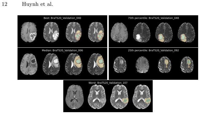
Reference graph
Works this paper leans on
-
[12]
Isensee, F., Kickingereder, P., Wick, W., Bendszus, M., Maier-Hein, K.H.: Brain tumor segmentation and radiomics survival prediction: Contribution to the brats 2017 challenge. In: Brainlesion: Glioma, Multiple Sclerosis, Stroke and Traumatic Brain Injuries: Third International Workshop, BrainLes 2017, Held in Conjunc- tion with MICCAI 2017, Quebec City, Q...
work page 2018
-
[1]
arXiv preprint arXiv:2107.02314 (2021)
Baid, U., Ghodasara, S., Mohan, S., Bilello, M., Calabrese, E., Colak, E., Farahani, K., Kalpathy-Cramer, J., Kitamura, F.C., Pati, S., et al.: The rsna-asnr-miccai brats 2021 benchmark on brain tumor segmentation and radiogenomic classifica- tion. arXiv preprint arXiv:2107.02314 (2021)
arXiv 2021
-
[2]
Scientific data 4(1), 1–13 (2017)
Bakas, S., Akbari, H., Sotiras, A., Bilello, M., Rozycki, M., Kirby, J.S., Freymann, J.B., Farahani, K., Davatzikos, C.: Advancing the cancer genome atlas glioma mri collections with expert segmentation labels and radiomic features. Scientific data 4(1), 1–13 (2017)
2017
-
[3]
arXiv preprint arXiv:1811.02629 (2018)
Bakas, S., Reyes, M., Jakab, A., Bauer, S., Rempfler, M., Crimi, A., Shinohara, R.T., Berger, C., Ha, S.M., Rozycki, M., et al.: Identifying the best machine learn- ing algorithms for brain tumor segmentation, progression assessment, and overall survival prediction in the brats challenge. arXiv preprint arXiv:1811.02629 (2018)
arXiv 2018
-
[4]
arXiv preprint arXiv:1904.00625 (2019)
Chen, S., Ma, K., Zheng, Y.: Med3d: Transfer learning for 3d medical image anal- ysis. arXiv preprint arXiv:1904.00625 (2019)
arXiv 2019
-
[5]
Deng, J., Dong, W., Socher, R., Li, L.J., Li, K., Fei-Fei, L.: Imagenet: A large-scale hierarchical image database. In: 2009 IEEE CVPR. pp. 248–255. Ieee (2009)
work page 2009
-
[6]
In: International MICCAI Brainlesion Workshop
Hatamizadeh, A., Nath, V., Tang, Y., Yang, D., Roth, H.R., Xu, D.: Swin unetr: Swin transformers for semantic segmentation of brain tumors in mri images. In: International MICCAI Brainlesion Workshop. pp. 272–284. Springer (2021)
work page 2021
-
[7]
In: Proceedings of the IEEE ICCV
He, K., Zhang, X., Ren, S., Sun, J.: Delving deep into rectifiers: Surpassing human- level performance on imagenet classification. In: Proceedings of the IEEE ICCV. pp. 1026–1034 (2015)
work page 2015
Show all 27 references
-
[8]
In: Proceedings of the IEEE CVPR
He, K., Zhang, X., Ren, S., Sun, J.: Deep residual learning for image recognition. In: Proceedings of the IEEE CVPR. pp. 770–778 (2016)
2016
-
[9]
In: Proceedings of the IEEE CVPR
Hu, J., Shen, L., Sun, G.: Squeeze-and-Excitation Networks. In: Proceedings of the IEEE CVPR. pp. 7132–7141 (June 2018)
2018
-
[10]
Nature methods18(2), 203–211 (2021)
Isensee, F., Jaeger, P.F., Kohl, S.A., Petersen, J., Maier-Hein, K.H.: nnu-net: a self-configuring method for deep learning-based biomedical image segmentation. Nature methods18(2), 203–211 (2021)
2021
-
[11]
In: Brainlesion: Glioma, Multiple Sclerosis, Stroke and Traumatic Brain Injuries: 6th International Workshop, BrainLes 2020, Held in Conjunction with MICCAI 2020
Isensee, F., J¨ ager, P.F., Full, P.M., Vollmuth, P., Maier-Hein, K.H.: nnu-net for brain tumor segmentation. In: Brainlesion: Glioma, Multiple Sclerosis, Stroke and Traumatic Brain Injuries: 6th International Workshop, BrainLes 2020, Held in Conjunction with MICCAI 2020. pp. ...
2021
-
[13]
Isensee, F., Kickingereder, P., Wick, W., Bendszus, M., Maier-Hein, K.H.: No new- net. In: Brainlesion: Glioma, Multiple Sclerosis, Stroke and Traumatic Brain In- juries: 4th International Workshop, BrainLes 2018, Held in Conjunction with MIC- CAI 2018, Granada, Spain, Septemb...
2018
-
[14]
pp. 234–244. Springer (2019)
2019
-
[15]
Jiang, Z., Ding, C., Liu, M., Tao, D.: Two-stage cascaded u-net: 1st place solution to brats challenge 2019 segmentation task. In: Brainlesion: Glioma, Multiple Scle- rosis, Stroke and Traumatic Brain Injuries: 5th International Workshop, BrainLes 2019, Held in Conjunction wit...
2020
-
[16]
In: In- ternational MICCAI Brainlesion Workshop
Luu, H.M., Park, S.H.: Extending nn-unet for brain tumor segmentation. In: In- ternational MICCAI Brainlesion Workshop. pp. 173–186. Springer (2021)
2021
-
[17]
IEEE TMI34(10), 1993–2024 (Oct 2015)
Menze, B.H., Jakab, A., Bauer, S., Kalpathy-Cramer, J., Farahani, K., Kirby, J., Burren, Y., Porz, N., Slotboom, J., Wiest, R., et al.: The Multimodal Brain Tumor Image Segmentation Benchmark (BRATS). IEEE TMI34(10), 1993–2024 (Oct 2015)
1993
-
[18]
MIC-DKFZ: nnUNet.https://github.com/MIC-DKFZ/nnUNet(2023)
2023
-
[19]
In: Brainlesion: Glioma, Multiple Sclerosis, Stroke and Traumatic Brain Injuries: 4th International Workshop, BrainLes 2018, Held in Conjunction with MICCAI
Myronenko, A.: 3d mri brain tumor segmentation using autoencoder regularization. In: Brainlesion: Glioma, Multiple Sclerosis, Stroke and Traumatic Brain Injuries: 4th International Workshop, BrainLes 2018, Held in Conjunction with MICCAI
2018
-
[20]
In: MICCAI 2015: 18th International Conference, Munich, Germany, October 5-9, 2015, Proceedings, Part III 18
Ronneberger, O., Fischer, P., Brox, T.: U-net: Convolutional networks for biomedi- cal image segmentation. In: MICCAI 2015: 18th International Conference, Munich, Germany, October 5-9, 2015, Proceedings, Part III 18. pp. 234–241. Springer (2015)
2015
-
[21]
Communications Engineering2(1), 23 (2023)
Pati, S., Thakur, S.P., Hamamcı, ˙I.E., Baid, U., Baheti, B., Bhalerao, M., G¨ uley, O., Mouchtaris, S., Lang, D., Thermos, S., et al.: Gandlf: the generally nuanced deep learning framework for scalable end-to-end clinical workflows. Communications Engineering2(1), 23 (2023)
2023
-
[22]
IEEE TIP30, 3113–3126 (2021)
Wu, Y.H., Gao, S.H., Mei, J., Xu, J., Fan, D.P., Zhang, R.G., Cheng, M.M.: Jcs: An explainable covid-19 diagnosis system by joint classification and segmentation. IEEE TIP30, 3113–3126 (2021)
2021
-
[23]
In: MICCAI 2021: 24th International Conference, Strasbourg, France, September 27–October 1, 2021, Proceedings, Part I 24
Wang, W., Chen, C., Ding, M., Yu, H., Zha, S., Li, J.: Transbts: Multimodal brain tumor segmentation using transformer. In: MICCAI 2021: 24th International Conference, Strasbourg, France, September 27–October 1, 2021, Proceedings, Part I 24. pp. 109–119. Springer (2021)
2021
-
[24]
arXiv preprint arXiv:2212.09310 (2022)
Zeineldin, R.A., Karar, M.E., Burgert, O., Mathis-Ullrich, F.: Multimodal cnn networks for brain tumor segmentation in mri: a brats 2022 challenge solution. arXiv preprint arXiv:2212.09310 (2022)
2022 arXiv
-
[25]
IEEE JBHI25(8), 3009–3018 (2021)
Yang, J., Huang, X., He, Y., Xu, J., Yang, C., Xu, G., Ni, B.: Reinventing 2d convolutions for 3d images. IEEE JBHI25(8), 3009–3018 (2021)
2021
-
[27]
Medical image analysis67, 101840 (2021)
Zhou, Z., Sodha, V., Pang, J., Gotway, M.B., Liang, J.: Models genesis. Medical image analysis67, 101840 (2021)
2021
-
[2018]
pp. 311–320. Springer (2019)
2019
Reviewed August 7, 2026 · model on record in the stance chip above.
Discussion (0). Sign in to comment.