REVIEW 5 major objections 4 minor 35 references
MR2US-Pro: Prostate MR to Ultrasound Image Translation and Registration Based on Diffusion Models
T0 review · 5 major / 4 minor · reviewed 2026-08-07 · deepseek-v4-flash
Pith's one-line read A single untracked, unsupervised pipeline registers prostate MRI to live 2D ultrasound by reconstructing the ultrasound volume and translating both modalities into one anatomy-only space.
desk verdict Genuinely new intermediate-modality translation idea, but the probe-free reconstruction rests on an unvalidated rigid-motion assumption and only 5 patients, so the strong claims are conditional. read the letter →
The pith
A machine-rendered reading of the paper's core claim, the machinery that carries it, and where it could break.
The reading
What carries the argument
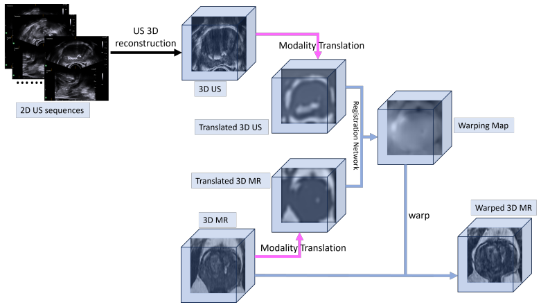
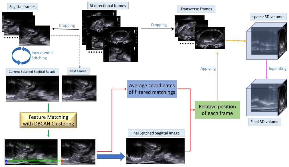
The load-bearing mechanism is the pseudo-intermediate modality built by a Schrödinger Bridge diffusion model (an optimal-transport stochastic process interpolating between two image distributions) with hierarchical feature disentanglement. Shallow network features passed through 7x7 kernels drive a texture-consistency loss that smooths modality-specific prostate-interior detail, while deep features passed through 3x3 kernels and a Sobel filter drive a boundary-preservation loss; together they map both MRI and ultrasound into a common anatomy-only representation. On the reconstruction side, the mechanism is stitching-first localization: each sagittal frame is matched against the growing composite sagittal map, a density-based clustering step groups the coordinate-difference vectors to reject outlier matches, and the orthogonal relationship between sagittal and transverse views transfers each frame's position to the third dimension before an inpainting network fills the sparse volume. Registration is carried by an anatomy-aware reweighting function $F_{\mathrm{Ana}}(x)=\mathrm{sigmoid}(-(x-\mathrm{mean}(x)))$ in a soft Dice similarity loss, which assigns higher weight to dark prostate interior and lower weight to bright boundary areas, together with a deformation smoothness loss and a diffusion score loss.
What would settle it
Perform a freehand TRUS sweep in which the probe is intentionally tilted or rotated by a few degrees between frames while an independent tracker records true positions; feed the same video to the untracked reconstruction, compare inferred frame locations and the final MRI-registration Dice and surface distance against the tracker-based reconstruction. If a few degrees of rotation or scale drift causes the stitched sagittal map to break or the registration metrics to fall materially, the rotational-fixed assumption fails.
Extended reading notes
Core claim
The paper's central claim is that cross-dimensional, cross-modal prostate MR-to-US registration can be solved in a fully untracked and unsupervised way by reconstructing the 3D ultrasound volume from the scanner's own sagittal-transverse geometry, then registering in a custom intermediate representation rather than in either native modality. The empirical claim is that this two-stage design beats the compared registration pipelines on every reported metric, with the full MR2US-Pro pipeline reaching DSC 0.97, IoU 0.94, ASD 4.45, and HE 7.35e4 while producing visibly smoother deformation fields. The authors attribute the gain to three interconnected choices: stitching each new sagittal frame against the already-built composite map instead of comparing consecutive frames, filtering feature matches by clustering their coordinate differences so only consistent translations survive; translating both modalities into an intermediate domain that preserves boundaries but homogenizes prostate interiors; and using an anatomy-aware soft Dice loss that concentrates alignment on the coherent low-intensity prostate interior. During inference the deformation field is computed from the translated volumes but applied to the original MRI, so the warped output keeps native intensity information for clinical use.
Load-bearing premise
The load-bearing premise is that the probe remains rotationally fixed during the initial scan, so every sagittal frame is a translated patch of one continuous sagittal plane and adjacent-frame motion is pure translation; if the operator's hand introduces even slight rotation or scale drift, the stitched map and the inferred transverse-frame positions are corrupted.
Editorial extensions
If this is right
- A biopsy or brachytherapy session begins with one brief rotational-fixed sweep; the rest of the procedure can proceed with freehand probe motion because the 3D ultrasound volume and its registration to MRI are already established.
- The warped MRI retains native intensities because the deformation field is estimated in the intermediate modality but applied to the original volume, so downstream fusion and interpretation keep the original contrast.
- The registration network needs no expert-annotated deformation fields or segmentation labels, removing a major barrier to clinical adoption.
- The same translation-then-register recipe is claimed to transfer to other cross-modal pairs such as brain CT-MR and cardiac PET-MR.
- Injecting the anatomy-aware reweighting into the loss is presented as the reason the final deformation fields are smoother and more physically plausible than those of the compared methods.
Reading between the lines
- If the rotational-fixed assumption survives a larger tracked-validation study, the stitching-first reconstruction could generalize to any bi-plane probe that acquires orthogonal views, including thyroid or breast ultrasound.
- The anatomy-aware reweighting function could be lifted out of the diffusion registration and applied to standard intensity-based registration losses; an ablation that swaps only that term would isolate how much of the reported gain comes from the reweighting versus the translation.
- A direct comparison against the same pipeline using tracker-based reconstruction would quantify the price paid for dropping the probe tracker, separating reconstruction error from registration error.
- The reported results are on five clinical cases; a multi-reader study on a larger cohort would show whether the achieved surface error translates into measurable benefit for targeting during biopsy.
Editorial analysis
A structured set of objections, weighed in public.
Referee Report
Summary. The paper proposes MR2US-Pro, a two-stage pipeline for registering 3D prostate MR images to 2D TRUS video streams without using external probe tracking. Stage 1 reconstructs a 3D US volume by stitching sagittal frames using feature matching with a DBSCAN-based inlier filter, transferring the inferred frame positions to the orthogonal transverse frames, and filling missing voxels with Deep Image Prior. Stage 2 maps both MR and US volumes into a pseudo-intermediate modality via a Schrödinger-bridge diffusion model trained with boundary-preservation and texture-consistency losses, and then performs an anatomy-aware diffusion-based registration that downweights boundary regions while emphasizing the prostate interior. Experiments on five patient pairs report improved translation quality (FID/KID) and registration accuracy (DSC, IoU, ASD, HE) relative to several diffusion-based baselines.
Significance. If the reported results were robust, the paper would offer a meaningful step toward probe-free TRUS 3D reconstruction and MR-US registration, with a novel intermediate-modality design that is plausible and potentially useful for biopsy guidance. The strengths include the explicit formulation of an end-to-end pipeline from raw video frames to a registered 3D MR volume, the use of a Schrödinger-bridge diffusion model for translation, the anatomy-aware weighting idea, and the inclusion of deformation-smoothness metrics. However, the current evidence is insufficient to support the paper's strong claims: the evaluation rests on only five patients with no error bars or significance tests, the reconstruction relies on an unvalidated pure-translation assumption, and the per-patient selection of the feature matcher introduces a form of test-set bias. The central idea is defensible, but the validation needs substantial strengthening.
major comments (5)
- [Section IV-A / Table II] The entire quantitative case for the method rests on five patient pairs, yet no error bars, per-fold results, or significance tests are reported. With n=5, the differences in Table II (e.g., DSC 0.97 vs. 0.92, ASD 4.45 vs. 6.82) could be dominated by patient-specific variation, so the claim that MR2US-Pro 'outperforms state-of-the-art methods' is not statistically supported. Please provide per-patient results, standard deviations, and pairwise tests or equivalent, and specify how the training/test split was constructed for each cross-validation fold.
- [Section III-A] The reconstruction assumes that the probe is 'rotationally fixed' during the initial scan and that adjacent-frame motion is 'simple translation, with negligible rotation or scale changes.' This assumption is load-bearing because DBSCAN retains the largest cluster of displacement vectors, which is only a valid inlier filter under pure translation. The paper does not quantify how much rotation or scale drift can be tolerated, nor does it validate the assumption against tracked acquisitions, synthetic freehand motion, or phantom data. Because the downstream registration metrics are computed on the final registered volumes, a geometrically incorrect reconstruction could be masked by the deformation field. Please add a validation of the reconstruction geometry, such as a comparison with a tracked sweep or a synthetic motion experiment, and report sensitivity to small rotations and scale changes.
- [Section III-B, Eq. (13)] The texture consistency loss explicitly minimizes the L2 difference between the translated MR and US shallow features, so the two inputs to the registration network are made similar by construction. This co-design explains part of the registration gain of ACMT+FSDiffReg over UNSB+FSDiffReg in Table II and should be acknowledged as an inductive bias rather than an emergent property. Please provide an ablation that registers the original MR/US volumes with the same registration network, and one that uses a simpler translation baseline (e.g., intensity normalization or histogram matching) to show what aspect of the diffusion-based translation is responsible for the improvement.
- [Section III-A] For each patient, the authors evaluated SIFT+clustering, ORB+clustering, and LoFTR+clustering and 'selected the best-performing approach.' If selection is based on the final registration outcome on the test set, this is a form of test-set selection bias, and the reported reconstruction/registration accuracy is not that of a fixed algorithm. Please specify the selection criterion (e.g., visual stitching quality or a validation set), report the results for each matcher on all patients, and, if possible, freeze the matcher choice before evaluation.
- [Section IV-A / IV-B] Baseline training details and evaluation details are missing. It is unclear whether DiffusionMorph and FSDiffReg were retrained on the same data or used with pretrained weights, what hyperparameters were used, and how the 'expert-guided manual segmentation ... on several key frames' was converted to a volumetric ground truth for DSC/IoU/ASD. Please clarify whether the metrics are computed on the full reconstructed volume or on selected 2D slices, and provide per-slice results if the latter.
minor comments (4)
- [Section III-A] There is a typo in 'beacuse' which should be 'because'.
- [Section IV-B / Figure 7] The method name is inconsistent: 'DiffuseMorph' appears in Figure 7 and in the text, while 'DiffusionMorph' is used in Table II and elsewhere. Please unify the terminology.
- [References] Reference [23] for harmonic energy cites the FSL software paper; please cite the original definition of harmonic energy for deformation fields or a standard medical image registration review instead.
- [Section IV-A] The paper does not state whether code, trained models, or the five-patient dataset will be made available; a reproducibility statement would strengthen the work.
Circularity Check
No significant circularity: final registration metrics are evaluated on independent manual masks, and the translation–registration co-design is an explicit objective rather than a hidden input.
full rationale
The paper's central claims are not derived from their own outputs by definition. Registration accuracy is quantified in Section IV-B2 by applying the predicted deformation fields to expert-guided manual segmentation masks of the original MR and US data, so the reported DSC, IoU, ASD, and HE are external to all training losses. The modality translation network is trained with a texture consistency loss (Eq. 13) that directly reduces feature differences between the translated MR and US images, and the registration network then aligns these translated volumes with an anatomy-aware soft Dice loss (Eq. 16). This is a deliberate co-design intended to ease registration, but it is not circular: the deformation field is still estimated by an unsupervised network, and the final evaluation uses original-image masks rather than the translated features or the training losses themselves. The self-citation to the authors' earlier work PMT [17] appears as a baseline and as motivation for moving beyond partial-modality translation, but it is not invoked as load-bearing evidence for the current method's correctness; the current method is compared against external baselines such as DiffusionMorph and FSDiffReg. The stated acquisition assumption that the probe is rotationally fixed and that adjacent-frame motion is simple translation is an explicit input to the 3D reconstruction pipeline, not a conclusion derived from it; whether that assumption holds in real freehand scanning is a robustness and validation concern, not a circularity. Likewise, per-patient selection among feature matchers is a potential evaluation-protocol weakness, but it is not an equivalence between an input and a claimed prediction. Overall, the derivation chain is self-contained: assumptions and losses are stated, and the headline metrics are grounded in independent manual annotations.
Assumptions & free parameters
free parameters (5)
- Loss weights λSB, λboundary, λtexture =
Not reported
- Loss weights λsim, λsmooth, λdiff =
Not reported
- Schrödinger bridge variance σ =
Not reported
- Feature matcher choice per patient =
SIFT, ORB, or LoFTR selected per patient
- DBSCAN parameters (eps, min_samples) =
Not reported
assumptions (5)
- domain assumption The probe is kept rotationally fixed during the initial scan, so sagittal frames lie in one continuous plane.
- domain assumption Adjacent-frame transformation is simple translation with negligible rotation or scale changes.
- ad hoc to paper Both MR and US can be mapped to a shared intermediate modality that preserves only boundary information.
- ad hoc to paper In the intermediate modality, prostate interior is darker than boundaries.
- domain assumption Deep Image Prior inpainting can fill missing voxels coherently.
invented entities (1)
-
Pseudo intermediate modality P1
Cite this review
Pith. "Pith review of MR2US-Pro: Prostate MR to Ultrasound Image Translation and Registration Based on Diffusion Models." pith.science (2026). https://pith.science/paper/VXAVGDMY
@misc{pith2026250600591,
  author       = {Pith},
  title        = {Pith review of: MR2US-Pro: Prostate MR to Ultrasound Image Translation and Registration Based on Diffusion Models},
  year         = {2026},
  howpublished = {\url{https://pith.science/paper/VXAVGDMY}},
  note         = {Machine review of arXiv:2506.00591}
}
read the original abstract
The diagnosis of prostate cancer increasingly depends on multimodal imaging, particularly magnetic resonance imaging (MRI) and transrectal ultrasound (TRUS). However, accurate registration between these modalities remains a fundamental challenge due to the differences in dimensionality and anatomical representations. In this work, we present a novel framework that addresses these challenges through a two-stage process: TRUS 3D reconstruction followed by cross-modal registration. Unlike existing TRUS 3D reconstruction methods that rely heavily on external probe tracking information, we propose a totally probe-location-independent approach that leverages the natural correlation between sagittal and transverse TRUS views. With the help of our clustering-based feature matching method, we enable the spatial localization of 2D frames without any additional probe tracking information. For the registration stage, we introduce an unsupervised diffusion-based framework guided by modality translation. Unlike existing methods that translate one modality into another, we map both MR and US into a pseudo intermediate modality. This design enables us to customize it to retain only registration-critical features, greatly easing registration. To further enhance anatomical alignment, we incorporate an anatomy-aware registration strategy that prioritizes internal structural coherence while adaptively reducing the influence of boundary inconsistencies. Extensive validation demonstrates that our approach outperforms state-of-the-art methods by achieving superior registration accuracy with physically realistic deformations in a completely unsupervised fashion.
Figures

Figures from the paper (7 more)


Reference graph
Works this paper leans on
-
[1]
IEEE transactions on medical imaging 38(8), 1788–1800 (2019)
Balakrishnan, G., Zhao, A., Sabuncu, M.R., Guttag, J., Dalca, A.V .: V oxelmorph: a learning framework for deformable medical image regis- tration. IEEE transactions on medical imaging 38(8), 1788–1800 (2019)
work page 2019
-
[2]
: Population-based organised prostate cancer testing: results from the first invitation of 50-year-old men
Bratt, O., et al. : Population-based organised prostate cancer testing: results from the first invitation of 50-year-old men. European Urology 85(3), 207–214 (2024)
2024
-
[3]
In: Proceedings of the IEEE/CVF conference on computer vision and pattern recognition
Chen, R., Huang, W., Huang, B., Sun, F., Fang, B.: Reusing discrimi- nators for encoding: Towards unsupervised image-to-image translation. In: Proceedings of the IEEE/CVF conference on computer vision and pattern recognition. pp. 8168–8177 (2020)
work page 2020
-
[4]
Medical physics 48(6), 3074–3083 (2021)
Chen, Y ., Xing, L., Yu, L., Liu, W., Pooya Fahimian, B., Niedermayr, T., Bagshaw, H.P., Buyyounouski, M., Han, B.: Mr to ultrasound image registration with segmentation-based learning for hdr prostate brachytherapy. Medical physics 48(6), 3074–3083 (2021)
2021
-
[5]
In: 2015 International Conference on Open Source Software Computing (OSS- COM)
Daoud, M.I., Alshalalfah, A.L., Awwad, F., Al-Najar, M.: Freehand 3d ultrasound imaging system using electromagnetic tracking. In: 2015 International Conference on Open Source Software Computing (OSS- COM). pp. 1–5. IEEE (2015)
work page 2015
-
[6]
In: 2020 7th international forum on electrical engineering and automation (IFEEA)
Deng, D.: Dbscan clustering algorithm based on density. In: 2020 7th international forum on electrical engineering and automation (IFEEA). pp. 949–953. IEEE (2020)
work page 2020
-
[7]
IEEE Transactions on Biomedical Engineering 70(3), 970–979 (2022)
Guo, H., Chao, H., Xu, S., Wood, B.J., Wang, J., Yan, P.: Ultrasound volume reconstruction from freehand scans without tracking. IEEE Transactions on Biomedical Engineering 70(3), 970–979 (2022)
work page 2022
-
[8]
WSEAS transactions on signal processing 6(4), 145–154 (2010)
Hafizah, M., Kok, T., Supriyanto, E.: Development of 3d image recon- struction based on untracked 2d fetal phantom ultrasound images using vtk. WSEAS transactions on signal processing 6(4), 145–154 (2010)
work page 2010
Show all 35 references
-
[9]
Advances in neural information processing systems 33, 6840–6851 (2020)
Ho, J., Jain, A., Abbeel, P.: Denoising diffusion probabilistic models. Advances in neural information processing systems 33, 6840–6851 (2020)
2020
-
[10]
Artificial Intelligence Review 56(1), 615– 651 (2023)
Jiang, J., Guo, Y ., Bi, Z., Huang, Z., Yu, G., Wang, J.: Segmentation of prostate ultrasound images: the state of the art and the future directions of segmentation algorithms. Artificial Intelligence Review 56(1), 615– 651 (2023)
2023
-
[11]
IEEE Trans- actions on Medical Imaging 39(12), 4413–4424 (2020)
Jiao, J., Namburete, A.I., Papageorghiou, A.T., Noble, J.A.: Self- supervised ultrasound to mri fetal brain image synthesis. IEEE Trans- actions on Medical Imaging 39(12), 4413–4424 (2020)
2020
-
[12]
arXiv preprint arXiv:2305.15086 (2023)
Kim, B., Kwon, G., Kim, K., Ye, J.C.: Unpaired image-to-image transla- tion via neural schr \” odinger bridge. arXiv preprint arXiv:2305.15086 (2023)
2023 arXiv
-
[13]
In: ICLR (2024)
Kim, B., Kwon, G., Kim, K., Ye, J.C.: Unpaired image-to-image translation via neural schr ¨odinger bridge. In: ICLR (2024)
2024
-
[14]
In: European conference on computer vision
Kim, B., Han, I., Ye, J.C.: Diffusemorph: Unsupervised deformable image registration using diffusion model. In: European conference on computer vision. pp. 347–364. Springer (2022)
2022
-
[15]
Dynamical Systems 34(4), 1533– 1574 (2014)
L ´eonard, C.: A survey of the schrodinger problem and some of its connections with optimal transport. Dynamical Systems 34(4), 1533– 1574 (2014)
2014
-
[16]
Advances in neural information processing systems 30 (2017)
Liu, M.Y ., Breuel, T., Kautz, J.: Unsupervised image-to-image transla- tion networks. Advances in neural information processing systems 30 (2017)
2017
-
[17]
In: Annual Conference on Medical Image Understanding and Analysis
Ma, X., Anantrasirichai, N., Bolomytis, S., Achim, A.: Pmt: Partial- modality translation based on diffusion models for prostate magnetic resonance and ultrasound image registration. In: Annual Conference on Medical Image Understanding and Analysis. pp. 285–297. Springer (2024)
2024
-
[18]
IEEE transactions on medical imaging 22(8), 986–1004 (2003)
Pluim, J.P., Maintz, J.A., Viergever, M.A.: Mutual-information-based registration of medical images: a survey. IEEE transactions on medical imaging 22(8), 986–1004 (2003)
2003
-
[19]
In: International Conference on Medical Image Computing and Computer- Assisted Intervention
Qin, Y ., Li, X.: Fsdiffreg: Feature-wise and score-wise diffusion-guided unsupervised deformable image registration for cardiac images. In: International Conference on Medical Image Computing and Computer- Assisted Intervention. pp. 655–665. Springer (2023)
2023
-
[20]
Medical image anal- ysis 3(4), 339–359 (1999)
Rohling, R., Gee, A., Berman, L.: A comparison of freehand three- dimensional ultrasound reconstruction techniques. Medical image anal- ysis 3(4), 339–359 (1999)
1999
-
[21]
In: 2011 International conference on computer vision
Rublee, E., Rabaud, V ., Konolige, K., Bradski, G.: Orb: An efficient alternative to sift or surf. In: 2011 International conference on computer vision. pp. 2564–2571. Ieee (2011)
2011
-
[22]
IEEE transactions on medical imaging 18(8), 712–721 (1999)
Rueckert, D., Sonoda, L.I., Hayes, C., Hill, D.L., Leach, M.O., Hawkes, D.J.: Nonrigid registration using free-form deformations: application to breast mr images. IEEE transactions on medical imaging 18(8), 712–721 (1999)
1999
-
[23]
Neuroimage 23, S208–S219 (2004)
Smith, S.M., Jenkinson, M., Woolrich, M.W., Beckmann, C.F., Behrens, T.E., Johansen-Berg, H., Bannister, P.R., De Luca, M., Drobnjak, I., Flitney, D.E., et al.: Advances in functional and structural mr image analysis and implementation as fsl. Neuroimage 23, S208–S219 (2004)
2004
-
[24]
In: Proceedings of the IEEE/CVF conference on computer vision and pattern recognition
Sun, J., Shen, Z., Wang, Y ., Bao, H., Zhou, X.: Loftr: Detector-free local feature matching with transformers. In: Proceedings of the IEEE/CVF conference on computer vision and pattern recognition. pp. 8922–8931 (2021)
2021
-
[25]
Plos one 17(7), e0271064 (2022)
Tasc ´on-Vidarte, J.D., Stick, L.B., Josipovic, M., Risum, S., Jomier, J., Erleben, K., V ogelius, I.R., Darkner, S.: Accuracy and consistency of intensity-based deformable image registration in 4dct for tumor motion estimation in liver radiotherapy planning. Plos one 17(7), e...
2022
-
[26]
In: 2024 IEEE International Symposium on Biomedical Imag- ing (ISBI)
Tian, X., Anantrasirichai, N., Nicholson, L., Achim, A.: Oct2confocal: 3d cyclegan based translation of retinal oct images to confocal mi- croscopy. In: 2024 IEEE International Symposium on Biomedical Imag- ing (ISBI). pp. 1–4. IEEE (2024)
2024
-
[27]
: Conditional flow matching: Simulation-free dynamic optimal transport
Tong, A., et al. : Conditional flow matching: Simulation-free dynamic optimal transport. arXiv preprint arXiv:2302.00482 2(3) (2023)
2023 arXiv
-
[28]
In: Proceed- ings of the IEEE conference on computer vision and pattern recognition
Ulyanov, D., Vedaldi, A., Lempitsky, V .: Deep image prior. In: Proceed- ings of the IEEE conference on computer vision and pattern recognition. pp. 9446–9454 (2018)
2018
-
[29]
European urology 66(4), 732–751 (2014)
Valerio, M., Ahmed, H.U., Emberton, M., Lawrentschuk, N., Lazzeri, M., Montironi, R., Nguyen, P.L., Trachtenberg, J., Polascik, T.J.: The role of focal therapy in the management of localised prostate cancer: a systematic review. European urology 66(4), 732–751 (2014)
2014
-
[30]
: Deep generative learning via schr ¨odinger bridge
Wang, G., et al. : Deep generative learning via schr ¨odinger bridge. In: International conference on machine learning. pp. 10794–10804. PMLR (2021)
2021
-
[31]
In: International Conference on Medical Image Computing and Computer-Assisted Intervention
Wein, W., Lupetti, M., Zettinig, O., Jagoda, S., Salehi, M., Markova, V ., Zonoobi, D., Prevost, R.: Three-dimensional thyroid assessment from untracked 2d ultrasound clips. In: International Conference on Medical Image Computing and Computer-Assisted Intervention. pp. 514–523...
2020
-
[32]
Weinreb, J.C., Barentsz, J.O., Choyke, P.L., Cornud, F., Haider, M.A., Macura, K.J., Margolis, D., Schnall, M.D., Shtern, F., Tempany, C.M., et al.: Pi-rads prostate imaging–reporting and data system: 2015, version
2015
-
[33]
European urology 69(1), 16–40 (2016)
2016
-
[34]
Biomedical Signal Processing and Control 8(6), 645–656 (2013)
Wen, T., Zhu, Q., Qin, W., Li, L., Yang, F., Xie, Y ., Gu, J.: An accurate and effective fmm-based approach for freehand 3d ultrasound reconstruction. Biomedical Signal Processing and Control 8(6), 645–656 (2013)
2013
-
[35]
In: International Conference on Medical im- age computing and computer-assisted intervention
Wolleb, J., Bieder, F., Sandk ¨uhler, R., Cattin, P.C.: Diffusion models for medical anomaly detection. In: International Conference on Medical im- age computing and computer-assisted intervention. pp. 35–45. Springer (2022)
2022
Reviewed August 7, 2026 · model on record in the stance chip above.
Discussion (0). Sign in to comment.