REVIEW 4 major objections 5 minor 40 references
Personalized MR-Informed Diffusion Models for 3D PET Image Reconstruction
T0 review · 4 major / 5 minor · reviewed 2026-08-07 · deepseek-v4-flash
Pith's one-line read Warping other patients' PET scans into a target's MR space yields a personalized diffusion prior that reconstructs low-count PET better than generic priors
desk verdict The pseudo-PET registration idea is a genuine, useful contribution; the evaluation is honest but has enough selection bias and weak-reference issues that the headline claim is supported rather than proven. read the letter →
The pith
A machine-rendered reading of the paper's core claim, the machinery that carries it, and where it could break.
The reading
What carries the argument
The central object is the pseudo-PET image: another subject's measured PET volume deformed into the target brain's coordinate frame by an affine alignment followed by a deep-learning deformable registration field learned from MR-to-MR alignment [24]. A random weighted sum of one or more such deformed images forms each training sample, so a small library of paired PET-MR scans yields a large, diverse, subject-specific training set without any generative model. The diffusion model is trained on these volumes with slice conditioning, since all volumes already share the target's slice geometry. Reconstruction then runs PET-DDS (Decomposed Diffusion Sampling), which alternates the diffusion denoiser with Poisson log-likelihood gradient steps on sinogram subsets, and the personalized prior is what anchors the iterate. The registration step is the load-bearing mechanism: it transfers anatomical context from MR into the PET training distribution while leaving the PET intensities measured, not hallucinated.
What would settle it
On a cohort whose FDG-avid lesions are partly invisible or misaligned on T1 MR, generate pseudo-PET by MR-to-MR registration and reconstruct 2.5% count data; if lesion contrast recovery falls below that of PET-DDS trained on unregistered PET, the MR-transfer assumption is the cause.
Extended reading notes
Core claim
The central claim is that a diffusion prior personalized to one subject's anatomy can be built by registering other subjects' measured PET images into that subject's MR space, and that this prior beats both a generic PET prior and an MR-conditioned prior for low-count 3D PET reconstruction. The pseudo-PET images mix real tracer measurements with MR-visible anatomy, so the learned score function becomes a joint anatomical-functional prior rather than an MR-imposing one. Using PET-DDS, the paper reports lower normalized root-mean-square error in mismatch regions, unchanged or better accuracy in agreement regions, and better lesion contrast versus background noise on simulated data with out-of-distribution lesions. On real FDG brain scans at 2.5% of full counts, it reports the best bias-variance trade-off among diffusion-based methods and improved agreement with full-count OSEM. The paper also reports that direct MR conditioning, either as an extra channel or through classifier-free guidance, does not consistently deliver these gains on small real datasets.
Load-bearing premise
The load-bearing premise is that warping another subject's brain into the target's MR space also carries that subject's PET tracer distribution into a plausible PET image for the target; if PET uptake does not track MR anatomy, the personalized prior is biased rather than helpful.
Editorial extensions
If this is right
- If the central claim holds, low-count brain PET can be reconstructed with a prior built from tens of paired scans instead of large corpora of high-quality images.
- Out-of-distribution lesions should remain detectable: lesion contrast recovery per unit background noise improves without sacrificing accuracy in MR-PET agreement regions.
- MR guidance becomes softer: the reconstruction can use anatomical information without requiring the tracer to match every MR boundary, reducing the blending artifacts seen with stronger MR priors.
- The improvement should transfer across tracers and dose levels, because the registration uses MR anatomy and is therefore tracer- and dose-invariant.
- On real data, the approach should dominate classifier-free MR conditioning when only limited paired training data are available, since the registration-based prior was the only diffusion variant that improved the bias-variance frontier.
Reading between the lines
- We infer that the random weighted summing step is itself a lightweight generative mechanism: it can synthesize an unbounded supply of plausible pseudo-PET volumes from a few registered scans, an idea the paper notes but leaves for future work.
- We infer a boundary condition: any tracer distribution that is invisible on MR, such as early amyloid deposition or tumors without structural correlate, will stress the MR-transfer assumption, and mixing in unregistered native PET training images may hedge against that failure.
- We infer that the shared target coordinate frame makes slice conditioning meaningful, so a patch-based or slab-wise training variant could cut the per-subject training cost while preserving the personalization benefit.
- We infer that the deformation fields, being MR-driven, are tracer-independent; a library of precomputed fields could be reused for new tracers without retraining the registration network.
Editorial analysis
A structured set of objections, weighed in public.
Referee Report
Summary. The paper proposes a method for generating subject-specific 'pseudo-PET' training images by registering other subjects' paired PET-MR images into the target subject's MR space using VoxelMorph MR-to-MR deformation fields, optionally summing several warped PET images. A diffusion model is trained on these pseudo-PET images and used as the prior within the PET-DDS reconstruction algorithm, which is then applied to low-count (2.5%) simulated and real [18F]FDG PET sinograms. The authors evaluate the method against OSEM, Bowsher, PET-DDS, and PET-DDS+MR, reporting improved NRMSE/SSIM in PET-MR mismatch regions, better lesion CRC versus background noise in simulations, and improved bias-variance and NRMSE on real data. The central claim is that personalizing the diffusion prior with MR-registered pseudo-PET images improves low-count PET reconstruction accuracy and the trade-off between PET-specific and MR-shared features.
Significance. If the quantitative claims hold, the method is a simple and practical way to inject MR information into diffusion-based PET reconstruction without training a conditional model, and it addresses the PET-MR mismatch problem by learning a softer MR-guided prior. The approach is original in its use of registration-based pseudo-PET synthesis rather than deep generative synthesis, and the authors show that it can be useful in low-training-data regimes. The scope of the experiments is commendable: both simulated and real FDG data, multiple baseline algorithms, and a bias-variance analysis. However, the evaluation has several methodological weaknesses—test-set hyperparameter selection, missing error bars in the simulated lesion experiments, and the use of full-count OSEM as the real-data reference—that currently prevent the quantitative claims from being fully established. The paper does not provide code, but the method is described in sufficient detail to be reproduced.
major comments (4)
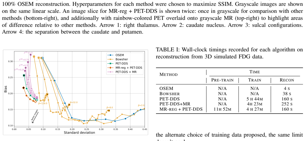
- [V-D, Fig. 9 and Fig. 10] The real-data comparison in Fig. 9 reports the 'best NRMSE' for each method across hyperparameter choices, and Fig. 10 selects hyperparameters to maximize SSIM on the test reconstruction. Because the regularization strength lambda_DDS and the number of iterations were chosen after seeing the test data, the reported improvements may reflect post-hoc selection rather than a reproducible operating point. Please report hyperparameters chosen by a validation set or a pre-specified rule, and show the full trade-off curves with the selected operating point marked.
- [IV-D5 and V-D] The bias-variance analysis and the real-data NRMSE in Figs. 9 and 11 use the 100%-count OSEM reconstruction as the ground truth. This is not an independent ground truth, and because all diffusion models are trained on OSEM images, the metric may favor reconstructions that are close to OSEM rather than accurate to the true tracer distribution. An independent reference (for example, a phantom with a known activity distribution, or a high-count reconstruction with a different algorithm) is needed to support the real-data claims.
- [IV-A1 and IV-B1] The central premise that an MR-derived deformation field is a valid transformation for PET tracer distributions is not directly tested. In the simulated data the ground-truth PET is analytically derived from MR segmentations, so the premise holds by construction; in the real data there is no independent ground truth and the subject population is not described with respect to pathology. The lesion experiments in Section V-B insert lesions only at test time, so the learned training distribution never contains PET-only structures. Please add a direct validation of the deformation assumption, for example by comparing warped PET images to native PET images of the same subject when available, or by reporting quantitative agreement between MR-registered and PET-registered pseudo-PET.
- [V-B, Figs. 5-7] Figures 5-7 report trade-off curves for the simulated lesion study without error bars or significance tests. It is not stated whether the metrics are averaged over multiple Poisson noise realizations, and several of the curves are close. Please provide means and standard deviations over at least 10 independent noise realizations (or an equivalent statistical summary) and a significance test for the claimed differences.
minor comments (5)
- [IV-D3] The sentence 'Over-fitting is not a concern for this task, as the test data is available during training' is confusing and, taken literally, describes a circular evaluation of the registration network. Please rephrase to clarify what is being personalized and why using the target MR for registration training is valid despite using that same MR for evaluation.
- [IV-A1] The heuristic for summing pseudo-PET images (P(N=n) proportional to 1/n and weights w_i ~ U[0,1]) is not justified, and since MR-reg & sum + PET-DDS 'performed similarly to MR-reg + PET-DDS' in Section V-B, the benefit of the summing step is not demonstrated empirically. Please provide supporting evidence or present the summing as an optional variant with limited effect.
- [IV-C3] The hyperparameters for PET-DDS+MR are not fully specified: lambda_MR is introduced as the guidance strength, but the text does not state which values were used or how it was varied. Please report the values or state explicitly that it was fixed.
- [II-E and IV-A1] There are typographical and formatting issues, including 'V oxelMorph' with extra spaces, 'bourne out' in Section V-C1, and the caption of Fig. 2 using 'blues' instead of a consistent term. These should be corrected.
- [V-D] The real-data evaluation omits MR-reg & sum + PET-DDS, even though random summing is part of the proposed method as described in Section IV-A1. The omission should be noted in the main text or the method should be evaluated on real data as well.
Circularity Check
Simulated validation of the MR-to-PET deformation assumption is self-fulfilling; real [18F]FDG results are the only non-circular support.
-
self definitional
[Section IV-A1 (pseudo-PET generation) and Section IV-B1 (simulated data generation)]
"we simulated ground truth [18F]FDG PET scans (and corresponding attenuation maps) from 39 real T1 MR images (following [26]) using representative activity values for segmented tissues. ... we compute the VoxelMorph registration map for image i as phi_i := phi(B_target xMR_target, B_i xMR_i), and use it to transform PET image xPET_i to the target reference space."
In the simulation, ground-truth PET is generated from MR tissue segmentations, and pseudo-PET is generated by warping other subjects' PET with MR-to-MR deformation fields. Because each simulated PET is a deterministic function of its own MR segmentation, aligning source MR to target MR also aligns the simulated source PET to the target's simulated PET (up to discretization). The pseudo-PET training set is therefore drawn from the target's true distribution by construction, so the simulated finding that personalized pseudo-PET pre-training improves reconstruction is an artifact of the experiment design: it asserts the MR-to-PET deformation assumption rather than testing it.
full rationale
The core reconstruction pipeline is not circular: the target low-count sinogram is never used to create pseudo-PET training images, and the target MR is legitimately available at test time for registration and guidance. PET-DDS data consistency enforces the measured sinogram, so the method does not simply return its training inputs. The one significant circularity is confined to the simulated validation: because the simulated ground-truth PET is derived from the same T1 MR segmentations used for MR-to-MR registration, the pseudo-PET training images are near copies of the target ground truth by construction, making the simulated improvement in NRMSE/CRC and bias-variance partly self-fulfilling. The real-data study provides non-circular support, though its force is tempered by using a 100%-count OSEM reference of the same kind used to create training images, and by reporting the minimum NRMSE over hyperparameters chosen on the test set. No load-bearing self-citation or imported uniqueness claim was found; the central method rests on external tools (VoxelMorph, PET-DDS) and its own real-data evaluation.
Assumptions & free parameters
free parameters (5)
- lambda_DDS (diffusion anchoring strength) =
varied per method, best chosen on test data
- lambda_RDP (axial RDP penalty strength) =
5e-4
- delta (gradient descent step size) =
0.2
- number of diffusion steps and data-consistency steps =
100 and 10
- pseudo-PET summing distribution =
P(N=n) proportional to 1/n; weights w_i ~ U[0,1]
assumptions (5)
- domain assumption MR-to-MR deformation fields are valid for warping PET images
- domain assumption OSEM reconstruction from 100% counts is a valid reference for real data
- domain assumption Training on lesion-free pseudo-PET generalizes to out-of-distribution lesions
- domain assumption Poisson thinning of prompt sinograms simulates low-count data
- standard math Standard score-matching diffusion training is valid for 3D medical images
Cite this review
Pith. "Pith review of Personalized MR-Informed Diffusion Models for 3D PET Image Reconstruction." pith.science (2026). https://pith.science/paper/M45OFRY7
@misc{pith2026250603804,
  author       = {Pith},
  title        = {Pith review of: Personalized MR-Informed Diffusion Models for 3D PET Image Reconstruction},
  year         = {2026},
  howpublished = {\url{https://pith.science/paper/M45OFRY7}},
  note         = {Machine review of arXiv:2506.03804}
}
abstract
Recent work has shown improved lesion detectability and flexibility to reconstruction hyperparameters (e.g. scanner geometry or dose level) when PET images are reconstructed by leveraging pre-trained diffusion models. Such methods train a diffusion model (without sinogram data) on high-quality, but still noisy, PET images. In this work, we propose a simple method for generating subject-specific PET images from a dataset of multi-subject PET-MR scans, synthesizing "pseudo-PET" images by transforming between different patients' anatomy using image registration. The images we synthesize retain information from the subject's MR scan, leading to higher resolution and the retention of anatomical features compared to the original set of PET images. With simulated and real [$^{18}$F]FDG datasets, we show that pre-training a personalized diffusion model with subject-specific "pseudo-PET" images improves reconstruction accuracy with low-count data. In particular, the method shows promise in combining information from a guidance MR scan without overly imposing anatomical features, demonstrating an improved trade-off between reconstructing PET-unique image features versus features present in both PET and MR. We believe this approach for generating and utilizing synthetic data has further applications to medical imaging tasks, particularly because patient-specific PET images can be generated without resorting to generative deep learning or large training datasets.
Figures


Figures from the paper (7 more)


Reference graph
Works this paper leans on
-
[1]
Positron emission tomography: An overview,
A. K. Shukla and U. Kumar, “Positron emission tomography: An overview,” Journal of Medical Physics , vol. 31, no. 1, pp. 13–21, 2006. doi:10.4103/0971-6203.25665
-
[2]
Radiation exposure and mortality risk from CT and PET imaging of patients with malignant lymphoma,
R. A. J. Nievelstein, H. M. E. Quarles van Ufford, T. C. Kwee, M. B. Bierings, I. Ludwig, F. J. A. Beek, J. M. H. de Klerk, W. P. T. M. Mali, P. W. de Bruin, and J. Geleijns, “Radiation exposure and mortality risk from CT and PET imaging of patients with malignant lymphoma,” European Radiology, vol. 22, no. 9, pp. 1946–1954, 2012. doi:10.1007/s00330-012-2447-9
-
[3]
PET/CT: Challenge for nuclear cardiology,
M. Schwaiger, S. Ziegler, and S. G. Nekolla, “PET/CT: Challenge for nuclear cardiology,” Journal of Nuclear Medicine , vol. 46, no. 10, pp. 1664–1678, Oct. 2005
work page 2005
-
[4]
Image reconstruction for PET/CT scanners: Past achievements and future challenges,
S. Tong, A. M. Alessio, and P. E. Kinahan, “Image reconstruction for PET/CT scanners: Past achievements and future challenges,” Imaging in Medicine, vol. 2, no. 5, pp. 529–545, Oct. 2010. doi:10.2217/iim.10.49
-
[5]
MR guided PET image reconstruction,
B. Bai, Q. Li, and R. M. Leahy, “MR guided PET image reconstruction,” Seminars in Nuclear Medicine , vol. 43, no. 1, pp. 30–44, Jan. 2013. doi:10.1053/j.semnuclmed.2012.08.006
-
[6]
Syn-Net for syn- ergistic deep-learned PET-MR reconstruction,
G. Corda-D’Incan, J. A. Schnabel, and A. J. Reader, “Syn-Net for syn- ergistic deep-learned PET-MR reconstruction,” in 2020 IEEE Nuclear Science Symposium and Medical Imaging Conference (NSS/MIC), pp. 1– 5, Oct. 2020. doi:10.1109/NSS/MIC42677.2020.9508086
arXiv 2020
-
[7]
Score-based diffusion models for accelerated MRI,
H. Chung and J. C. Ye, “Score-based diffusion models for accelerated MRI,” Medical Image Analysis , vol. 80, p. 102479, Aug. 2022. doi:10.1016/j.media.2022.102479
arXiv 2022
-
[8]
Decomposed diffusion sampler for accelerating large-scale inverse problems,
H. Chung, S. Lee, and J. C. Ye, “Decomposed diffusion sampler for accelerating large-scale inverse problems,” in Proc. ICLR, 2024. (ICLR 2024 proceedings; also arXiv:2303.05754)
arXiv 2024
Show all 40 references
-
[9]
Score-based generative models for PET image reconstruction,
I. R. Singh et al. , “Score-based generative models for PET image reconstruction,” MELBA, vol. 2, pp. 547–585, Jan. 2024. doi:10.59275/j.melba.2024-5d51
2024 doi
-
[10]
Likelihood-scheduled score-based generative modeling for fully 3D PET image reconstruction,
G. Webber, Y . Mizuno, O. D. Howes, A. Hammers, A. P. King, and A. J. Reader, “Likelihood-scheduled score-based generative modeling for fully 3D PET image reconstruction,” IEEE Transactions on Medical Imaging, early access, 2025. doi:10.1109/TMI.2025.3576483
2025
-
[11]
Deep learning for PET image reconstruction,
A. J. Reader, G. Corda, A. Mehranian, C. da Costa-Luis, S. Ellis, and J. A. Schnabel, “Deep learning for PET image reconstruction,” IEEE Transactions on Radiation and Plasma Medical Sciences , vol. 5, no. 1, pp. 1–25, Jan. 2021. doi:10.1109/TRPMS.2020.3014786
2021
-
[12]
Maximum likelihood reconstruction for emission tomography,
L. A. Shepp and Y . Vardi, “Maximum likelihood reconstruction for emission tomography,” IEEE Transactions on Medical Imaging , vol. 1, no. 2, pp. 113–122, Oct. 1982. doi:10.1109/TMI.1982.4307558
1982
-
[13]
A maximum a posteriori probability ex- pectation maximization algorithm for image reconstruction in emission tomography,
E. Levitan and G. T. Herman, “A maximum a posteriori probability ex- pectation maximization algorithm for image reconstruction in emission tomography,” IEEE Transactions on Medical Imaging , vol. 6, no. 3, pp. 185–192, Sept. 1987. doi:10.1109/TMI.1987.4307826
1987
-
[14]
Accelerated image reconstruction using ordered subsets of projection data,
H. M. Hudson and R. S. Larkin, “Accelerated image reconstruction using ordered subsets of projection data,” IEEE Transactions on Medical Imaging, vol. 13, no. 4, pp. 601–609, 1994. doi:10.1109/42.363108
1994 doi
-
[15]
Fast EM-like methods for maximum
A. De Pierro and M. Yamagishi, “Fast EM-like methods for maximum ”a posteriori” estimates in emission tomography,” IEEE Transactions on Medical Imaging, vol. 20, no. 4, pp. 280–288, Apr. 2001. doi:10.1109/42.921477
2001 doi
-
[16]
Utilizing MRI information to estimate F18-FDG distributions in rat flank tumors,
J. E. Bowsher, H. Yuan, L. W. Hedlund, T. G. Turkington, G. Akabani, A. Badea, W. C. Kurylo, C. T. Wheeler, G. P. Cofer, M. W. Dewhirst, and G. A. Johnson, “Utilizing MRI information to estimate F18-FDG distributions in rat flank tumors,” in Proc. IEEE Nuclear Science Symposiu...
2004 arXiv
-
[17]
Evaluation of parallel level sets and Bowsher’s method as segmentation-free anatomical priors for time-of-flight PET reconstruction,
G. Schramm, M. Holler, A. Rezaei, K. Vunckx, F. Knoll, K. Bredies, F. Boada, and J. Nuyts, “Evaluation of parallel level sets and Bowsher’s method as segmentation-free anatomical priors for time-of-flight PET reconstruction,” IEEE Transactions on Medical Imaging , vol. 37, no....
2018
-
[18]
Denoising diffusion probabilistic models,
J. Ho, A. Jain, and P. Abbeel, “Denoising diffusion probabilistic models,” in Advances in Neural Information Processing Systems 33 (NeurIPS 2020), H. Larochelle, M. Ranzato, R. Hadsell, M. F. Balcan, and H. Lin (eds.), Curran Associates, Inc., 2020, pp. 6840–6851. doi:10.5555/...
2020
-
[19]
Deep unsupervised learning using nonequilibrium thermodynamics,
J. Sohl-Dickstein, E. Weiss, N. Maheswaranathan, and S. Ganguli, “Deep unsupervised learning using nonequilibrium thermodynamics,” in Proceedings of the 32nd International Conference on Machine Learning (ICML), F. Bach and D. Blei, Eds., vol. 37 of Proceedings of Machine Learn...
2015
-
[20]
Improved techniques for training score-based generative models,
Y . Song and S. Ermon, “Improved techniques for training score-based generative models,” in Advances in Neural Information Processing Systems 33 (NeurIPS 2020) , H. Larochelle, M. Ranzato, R. Had- sell, M. F. Balcan, and H. Lin (eds.), Curran Associates, Inc., 2020, pp. 12438–...
2020
-
[21]
Solving 3D inverse problems using pre-trained 2D diffusion models,
H. Chung, D. Ryu, M. T. McCann, M. L. Klasky, and J. C. Ye, “Solving 3D inverse problems using pre-trained 2D diffusion models,” in Pro- ceedings of the IEEE/CVF Conference on Computer Vision and Pattern Recognition (CVPR) , Vancouver, BC, Canada, June 2023, pp. 22542– 22551. ...
2023
-
[22]
Diffusion models for medical image reconstruction,
G. Webber and A. J. Reader, “Diffusion models for medical image reconstruction,” BJR|Artificial Intelligence, p. ubae013, Aug. 2024. doi:10.1093/bjrai/ubae013
2024 doi
- [23]
-
[24]
V oxelMorph: A learning framework for deformable medical image registration,
G. Balakrishnan, A. Zhao, M. R. Sabuncu, J. V . Guttag, and A. V . Dalca, “V oxelMorph: A learning framework for deformable medical image registration,” IEEE Transactions on Medical Imaging , vol. 38, no. 8, pp. 1788–1800, Aug. 2019. doi:10.1109/TMI.2019.2897538
2019
-
[25]
MR-guided kernel EM reconstruction for reduced dose PET imaging,
J. Bland, A. Mehranian, M. A. Belzunce, S. Ellis, C. J. McGinnity, A. Hammers, and A. J. Reader, “MR-guided kernel EM reconstruction for reduced dose PET imaging,” IEEE Transactions on Radiation and Plasma Medical Sciences , vol. 2, no. 3, pp. 235–243, May 2018. doi:10.1109/TR...
2018
-
[26]
Model-based deep learning PET image reconstruction using forward–backward splitting expec- tation–maximization,
A. Mehranian and A. J. Reader, “Model-based deep learning PET image reconstruction using forward–backward splitting expec- tation–maximization,” IEEE Transactions on Radiation and Plasma Medical Sciences, vol. 5, no. 1, pp. 54–64, June 2020. doi:10.1109/TRPMS.2020.3004408
2020
-
[27]
Anatomically aided PET image reconstruction using deep neural networks,
Z. Xie, T. Li, X. Zhang, W. Qi, E. Asma, and J. Qi, “Anatomically aided PET image reconstruction using deep neural networks,” Medical Physics, vol. 48, no. 9, pp. 5244–5258, Sept. 2021. doi:10.1002/mp.15051
2021 doi
-
[28]
PET image reconstruc- tion using multi-parametric anato-functional priors,
A. Mehranian, M. A. Belzunce, F. Niccolini, M. Politis, C. Prieto, F. Turkheimer, A. Hammers, and A. J. Reader, “PET image reconstruc- tion using multi-parametric anato-functional priors,” Physics in Medicine and Biology, vol. 62, no. 15, p. 5975-6007, July 2017. doi: 10.1088/...
2017 doi
-
[29]
Micro-networks for robust MR- guided low count PET imaging,
C. da Costa-Luis and A. J. Reader, “Micro-networks for robust MR- guided low count PET imaging,” IEEE Transactions on Radiation and Plasma Medical Sciences , vol. 5, no. 2, pp. 202–212, Mar. 2021. doi:10.1109/TRPMS.2020.2986414
2021
-
[30]
PET image reconstruction using deep image prior,
K. Gong, C. Catana, J. Qi, and Q. Li, “PET image reconstruction using deep image prior,” IEEE Transactions on Medical Imaging , vol. 38, no. 7, pp. 1655–1665, July 2019. doi:10.1109/TMI.2018.2888491
2019
-
[31]
Subject-specific models for the analysis of pathological FDG PET data,
N. Burgos, M. Jorge Cardoso, A. F. Mendelson, J. M. Schott, D. Atkin- son, S. R. Arridge, B. F. Hutton, and S. Ourselin, “Subject-specific models for the analysis of pathological FDG PET data,” in Medical Image Computing and Computer-Assisted Intervention – MICCAI 2015 , N. Na...
2015 doi
-
[32]
Multi-subject image synthesis as a generative prior for single-subject PET image reconstruction,
G. Webber, Y . Mizuno, O. D. Howes, A. Hammers, A. P. King, and A. J. Reader, “Multi-subject image synthesis as a generative prior for single-subject PET image reconstruction,” in 2024 IEEE Nuclear Science Symposium (NSS), Medical Imaging Conference (MIC) and Room Temperature ...
2024
-
[33]
Global image registration using a symmetric block- matching approach,
M. Modat, D. M. Cash, P. Daga, G. P. Winston, J. S. Duncan, and S. Ourselin, “Global image registration using a symmetric block- matching approach,” Journal of Medical Imaging , vol. 1, no. 2, p. 024003, Sept. 2014. doi: 10.1117/1.JMI.1.2.024003
2014 doi
-
[34]
Diffusion models beat GANs on image synthesis,
P. Dhariwal and A. Nichol, “Diffusion models beat GANs on image synthesis,” in Advances in Neural Information Processing Systems 34 (NeurIPS 2021), M. Ranzato, A. Beygelzimer, Y . Dauphin, P. S. Liang, and J. W. Vaughan (eds.), Curran Associates, Inc., 2021, pp. 8780–8794. doi...
2021
-
[35]
U-Net: Convolutional net- works for biomedical image segmentation,
O. Ronneberger, P. Fischer, and T. Brox, “U-Net: Convolutional net- works for biomedical image segmentation,” in Medical Image Comput- ing and Computer-Assisted Intervention – MICCAI 2015 , N. Navab, J. Hornegger, W. M. Wells, and A. F. Frangi (eds.), Lecture Notes in Computer...
2015 doi
-
[36]
A connection between score matching and denoising au- toencoders,
P. Vincent, “A connection between score matching and denoising au- toencoders,” Neural Computation, vol. 23, pp. 1661–1674, July 2011. doi:10.1162/NECO a 00142
2011 doi
-
[37]
PARALLELPROJ—an open-source framework for fast calculation of projections in tomography,
G. Schramm and K. Thielemans, “PARALLELPROJ—an open-source framework for fast calculation of projections in tomography,” Frontiers in Nuclear Medicine , vol. 3, Jan. 2024. doi:10.3389/fnume.2023.1324562
2024
-
[38]
Bootstrap-optimised regularised image reconstruction for emission tomography,
A. J. Reader and S. Ellis, “Bootstrap-optimised regularised image reconstruction for emission tomography,” IEEE Transactions on Medical Imaging, vol. 39, no. 6, pp. 2163–2175, June 2020. doi: 10.1109/TMI.2019.2956878
2020
-
[39]
Clinical and deep-learned evaluation of MR-guided self-supervised PET reconstruction,
J. B. Hopson, S. Ellis, A. Flaus, C. J. McGinnity, R. Neji, A. J. Reader, and A. Hammers, “Clinical and deep-learned evaluation of MR-guided self-supervised PET reconstruction,” IEEE Transactions on Radiation and Plasma Medical Sciences , vol. 9, no. 3, pp. 337–346, Mar. 2025....
2025
-
[40]
Patch-based diffusion models beat whole-image models for mismatched distribution inverse problems,
J. Hu, B. Song, J. A. Fessler, and L. Shen, “Patch-based diffusion models beat whole-image models for mismatched distribution inverse problems,” arXiv:2410.11730, Oct. 2024. doi:10.48550/arXiv.2410.11730
Reviewed August 7, 2026 · model on record in the stance chip above.
Discussion (0). Sign in to comment.