REVIEW 4 major objections 7 minor 17 references
Beyond the Desktop: XR-Driven Segmentation with Meta Quest 3 and MX Ink
T0 review · 4 major / 7 minor · reviewed 2026-08-07 · deepseek-v4-flash
Pith's one-line read A Quest 3 plus MX Ink stylus reaches a standard usability score of 66 for clinical CT segmentation, a viable foundation for immersive annotation.
desk verdict A straightforward prototype usability study for a specific XR-stylus combo; the cognitive-load claim overreaches, but the rest holds up as a modest contribution. read the letter →
The pith
A machine-rendered reading of the paper's core claim, the machinery that carries it, and where it could break.
The reading
What carries the argument
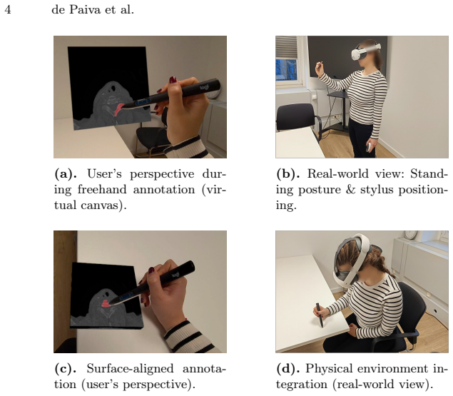
The mechanism that carries the argument is the stylus-to-image coordinate mapping: a raycast from the MX Ink stylus tip is projected onto a dynamic 3D canvas holding the medical image, and the intersection is translated into pixel coordinates so that where the user draws is exactly where the segmentation mask is written. This positional equivalence preserves pen-on-paper muscle memory inside VR. On top of it, the system renders a volumetric iso-surface with Marching Cubes and decimates the mesh to keep updates under 200 ms at 72 FPS, and it preloads TIFF slices so the user can switch between 2D annotation and 3D inspection without leaving the workspace.
What would settle it
Give a group of experienced clinicians the same craniofacial CT volumes to segment with the XR stylus and with a standard desktop contouring tool, scoring both against expert ground-truth masks with Dice overlap and recording task time: if the XR condition is not at least comparable on accuracy or time, the clinical-foundation claim is falsified.
Extended reading notes
Core claim
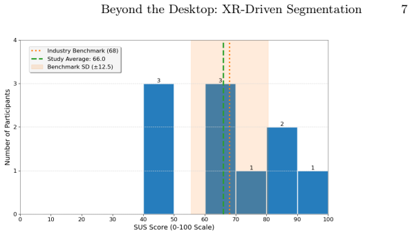
The paper's central claim is that the XR-stylus paradigm—Meta Quest 3 inside-out tracking plus the Logitech MX Ink stylus—can unify 2D slice annotation and 3D volumetric manipulation in one workspace and does so with usable ergonomics. The authors report a mean SUS of 66.0, a value they argue is inside the standard deviation of digital-health benchmarks even though it sits just below the general industry benchmark of 68, and ISO 9241-110 item ratings led by self-descriptiveness at 4.1/5. Qualitative feedback adds that the stylus "felt like annotating on paper, but in 3D space" and that the hybrid 2D/3D navigation reduced cognitive load. The study deliberately measured usability rather than segmentation accuracy, so the claim is about viability as a foundation, not about parity with established tools.
Load-bearing premise
The load-bearing premise, stated in the Evaluation Protocol and leaned on in the Discussion, is that ten participants—mostly medical students aged 20-27 and 60% without prior head-mounted display experience—plus self-reported SUS and ISO scores and qualitative comments are enough to establish clinical applicability and reduced cognitive load, since no desktop baseline or accuracy measurement was taken.
Editorial extensions
If this is right
- Clinical teams could annotate CT scans without a desktop workstation or externally mounted sensors, since the headset provides standalone inside-out tracking.
- The pen-on-paper feel of the stylus could shorten the learning curve for clinicians who already contour by drawing, lowering one barrier to XR adoption.
- The hybrid 2D/3D workspace is a candidate scaffold for teaching anatomical spatial reasoning, a use participants themselves raised.
- Because the platform already imports pre-annotated masks, the same interface could become a review-and-correction tool for AI-generated segmentations.
- A 66 SUS with a 4.1/5 self-descriptiveness score gives future iterations a concrete baseline: improve precision and error management without abandoning the interaction paradigm.
Reading between the lines
- Beyond the paper: because no accuracy or time metrics were collected, the strongest testable next step is a same-task comparison of XR stylus versus desktop mouse on Dice overlap and task duration; the current study's usability-only design cannot speak to segmentation quality.
- Beyond the paper: the raycast-based pixel mapping is not tied to CT or to craniofacial anatomy, so the same interaction layer could plausibly be reused for MRI, ultrasound, or radiation-therapy contouring with only the import pipeline changed; the paper does not demonstrate this.
- Beyond the paper: if a larger clinician sample reproduces the 66 SUS score, the economic case for XR contouring as a space-saving complement to desktop workstations becomes testable, weighing headset cost and training time against workflow continuity; the paper leaves that trade-off unquantified.
Editorial analysis
A structured set of objections, weighed in public.
Referee Report
Summary. The paper describes the design and a ten-participant usability evaluation of an XR-based CT segmentation platform built on the Meta Quest 3 headset and Logitech MX Ink stylus. The system supports freehand stylus annotation on 2D slices, real-time 3D volumetric rendering via marching cubes, and export of segmentation masks. Participants performed segmentation tasks on Open-Full-Jaw CT volumes and then completed a System Usability Scale (SUS) questionnaire, a custom ISO 9241-110-based questionnaire, and semi-structured interviews. The reported mean SUS score is 66.0, ISO item means range from 2.5 to 4.1, and qualitative feedback emphasizes stylus ergonomics and hybrid 2D/3D navigation. The authors claim the results demonstrate foundational viability for clinical segmentation and reduced cognitive load compared to desktop tools, with recommendations for future haptic and AI refinements.
Significance. If the claims are taken at face value, this is a modest but useful feasibility study: it provides real SUS and ISO questionnaire data for a specific commercial XR-stylus combination (Meta Quest 3 + MX Ink) that has not been widely evaluated in medical segmentation contexts, and it uses a public craniofacial CT dataset. The paper is transparent about its evaluation protocol, reports internal consistency (Cronbach's alpha = 0.89), and explicitly acknowledges in Section 2.2 that only CT volumes were used so participants focused on usability rather than segmentation accuracy. These are genuine strengths. However, the paper's broader significance is limited by the absence of any quantitative measure of segmentation output quality, the lack of a desktop comparison condition, and the small homogeneous sample; as a result, the comparative and clinical-viability claims in the abstract, Section 4, and Section 5 exceed what the evidence supports.
major comments (4)
- [Section 2.2] The evaluation protocol explicitly states that only CT volumes were used 'to ensure participants focused on the XR-stylus interface's usability over segmentation accuracy.' Because producing accurate masks is the core function of a segmentation tool, the study never tests whether the system produces usable segmentation output. The central claim of a 'viable foundation for clinical segmentation' is therefore unsupported at the level of task performance. The authors should either report quantitative agreement between participant-produced masks and the expert-validated labels available in the Open-Full-Jaw dataset (e.g., Dice coefficient or Hausdorff distance), or explicitly restrict all claims to 'usability feasibility' and remove the clinical-viability wording.
- [Section 4 and Abstract] The claim that the platform 'reduced cognitive load compared to desktop tools' is not supported by the study design. No desktop control condition was run, no task completion times or error rates were recorded, and no workload-specific instrument such as NASA-TLX was administered. The ISO self-descriptiveness score of 4.1/5, which the paper cites as evidence of reduced cognitive effort, measures whether an interface reveals its own purpose, not cognitive workload. The participant quote in Section 4 is illustrative but not evidential for a comparative claim. Either add a comparative desktop condition with workload and performance measures, or revise the abstract, Section 4, and Section 5 to state that participants perceived reduced cognitive load, not that the system reduces cognitive load.
- [Figure 3 and Section 4] The interpretation of the SUS score relies on a statistically unsupported equivalence: the paper states that SUS 66 is 'within the expected range for medical applications' because it lies within one standard deviation (12.5) of the benchmark of 68. Being within one standard deviation of a reference mean is not the same as meeting or being in the expected range for that benchmark; with n=10 and no confidence intervals, the difference between 66 and 68 cannot be interpreted as acceptable or unacceptable. Report the confidence interval for the SUS mean, state the comparison to the benchmark as descriptive only, or use an appropriate significance test if a claim of equivalence is intended.
- [Section 3] The sample of ten participants, aged 20-27 and mostly medical students with 60% having no HMD experience, is small and demographically narrow. No inferential statistics, effect sizes, or confidence intervals are reported for the SUS or ISO item means, so statements about generalizability to practicing clinicians in a clinical workflow are not supported. The paper should temper population-level conclusions and, where possible, report individual participant-level results or nonparametric confidence intervals.
minor comments (7)
- [Figure 4 caption] The caption contains the typo 'ISO 9241-110 S compliant'; remove the stray 'S'.
- [Section 2.2] The text says 'a 10-item questionnaire with nine items compliant with ISO 9241-110,' but then lists Q1 through Q10, leaving unclear which item is non-compliant; presumably Q10 is overall satisfaction, but this should be stated explicitly.
- [Abstract and Section 2.2] The abstract refers to 'ISONORM metrics' while Section 2.2 describes an 'adapted ISO instrument'; use consistent terminology for the ISO 9241-110 questionnaire.
- [References] Reference [4] has a garbled author string ('in Oncology Group+ 1-210-4501127+ 1-210-4505085 fullercd@ uthscsa. edu'); the citation needs to be corrected.
- [Section 4] In the participant quote, there is a space before the closing quotation mark ('"felt like annotating on paper, but in 3D space. "'), and the punctuation placement should be cleaned up.
- [Section 2.1] The Marching Cubes description says an iso-level of ι = 0.5 is applied to Hounsfield units; 0.5 is not a Hounsfield threshold, and the text should clarify whether the threshold applies to a normalized density grid and what anatomical boundary it selects.
- [Section 2.1] The performance claims of 'latencies below 200ms' and 'sustained 72 FPS' are stated without describing how they were measured; either specify the measurement method or label these as developer observations.
Circularity Check
No circular derivation: usability claims are supported by external instruments and benchmarks, not by self-referential fitting.
full rationale
The paper makes no fitted predictions and contains no derivation chain whose output is equivalent to its input. The central quantitative result, SUS = 66, is a directly measured questionnaire outcome interpreted against external benchmarks (Lewis and Sauro; Hyzy et al.), and the ISO 9241-110 item scores are measured responses to an externally validated instrument. The Open-Full-Jaw dataset is used as test material, and the sole self-authorship connection (Ref. 16; also Ref. 5 as related work) is data provenance or prior context rather than a load-bearing citation. The paper's stated limitation in Section 2.2 - 'only the CT volumes were used to ensure participants focused on the XR-stylus interface's usability over segmentation accuracy' - honestly discloses that segmentation accuracy was not evaluated; that is an evidentiary gap for the clinical-applicability claim, not circular reasoning. Similarly, the cognitive-load reduction claim in Section 4 is inferred from qualitative feedback and self-descriptiveness scores without a desktop baseline; this is unsupported interpretation, not circularity. No equation, fitted parameter, or cited uniqueness theorem is used to force a conclusion, so the appropriate finding is no significant circularity.
Assumptions & free parameters
free parameters (2)
- Iso-level threshold for marching cubes =
0.5
- Mesh decimation ratio =
70-80% triangle reduction
assumptions (4)
- domain assumption SUS and ISO 9241-110 questionnaire scores are valid proxies for clinical usability and ergonomic integration.
- domain assumption A sample of ten participants, mostly medical students aged 20-27, is representative enough to support conclusions about clinical applicability.
- domain assumption Participants' self-reported reductions in cognitive load correspond to actual cognitive workload changes.
- domain assumption The Open-Full-Jaw dataset CT volumes are an appropriate testbed for evaluating segmentation usability.
Cite this review
Pith. "Pith review of Beyond the Desktop: XR-Driven Segmentation with Meta Quest 3 and MX Ink." pith.science (2026). https://pith.science/paper/Y3VEWO3O
@misc{pith2026250604858,
  author       = {Pith},
  title        = {Pith review of: Beyond the Desktop: XR-Driven Segmentation with Meta Quest 3 and MX Ink},
  year         = {2026},
  howpublished = {\url{https://pith.science/paper/Y3VEWO3O}},
  note         = {Machine review of arXiv:2506.04858}
}
read the original abstract
Medical imaging segmentation is essential in clinical settings for diagnosing diseases, planning surgeries, and other procedures. However, manual annotation is a cumbersome and effortful task. To mitigate these aspects, this study implements and evaluates the usability and clinical applicability of an extended reality (XR)-based segmentation tool for anatomical CT scans, using the Meta Quest 3 headset and Logitech MX Ink stylus. We develop an immersive interface enabling real-time interaction with 2D and 3D medical imaging data in a customizable workspace designed to mitigate workflow fragmentation and cognitive demands inherent to conventional manual segmentation tools. The platform combines stylus-driven annotation, mirroring traditional pen-on-paper workflows, with instant 3D volumetric rendering. A user study with a public craniofacial CT dataset demonstrated the tool's foundational viability, achieving a System Usability Scale (SUS) score of 66, within the expected range for medical applications. Participants highlighted the system's intuitive controls (scoring 4.1/5 for self-descriptiveness on ISONORM metrics) and spatial interaction design, with qualitative feedback highlighting strengths in hybrid 2D/3D navigation and realistic stylus ergonomics. While users identified opportunities to enhance task-specific precision and error management, the platform's core workflow enabled dynamic slice adjustment, reducing cognitive load compared to desktop tools. Results position the XR-stylus paradigm as a promising foundation for immersive segmentation tools, with iterative refinements targeting haptic feedback calibration and workflow personalization to advance adoption in preoperative planning.
Figures

Figures from the paper (1 more)

Reference graph
Works this paper leans on
-
[1]
Current treatment options in cardiovascular medicine21, 1–12 (2019)
Andrews,C.,Southworth,M.K.,Silva,J.N.,Silva,J.R.:Extendedrealityinmedical practice. Current treatment options in cardiovascular medicine21, 1–12 (2019)
work page 2019
-
[2]
Usability evaluation in industry 189(194), 4–7 (1996)
Brooke, J., et al.: Sus-a quick and dirty usability scale. Usability evaluation in industry 189(194), 4–7 (1996)
work page 1996
-
[3]
Segmentation and Interactive Mask Editing (2024)
Charalampidis, V.L., Trakos, D., Tsiobra, M., Bizopoulos, P., Votis, K.: Ar-visem: Ar tool for visualization, segmentation and interactive mask editing. Segmentation and Interactive Mask Editing (2024)
work page 2024
-
[4]
in Oncology Group+ 1-210-4501127+ 1-210-4505085 fullercd@ uthscsa. edu, M.I.T.D.: Human–computer interaction in radiotherapy target volume delineation: a prospective, multi-institutional comparison of user input devices. Journal of dig- ital imaging 24, 794–803 (2011)
work page 2011
-
[5]
Expert Systems with Applications239, 122275 (2024)
Gruber, L.J., Egger, J., Bönsch, A., Kraeima, J., Ulbrich, M., van den Bosch, V., Motmaen, I., Wilpert, C., Ooms, M., Isfort, P., et al.: Accuracy and precision of mandible segmentation and its clinical implications: virtual reality, desktop screen and artificial intelligence. Expert Systems with Applications239, 122275 (2024)
work page 2024
-
[6]
Ad- vances in Ergonomics In Design, Usability & Special Populations: Part 3, 18 (2014)
Heimgärtner, R.: Human factors of iso 9241-110 in the intercultural context. Ad- vances in Ergonomics In Design, Usability & Special Populations: Part 3, 18 (2014)
work page 2014
-
[7]
IEEE Robotics and Automation Letters (2025)
Heinold, E., Rosen, P., Wischniewski, S.: Usability questionnaire for robotic sys- tems based on the iso 9241-110. IEEE Robotics and Automation Letters (2025)
work page 2025
-
[8]
JMIR mHealth and uHealth10(8), e37290 (2022) 10 de Paiva et al
Hyzy, M., Bond, R., Mulvenna, M., Bai, L., Dix, A., Leigh, S., Hunt, S., et al.: System usability scale benchmarking for digital health apps: meta-analysis. JMIR mHealth and uHealth10(8), e37290 (2022) 10 de Paiva et al
work page 2022
Show all 17 references
-
[9]
Health Data Science (2022)
JohnsonChris,R.,etal.:Areviewofthree-dimensionalmedicalimagevisualization. Health Data Science (2022)
2022
-
[10]
Tomography 9(3), 1071–1082 (2023)
Kukla, P., Maciejewska, K., Strojna, I., Zapał, M., Zwierzchowski, G., Bąk, B.: Extended reality in diagnostic imaging—a literature review. Tomography 9(3), 1071–1082 (2023)
2023
-
[11]
Journal of Usability studies 13(3) (2018)
Lewis, J.R., Sauro, J.: Item benchmarks for the system usability scale. Journal of Usability studies 13(3) (2018)
2018
-
[12]
In: Seminal graphics: pioneering efforts that shaped the field, pp
Lorensen, W.E., Cline, H.E.: Marching cubes: A high resolution 3d surface con- struction algorithm. In: Seminal graphics: pioneering efforts that shaped the field, pp. 347–353 (1998)
1998
-
[13]
Nature Communications15(1), 654 (2024)
Ma, J., He, Y., Li, F., Han, L., You, C., Wang, B.: Segment anything in medical images. Nature Communications15(1), 654 (2024)
2024
-
[14]
In: 2024 4th International Conference of Science and Information Technology in Smart Administration (ICSINTESA)
Nagendra, R., Iyer, S.V., Dinakar, P., Repalle, S.S.K., Likitha, P.: Multi-organ 3d reconstruction and virtual reality visualization using graph-based segmentation. In: 2024 4th International Conference of Science and Information Technology in Smart Administration (ICSINTESA)....
2024
-
[15]
The British Journal of Radiology78(931), 582–586 (2005)
Prabhu, S., Gandhi, S., Goddard, P.: Ergonomics of digital imaging. The British Journal of Radiology78(931), 582–586 (2005)
2005
-
[16]
Scientific data 6(1), 1–14 (2019)
Wallner, J., Mischak, I., Egger, J.: Computed tomography data collection of the complete human mandible and valid clinical ground truth models. Scientific data 6(1), 1–14 (2019)
2019
-
[17]
In: Proceedings of the 2024 ACM Designing Interactive Systems Conference
Yarmand, M., Chen, C., Sherer, M.V., Shah, Y.N., Liu, P., Wang, B., Hernandez, L., Murphy, J.D., Weibel, N.: Enhancing accuracy, time spent, and ubiquity in critical healthcare delineation via cross-device contouring. In: Proceedings of the 2024 ACM Designing Interactive Syste...
2024
Reviewed August 7, 2026 · model on record in the stance chip above.
Discussion (0). Sign in to comment.