REVIEW 4 major objections 5 minor 44 references
DeformCL: Learning Deformable Centerline Representation for Vessel Extraction in 3D Medical Image
T0 review · 4 major / 5 minor · reviewed 2026-08-07 · deepseek-v4-flash
Pith's one-line read Deformable centerline graphs outperform per-pixel masks for 3D vessel extraction.
desk verdict DeformCL is a well-specified method-level advance in deformable centerline representation, but its topology claims are overstated because the graph cannot add or remove branches; still worth a serious referee. read the letter →
The pith
A machine-rendered reading of the paper's core claim, the machinery that carries it, and where it could break.
The reading
What carries the argument
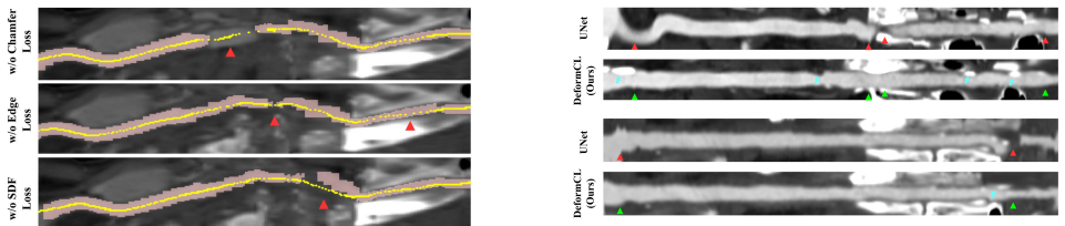
The machinery is the deformable centerline graph: a set of 3D points connected by edges, initialized per image from the coarse segmentation, and refined by a cascade of deformation stages. Each stage combines trilinear feature sampling, a Transformer over the graph for global context, an MLP with tanh activation for bounded point movement, and unpooling that inserts midpoints between connected points. Three auxiliary losses shape the deformation: a local Chamfer loss restricted to patches around ground-truth points, a signed-distance-function loss that pulls points toward the vessel center, and a regularization loss penalizing long edges. The segmentation head then converts the deformed centerlines into a mask through a distance map fused with the coarse prediction.
What would settle it
Compute per-branch centerline recall of the coarse segmentation and of DeformCL on a dataset with partially occluded vessels; if a branch entirely absent from the coarse mask appears in the final prediction, the deformation can create branches, whereas if none ever appear, the method's recall is capped by the coarse mask's completeness.
Extended reading notes
Core claim
The central claim is that vessel extraction should be posed as deforming a connected centerline graph rather than classifying voxels. The paper introduces DeformCL, where a graph G = {V, ε} has 3D centerline points as nodes and edges that encode adjacency. An adaptive template, built by skeletonizing a coarse segmentation, reconstructing a graph via minimum spanning tree, and subsampling to a few control points, is refined through L cascaded stages. Each stage samples hierarchical feature maps at the point locations, aggregates features along the graph with a Transformer, predicts a bounded offset, and unpooling doubles the point density. The final centerlines are converted to a segmentation by computing a voxel-to-centerline distance map and fusing it with the coarse mask. The paper reports that this continuous representation with natural connectivity reduces fractures, filters local false positives, and improves topological metrics across HaN-Seg, HNCTA, ASOCA, and ImageCAS datasets.
Load-bearing premise
Every vessel branch that matters must already be present in the coarse segmentation, because the deformation and unpooling layers move points along existing edges and never create new branches.
Editorial extensions
If this is right
- Adding DeformCL as an extra head improves Dice, clDice, and Betti number errors of softDice, clDice, and DSCNet baselines on all four datasets.
- The predicted centerlines are structurally complete and smooth enough to be used directly for curved planar reformation, without the usual mask-to-skeleton conversion.
- Because the representation is continuous, noise robustness follows from the top-down deformation, reducing scattered false positives compared with bottom-up per-pixel classification.
- The added computational cost is modest: about 0.12M extra parameters and 0.06 seconds per image over the 3D UNet backbone.
Reading between the lines
- A direct consequence not tested in the paper: since deformation never adds branches, centerline recall cannot exceed the recall of the coarse segmentation; a version that grows new edges during deformation would be needed for fully occluded vessels.
- The same deformable-centerline principle could be applied to other curvilinear structures—neuronal arbors, airways, or road networks—where connectivity, centrality, and noise tolerance matter more than voxel-wise accuracy.
- The paper's preference for rough initial templates (four control points) suggests the deformation is an active-contour-like process; measuring sensitivity to random template perturbations would reveal how much of the performance comes from the template prior versus the learned deformation.
Editorial analysis
A structured set of objections, weighed in public.
Referee Report
Summary. The paper introduces DeformCL, a continuous representation of 3D vessels as graphs of deformable centerline points. Starting from a coarse segmentation produced by the backbone, the method skeletonizes the coarse mask, builds a minimum-spanning-tree graph, selects k control points, and interpolates an initial centerline template. A cascade of Transformer feature aggregation, deformation, and unpooling layers then refines the point positions. The final centerline points are converted into a distance map that is fused with the coarse segmentation to produce a refined mask. The method is evaluated on HaN-Seg, HNCTA, ASOCA, and ImageCAS, with three base segmentors (softDice, clDice, DSCNet), reporting segmentation Dice/clDice, topological errors, HD95, and centerline F1/HD/Chamfer/beta0, plus qualitative CPR visualizations.
Significance. If its central claims are validated, DeformCL would be a practically useful representation: it outputs centerlines directly, supports feature aggregation along tubular curves, and improves segmentation connectivity. The manuscript has clear strengths: the code is released; the pipeline is specified in enough detail to reimplement; experiments cover four datasets and multiple backbones; ablations test template control-point count, loss components, interpolation, and interaction mechanism; and a Swin UNETR backbone is included. The clinical CPR visualization is a relevant and informative qualitative contribution. The main weakness is that the topology of the final centerline is fixed by the coarse-segmentation skeleton, which limits the claimed noise robustness and natural connectivity and makes the topological improvements in the main tables hard to attribute to the deformable representation.
major comments (4)
- [Sec. 3.2, Eqs. (4)-(5), Supplementary Fig. 13] The deformation and unpooling operations only move existing points and insert midpoints on existing edges; no operation adds or removes branches. Because the initial template is derived from the coarse segmentation skeleton, the final centerline topology is predetermined by the coarse segmentation. Therefore the method cannot recover a vessel branch that is absent from the coarse segmentation, nor can it prune a spurious branch that the coarse segmentation contains. This directly weakens the claims of 'noise robustness' and 'natural connectivity' and is consistent with the fragmented predictions on occluded vessels shown in Supplementary Fig. 13. To support the central claim, the authors should quantify, on the test sets, how often ground-truth branches are missing from the coarse segmentation and report branch-level recall/precision of the final centerlines relative to the coarse skeleton. Without such analysis, the topological gains in Table 1 cannot be separated from the quality of the initial skeleton.
- [Table 2] The centerline comparison is not apples-to-apples because the baseline methods are segmentation models that were not trained for centerline extraction, and the procedure used to derive their centerlines from the predicted masks is not described. A common protocol (same skeletonization algorithm, same tolerance, same matching criterion) is essential for F1, HD95, and Chamfer comparisons. Moreover, the claim of consistent centerline improvement is contradicted by the HaN-Seg row: Ours(DSCNet) reports HD95=8.222, which is worse than DSCNet's 7.462. The authors should specify the centerline generation procedure for all methods, release the evaluation script, and either train the baselines with a centerline-aware head or restrict the centerline claims to comparisons among methods that produce centerlines natively.
- [Tables 1 and 2] No standard deviations, confidence intervals, or significance tests are reported. This is particularly important because the test sets are small (11 images for HaN-Seg and 7 for ASOCA), and several reported improvements are around 1 Dice point or less (e.g., Ours(clDice) on ImageCAS improves Dice by only 0.07). Without variance estimates, the claim of 'effectiveness and superiority' is not statistically supported. The authors should report per-subject variability or fold-based intervals and apply paired tests to the Dice, clDice, Betti, and centerline metrics where small numbers of branches can drive the average.
- [Sec. 4.5, Table 4] The ablation of loss functions reports only segmentation Dice and clDice, yet the text argues that SDF loss and regularization loss improve centerline centrality and smoothness. These claims are qualitative and based on Fig. 7. The ablation should report the centerline metrics (F1, HD95, Chamfer, beta0) for each loss configuration, because the paper's central contribution is the centerline representation and its downstream effect on segmentation. Without these numbers, the contribution of L_sdf and L_reg to centerline quality is not quantitatively established.
minor comments (5)
- [Throughout] There are several typos: 'strcutures' in the SDF Loss paragraph, 'neightbour' in Eq. (8), 'analusis' in the caption of Table 5, and 'planner' should be 'planar' in Sec. 3.1.
- [Eq. (6)] The domain of the distance map is declared as R^{H*D*W}, but the input volume is described as R^{H*W*D}; please make the dimension order consistent.
- [Figure 3] The overview figure is dense and hard to read at print size; the stage weights and loss labels should be enlarged or split into a separate figure.
- [Sec. 4.4] The sentence 'We have not included PointScatter [31] and Topoloss [9] in the comparison as it is designed for 2D images' has an ambiguous pronoun; clarify that both methods are designed for 2D and are not applicable to 3D volumes under the reported GPU memory constraints.
- [Sec. 3.2, Eq. (2)-(4)] The Transformer architecture is not fully specified in the main text; please state the number of layers, heads, and hidden dimensions, or refer the reader to a precise supplementary section.
Circularity Check
No significant circularity: DeformCL is an empirically trained pipeline evaluated on held-out test sets; the centerline template is an architectural initialization, not a fitted parameter renamed as a prediction.
full rationale
The derivation chain is supervised end-to-end by ground-truth masks and centerlines: coarse segmentation, centerline deformation (Eqs. 1-5), and distance-map-based refined segmentation (Eq. 6) are trained with Dice, local Chamfer, SDF, and regularization losses (Eqs. 7-13), and evaluated on held-out test splits of four datasets. The initial template is generated from the coarse segmentation skeleton, but deformation and unpooling are learned and tested against GT centerlines; this is an architectural initialization, not a fitted parameter renamed as a prediction. The paper's own Supplementary Fig. 13 documents a failure mode for occluded vessels where fragmented coarse segmentation cannot be recovered; this is a limitation of the fixed-topology deformation, not evidence that a claimed result reduces to its input by construction. Self-citations ([31], [39], [40]) are present, but [39] is only used as a graph-reconstruction recipe in template generation and [31]/[40] are related work; none is invoked as an unverified uniqueness theorem or as the answer to the central claim. Therefore no circular step is exhibited.
Assumptions & free parameters
free parameters (5)
- Number of template control points k =
4
- Centerline loss weights (lambda_cha, lambda_sdf, lambda_reg) =
30, 0.5, 60
- Stage balance weights w_l =
0.05, 0.60, 0.95, 1.00
- Initial centerline point count N_c =
100 per category
- Local patch size M and patch count |Omega| =
M unspecified; |Omega| sampled from U(60,80)
assumptions (4)
- domain assumption Ground-truth centerlines obtained by 3D thinning of GT masks are valid supervision targets for vessel centerlines.
- domain assumption The coarse segmentation from the 3D UNet backbone contains the correct vessel topology, so the initial template can capture all relevant branches.
- domain assumption Deformation and unpooling never change graph topology; they only move points and insert points along existing edges.
- domain assumption Minimum spanning tree reconstruction of the thinning output preserves vessel branching topology.
Cite this review
Pith. "Pith review of DeformCL: Learning Deformable Centerline Representation for Vessel Extraction in 3D Medical Image." pith.science (2026). https://pith.science/paper/HQAI5JCA
@misc{pith2026250605820,
  author       = {Pith},
  title        = {Pith review of: DeformCL: Learning Deformable Centerline Representation for Vessel Extraction in 3D Medical Image},
  year         = {2026},
  howpublished = {\url{https://pith.science/paper/HQAI5JCA}},
  note         = {Machine review of arXiv:2506.05820}
}
read the original abstract
In the field of 3D medical imaging, accurately extracting and representing the blood vessels with curvilinear structures holds paramount importance for clinical diagnosis. Previous methods have commonly relied on discrete representation like mask, often resulting in local fractures or scattered fragments due to the inherent limitations of the per-pixel classification paradigm. In this work, we introduce DeformCL, a new continuous representation based on Deformable Centerlines, where centerline points act as nodes connected by edges that capture spatial relationships. Compared with previous representations, DeformCL offers three key advantages: natural connectivity, noise robustness, and interaction facility. We present a comprehensive training pipeline structured in a cascaded manner to fully exploit these favorable properties of DeformCL. Extensive experiments on four 3D vessel segmentation datasets demonstrate the effectiveness and superiority of our method. Furthermore, the visualization of curved planar reformation images validates the clinical significance of the proposed framework. We release the code in https://github.com/barry664/DeformCL
Figures


Figures from the paper (9 more)




Reference graph
Works this paper leans on
-
[40]
Zhao Zhang, Ziwei Zhao, Dong Wang, and Liwei Wang. Graphmorph: Tubular structure extraction by morphing pre- dicted graphs.Advances in Neural Information Processing Systems, 37:68472–68499, 2024. 2
work page 2024
-
[1]
Topological Similarity Index and Loss Function for Blood Vessel Segmentation
Ricardo J Ara ´ujo, Jaime S Cardoso, and H ´elder P Oliveira. Topological similarity index and loss function for blood ves- sel segmentation.arXiv preprint arXiv:2107.14531, 2021. 1, 2, 3
work page Pith review arXiv 2021
-
[2]
Fabian Bongratz, Anne-Marie Rickmann, Sebastian P ¨olsterl, and Christian Wachinger. V ox2cortex: fast explicit recon- struction of cortical surfaces from 3d mri scans with geomet- ric deep neural networks. InProceedings of the IEEE/CVF Conference on Computer Vision and Pattern Recognition, pages 20773–20783, 2022. 3
work page 2022
-
[3]
3d u-net: learn- ing dense volumetric segmentation from sparse annota- tion
¨Ozg¨un C ¸ ic ¸ek, Ahmed Abdulkadir, Soeren S Lienkamp, Thomas Brox, and Olaf Ronneberger. 3d u-net: learn- ing dense volumetric segmentation from sparse annota- tion. InMedical Image Computing and Computer-Assisted Intervention–MICCAI 2016: 19th International Conference, Athens, Greece, October 17-21, 2016, Proceedings, Part II 19, pages 424–432. Spring...
work page 2016
-
[4]
Deformable convolutional networks
Jifeng Dai, Haozhi Qi, Yuwen Xiong, Yi Li, Guodong Zhang, Han Hu, and Yichen Wei. Deformable convolutional networks. InProceedings of the IEEE international confer- ence on computer vision, pages 764–773, 2017. 3
work page 2017
-
[5]
Vilac ¸a, Xiyue Wang, Sen Yang, Arcot Sowmya, and Susann Beier
Ramtin Gharleghi, Dona Adikari, Katy Ellenberger, Sze- Yuan Ooi, Chris Ellis, Chung-Ming Chen, Ruochen Gao, Yuting He, Raabid Hussain, Chia-Yen Lee, Jun Li, Jun Ma, Ziwei Nie, Bruno Oliveira, Yaolei Qi, Youssef Skandarani, Jo˜ao L. Vilac ¸a, Xiyue Wang, Sen Yang, Arcot Sowmya, and Susann Beier. Automated segmentation of normal and dis- eased coronary arte...
work page 2022
-
[6]
R Gharleghi, D Adikari, K Ellenberger, M Webster, C El- lis, A Sowmya, S Ooi, and S Beier. Annotated computed tomography coronary angiogram images and associated data of normal and diseased arteries.Scientific Data, 10(1):128,
-
[7]
Ronald L Graham and Pavol Hell. On the history of the min- imum spanning tree problem.Annals of the History of Com- puting, 7(1):43–57, 1985. 4
work page 1985
Show all 44 references
-
[8]
A fast and efficient technique for the automatic tracing of corneal nerves in confocal microscopy.Translational vision science & technology, 5(5), 2016
Pedro Guimaraes, Jeffrey Wigdahl, and Alfredo Ruggeri. A fast and efficient technique for the automatic tracing of corneal nerves in confocal microscopy.Translational vision science & technology, 5(5), 2016. 6
2016
-
[9]
Topology-preserving deep image segmentation.Advances in neural information processing systems, 32, 2019
Xiaoling Hu, Fuxin Li, Dimitris Samaras, and Chao Chen. Topology-preserving deep image segmentation.Advances in neural information processing systems, 32, 2019. 1, 2, 7
2019
-
[10]
The vascular modeling toolkit: a python library for the analysis of tubular structures in medical images.Journal of Open Source Software, 3(25):745, 2018
Richard Izzo, David Steinman, Simone Manini, and Luca Antiga. The vascular modeling toolkit: a python library for the analysis of tubular structures in medical images.Journal of Open Source Software, 3(25):745, 2018. 2, 3, 8
2018
-
[11]
IEEE, 2002
Armin Kanitsar, Dominik Fleischmann, Rainer Wegenkittl, Petr Felkel, and Eduard Groller.CPR-curved planar refor- mation. IEEE, 2002. 2, 3, 8, 11
2002
-
[12]
Semi-supervised classi- fication with graph convolutional networks.arXiv preprint arXiv:1609.02907, 2016
Thomas N Kipf and Max Welling. Semi-supervised classi- fication with graph convolutional networks.arXiv preprint arXiv:1609.02907, 2016. 3, 12
2016 arXiv
-
[13]
Learning tree-structured representation for 3d coronary artery segmen- tation.Computerized Medical Imaging and Graphics, 80: 101688, 2020
Bin Kong, Xin Wang, Junjie Bai, Yi Lu, Feng Gao, Kun- lin Cao, Jun Xia, Qi Song, and Youbing Yin. Learning tree-structured representation for 3d coronary artery segmen- tation.Computerized Medical Imaging and Graphics, 80: 101688, 2020. 1
2020
-
[14]
A deep- learning approach for direct whole-heart mesh reconstruc- tion.Medical image analysis, 74:102222, 2021
Fanwei Kong, Nathan Wilson, and Shawn Shadden. A deep- learning approach for direct whole-heart mesh reconstruc- tion.Medical image analysis, 74:102222, 2021. 2, 3
2021
-
[15]
Building skeleton models via 3-d medial surface axis thin- ning algorithms.CVGIP: Graphical Models and Image Pro- cessing, 56(6):462–478, 1994
Ta-Chih Lee, Rangasami L Kashyap, and Chong-Nam Chu. Building skeleton models via 3-d medial surface axis thin- ning algorithms.CVGIP: Graphical Models and Image Pro- cessing, 56(6):462–478, 1994. 4, 5
1994
-
[16]
Jonathon Leipsic, Suhny Abbara, Stephan Achenbach, Ri- cardo Cury, James P Earls, GB John Mancini, Koen Nieman, Gianluca Pontone, and Gilbert L Raff. Scct guidelines for the interpretation and reporting of coronary ct angiography: a re- port of the society of cardiovascular co...
2014
-
[17]
Fully convolutional networks for semantic segmentation
Jonathan Long, Evan Shelhamer, and Trevor Darrell. Fully convolutional networks for semantic segmentation. InPro- ceedings of the IEEE conference on computer vision and pat- tern recognition, pages 3431–3440, 2015. 2
2015
-
[18]
Decoupled weight decay regularization.arXiv preprint arXiv:1711.05101, 2017
Ilya Loshchilov and Frank Hutter. Decoupled weight decay regularization.arXiv preprint arXiv:1711.05101, 2017. 11
2017 arXiv
-
[19]
V-net: Fully convolutional neural networks for volumetric medical image segmentation
Fausto Milletari, Nassir Navab, and Seyed-Ahmad Ahmadi. V-net: Fully convolutional neural networks for volumetric medical image segmentation. In2016 fourth international conference on 3D vision (3DV), pages 565–571. Ieee, 2016. 6, 7
2016
-
[20]
Zeming Lin
Adam Paszke, Sam Gross, Francisco Massa, Adam Lerer, James Bradbury, Gregory Chanan, Trevor Killeen, and et al. Zeming Lin. Pytorch: An imperative style, high- performance deep learning library. InNeurIPS, 2019. 11
2019
-
[21]
A framework for geo- metric analysis of vascular structures: application to cere- bral aneurysms.IEEE transactions on medical imaging, 28 (8):1141–1155, 2009
Marina Piccinelli, Alessandro Veneziani, David A Steinman, Andrea Remuzzi, and Luca Antiga. A framework for geo- metric analysis of vascular structures: application to cere- bral aneurysms.IEEE transactions on medical imaging, 28 (8):1141–1155, 2009. 3, 8
2009
-
[22]
Han-seg: The head and neck organ-at-risk ct and mr segmentation dataset.Medical physics, 50(3):1917–1927, 2023
Ga ˇsper Podobnik, Primo ˇz Strojan, Primo ˇz Peterlin, Bulat Ibragimov, and Toma ˇz Vrtovec. Han-seg: The head and neck organ-at-risk ct and mr segmentation dataset.Medical physics, 50(3):1917–1927, 2023. 6, 11
1917
-
[23]
Dynamic snake convolution based on topo- logical geometric constraints for tubular structure segmenta- tion
Yaolei Qi, Yuting He, Xiaoming Qi, Yuan Zhang, and Guanyu Yang. Dynamic snake convolution based on topo- logical geometric constraints for tubular structure segmenta- tion. InProceedings of the IEEE/CVF International Confer- ence on Computer Vision, pages 6070–6079, 2023. 1, 2...
2023
-
[24]
Heart disease and stroke statistics—2011 update: a re- port from the american heart association.Circulation, 123 (4):e18–e209, 2011
V ´eronique L Roger, Alan S Go, Donald M Lloyd-Jones, Robert J Adams, Jarett D Berry, Todd M Brown, Mercedes R Carnethon, Shifan Dai, Giovanni De Simone, Earl S Ford, et al. Heart disease and stroke statistics—2011 update: a re- port from the american heart association.Circula...
2011
-
[25]
U- net: Convolutional networks for biomedical image segmen- tation
Olaf Ronneberger, Philipp Fischer, and Thomas Brox. U- net: Convolutional networks for biomedical image segmen- tation. InMedical Image Computing and Computer-Assisted Intervention–MICCAI 2015: 18th International Conference, Munich, Germany, October 5-9, 2015, Proceedings, Par...
2015
-
[26]
Standardized evaluation methodology and reference database for evaluating coronary artery centerline extraction algorithms.Medical image analysis, 13(5):701–714, 2009
Michiel Schaap, Coert T Metz, Theo van Walsum, Alina G van der Giessen, Annick C Weustink, Nico R Mollet, Chris- tian Bauer, Hrvoje Bogunovi ´c, Carlos Castro, Xiang Deng, et al. Standardized evaluation methodology and reference database for evaluating coronary artery centerli...
2009
-
[27]
Deep vessel segmentation by learning graph- ical connectivity.Medical image analysis, 58:101556, 2019
Seung Yeon Shin, Soochahn Lee, Il Dong Yun, and Ky- oung Mu Lee. Deep vessel segmentation by learning graph- ical connectivity.Medical image analysis, 58:101556, 2019. 1, 2, 3
2019
-
[28]
cldice-a novel topology-preserving loss function for tubular structure seg- mentation
Suprosanna Shit, Johannes C Paetzold, Anjany Sekuboyina, Ivan Ezhov, Alexander Unger, Andrey Zhylka, Josien PW Pluim, Ulrich Bauer, and Bjoern H Menze. cldice-a novel topology-preserving loss function for tubular structure seg- mentation. InProceedings of the IEEE/CVF Conferen...
2021
-
[29]
Charles A Taylor and David A Steinman. Image-based mod- eling of blood flow and vessel wall dynamics: applications, methods and future directions: Sixth international bio-fluid mechanics symposium and workshop, march 28–30, 2008 pasadena, california.Annals of biomedical engine...
2008
-
[30]
Attention is all you need.Advances in neural information processing systems, 30, 2017
Ashish Vaswani, Noam Shazeer, Niki Parmar, Jakob Uszko- reit, Llion Jones, Aidan N Gomez, Łukasz Kaiser, and Illia Polosukhin. Attention is all you need.Advances in neural information processing systems, 30, 2017. 3, 4, 12
2017
-
[31]
Pointscatter: Point set representation for tubular structure extraction
Dong Wang, Zhao Zhang, Ziwei Zhao, Yuhang Liu, Yihong Chen, and Liwei Wang. Pointscatter: Point set representation for tubular structure extraction. InEuropean Conference on Computer Vision, pages 366–383. Springer, 2022. 1, 2, 3, 6, 7, 12
2022
-
[32]
Pixel2mesh: Generating 3d mesh models from single rgb images
Nanyang Wang, Yinda Zhang, Zhuwen Li, Yanwei Fu, Wei Liu, and Yu-Gang Jiang. Pixel2mesh: Generating 3d mesh models from single rgb images. InProceedings of the Euro- pean conference on computer vision (ECCV), pages 52–67,
-
[33]
Deep distance transform for tubular structure segmentation in ct scans
Yan Wang, Xu Wei, Fengze Liu, Jieneng Chen, Yuyin Zhou, Wei Shen, Elliot K Fishman, and Alan L Yuille. Deep distance transform for tubular structure segmentation in ct scans. InProceedings of the IEEE/CVF Conference on Com- puter Vision and Pattern Recognition, pages 3833–3842,
-
[34]
V oxel2mesh: 3d mesh model genera- tion from volumetric data
Udaranga Wickramasinghe, Edoardo Remelli, Graham Knott, and Pascal Fua. V oxel2mesh: 3d mesh model genera- tion from volumetric data. InMedical Image Computing and Computer Assisted Intervention–MICCAI 2020: 23rd Inter- national Conference, Lima, Peru, October 4–8, 2020, Pro- ...
2020
-
[35]
Deep closing: Enhancing topological connectiv- ity in medical tubular segmentation.IEEE Transactions on Medical Imaging, 2024
Qian Wu, Yufei Chen, Wei Liu, Xiaodong Yue, and Xiahai Zhuang. Deep closing: Enhancing topological connectiv- ity in medical tubular segmentation.IEEE Transactions on Medical Imaging, 2024. 2
2024
-
[36]
Implicitatlas: learning deformable shape templates in medical imaging
Jiancheng Yang, Udaranga Wickramasinghe, Bingbing Ni, and Pascal Fua. Implicitatlas: learning deformable shape templates in medical imaging. InProceedings of the IEEE/CVF Conference on Computer Vision and Pattern Recognition, pages 15861–15871, 2022. 3
2022
-
[37]
An Zeng, Chunbiao Wu, Guisen Lin, Wen Xie, Jin Hong, Meiping Huang, Jian Zhuang, Shanshan Bi, Dan Pan, Najeeb Ullah, et al. Imagecas: A large-scale dataset and benchmark for coronary artery segmentation based on computed tomog- raphy angiography images.Computerized Medical Ima...
2023
-
[38]
Pro- gressive deep segmentation of coronary artery via hierarchi- cal topology learning
Xiao Zhang, Jingyang Zhang, Lei Ma, Peng Xue, Yan Hu, Dijia Wu, Yiqiang Zhan, Jun Feng, and Dinggang Shen. Pro- gressive deep segmentation of coronary artery via hierarchi- cal topology learning. InInternational Conference on Med- ical Image Computing and Computer-Assisted Int...
2022
-
[39]
Topology-preserving automatic labeling of coronary arteries via anatomy-aware connection classifier
Zhixing Zhang, Ziwei Zhao, Dong Wang, Shishuang Zhao, Yuhang Liu, Jia Liu, and Liwei Wang. Topology-preserving automatic labeling of coronary arteries via anatomy-aware connection classifier. InInternational Conference on Med- ical Image Computing and Computer-Assisted Interve...
2023
-
[41]
Graph convolution based cross-network multiscale feature fusion for deep vessel segmentation.IEEE Transactions on Med- ical Imaging, 42(1):183–195, 2022
Gangming Zhao, Kongming Liang, Chengwei Pan, Fandong Zhang, Xianpeng Wu, Xinyang Hu, and Yizhou Yu. Graph convolution based cross-network multiscale feature fusion for deep vessel segmentation.IEEE Transactions on Med- ical Imaging, 42(1):183–195, 2022. 1, 2
2022
-
[42]
3d graph anatomy geometry-integrated network for pancre- atic mass segmentation, diagnosis, and quantitative patient management
Tianyi Zhao, Kai Cao, Jiawen Yao, Isabella Nogues, Le Lu, Lingyun Huang, Jing Xiao, Zhaozheng Yin, and Ling Zhang. 3d graph anatomy geometry-integrated network for pancre- atic mass segmentation, diagnosis, and quantitative patient management. InProceedings of the IEEE/CVF con...
2021
-
[43]
Deformable detr: Deformable trans- formers for end-to-end object detection.arXiv preprint arXiv:2010.04159, 2020
Xizhou Zhu, Weijie Su, Lewei Lu, Bin Li, Xiaogang Wang, and Jifeng Dai. Deformable detr: Deformable trans- formers for end-to-end object detection.arXiv preprint arXiv:2010.04159, 2020. 3 We will elaborate on the details about datasets, imple- mentation and more experimental a...
2010 arXiv
-
[2020]
1, 2, 3, 5, 6, 7, 12
Reviewed August 7, 2026 · model on record in the stance chip above.
Discussion (0). Sign in to comment.