REVIEW 5 major objections 5 minor 57 references
The Four Color Theorem for Cell Instance Segmentation
T0 review · 5 major / 5 minor · reviewed 2026-08-07 · deepseek-v4-flash
Pith's one-line read Cell instance segmentation reduces to a four-class semantic labeling problem when adjacent cells are colored differently, and the compact network that learns this encoding reports state-of-the-art accuracy on four microscopy benchmarks at…
desk verdict The four-color encoding is a fresh idea with consistent empirical gains, but the missing inference decoding step and flawed theory make the paper a major-revision case, not a clean accept. read the letter →
The pith
A machine-rendered reading of the paper's core claim, the machinery that carries it, and where it could break.
The reading
What carries the argument
The load-bearing object is the greedy four-color encoding of the cell adjacency graph $G=(V,E)$: vertices are cells, edges connect touching cells, and each vertex receives $C(v)=\min(C\setminus C_{\mathrm{used}})$ from the color set $\{1,2,3,4\}$, yielding a pixel-level semantic mask in which adjacent instances always differ. The four-color theorem supplies the existence guarantee that makes this a valid reformulation of instance segmentation. Two mechanisms carry the training-stability argument: the asymptotic training strategy, which first optimizes a binary foreground/background segmentation and only then supervises the four color classes inside the foreground, and the encoding-transformation module (two convolutional layers) together with a negative-sampling orthogonality loss that minimizes the cosine similarity between sampled feature vectors of adjacent cells, steering the network toward the canonical greedy encoding despite the non-uniqueness of valid colorings. The encoding matrix's sparsity (one nonzero entry per cell, rank at most four) is what makes the transformation a small correction rather than a second segmentation problem.
What would settle it
On a held-out set, tally how often two adjacent cells receive the same color under the greedy ground-truth encoding and how often connected-component decoding of a correct four-color prediction merges two distinct cells; even one such pair would falsify the claim that the four-color map can be decoded back into instances by color-connected grouping, and the overall rate across datasets would measure how close the empirical guarantee comes to a real proof.
Extended reading notes
Core claim
The paper's central claim is that instance segmentation of cells is equivalent to a four-class semantic segmentation problem. Treating each cell as a country and the background as the ocean, a greedy algorithm assigns each cell the smallest available color from $\{1,2,3,4\}$ that no already-colored neighbor uses, so adjacent cells always differ; the four-color theorem guarantees such a coloring exists for planar arrangements. The network learns to predict this four-color map together with a foreground/background channel, and instances are separated by the coloring itself. Because valid four-color encodings are non-unique — colors can be substituted, exchanged, or re-rule-modified — direct supervision is unstable, so the paper proposes an asymptotic training strategy (background first, then color within foreground), an encoding-transformation layer that maps the network output back to the greedy encoding, and a negative-sampling orthogonality loss over adjacent cells. Two theorems ground the design: greedy coloring is globally optimal for planar cell graphs with bounded degree and chain-like or rectangular arrangement (Theorem 1), and every predicted encoding related by substitution, exchange, or rule modification can be mapped to the greedy encoding (Theorem 2). With these components, FCIS reports state-of-the-art accuracy on DSB2018, PanNuke, BBBC006v1, and YeaZ.
Load-bearing premise
The load-bearing premise is that connected same-color pixels always decode into exactly the original cell and that the greedy coloring always separates touching cells within the four-color budget — the decoding half is assumed rather than demonstrated, and the coloring half is backed only by empirical color-use statistics on the four datasets, not a proof.
Editorial extensions
If this is right
- One compact semantic segmentation network with connected-component grouping of same-color foreground pixels can replace the detection heads, contour branches, and distance-map post-processing used by the compared approaches on the tested cell types.
- Results across fluorescence, H&E histopathology, bright-field, and phase-contrast images indicate the four-color formulation transfers across imaging modalities without architecture changes.
- Directly training on the raw four-color labels hurts accuracy; the ablation shows the asymptotic schedule, the transformation, and the sampling constraint are all needed, with the sampling constraint giving the largest gain.
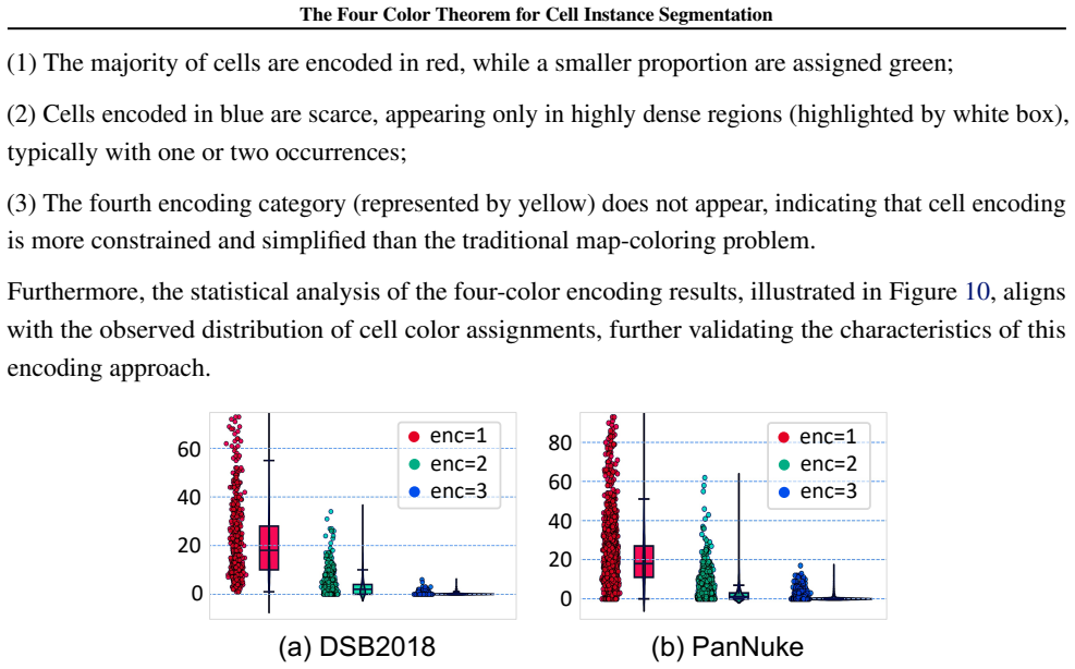
- The statistics that almost no image requires all four colors imply that real cell coloring is a strictly easier problem than general map coloring, so the four-class output space has unused capacity.
- If the complexity comparison holds, the method sits below most detection- and distance-based baselines in parameter count and FLOPs at $256\times256$ input, which matters for large-scale screening workloads.
Reading between the lines
- The decoding step — grouping connected same-color pixels into instances — is left implicit in the paper, and its correctness is assumed rather than demonstrated; this is the natural place for silent merge or split errors.
- The same greedy-coloring-plus-transformation recipe could apply to other adjacency-constrained labeling tasks, such as gland, tissue-region, or even non-biological planar-object segmentation, wherever the ground-truth label is a non-unique coloring.
- The observed color statistics suggest a two- or three-color encoding might suffice on these datasets; a testable extension is an adaptive encoding that uses fewer output channels when the local graph is sparse and reserves the fourth color for dense regions.
- Heavy overlap or projections of three-dimensional cell stacks strain the planar-map assumption behind the four-color guarantee, so the method's headroom on those images is an open question the paper does not address.
Editorial analysis
A structured set of objections, weighed in public.
Referee Report
Summary. This paper proposes FCIS, a cell instance segmentation method that converts the instance segmentation task into a four-class semantic segmentation problem. The authors construct a cell adjacency graph from instance labels and greedily color it with at most four colors so that adjacent cells receive different colors. The network predicts a background channel plus four color channels; training uses an asymptotic strategy, an orthogonality loss on sampled adjacent-cell features, and an 'encoding transformation' module intended to stabilize the non-unique color-label supervision. Experiments on DSB2018, PanNuke, BBBC006v1, and YeaZ report state-of-the-art or competitive results on DICE, AJI, DQ, SQ, and PQ with lower parameter/FLOP counts than many existing methods. The paper also states two theorems claiming global optimality of greedy coloring and existence of a mapping from predicted encodings to the greedy encoding.
Significance. If the central claims hold, the paper provides an appealingly simple and efficient alternative to detection-, contour-, and distance-based instance segmentation: a compact semantic segmentation network with a lightweight post-processing step. The empirical comparison is broad, covering four imaging modalities and multiple instance-level metrics, and the ablations isolate the contribution of each proposed module. The code release is a further strength. However, the current manuscript does not specify how four-class predictions are converted into instance masks, the data splits are inconsistent between the main text and the supplementary material, and the theoretical support (Theorems 1 and 2) is not valid as stated. The empirical results may be reproducible from the code, but the paper is not yet self-contained enough to support its stated claims.
major comments (5)
- [Section 4; Algorithm 1; Tables 2–5] The paper never defines the inference-time procedure that maps the network's five-channel output to the instance masks evaluated in Tables 2–5. Algorithm 1 only describes the ground-truth encoding from instance labels, and Section 4 defines losses. Without a stated decoding rule, instance-level metrics such as AJI and PQ are undefined from the paper alone. Please specify the exact decoding algorithm, including how to handle adjacent cells predicted with the same color, a single cell predicted with multiple colors, and the role of the background channel. Connected-component labeling per color class may be the intended rule, but it must be stated and its failure modes analyzed; the public code does not replace this specification.
- [Supplementary E, Theorem 1] Theorem 1 is false as stated. The proof asserts that for a path graph Pn, greedy coloring in any vertex order uses at most two colors; but for P4 with vertex order (v1, v4, v2, v3), greedy uses three colors. Similarly, for a 3×3 grid graph with the center vertex colored first, greedy uses three colors although the graph is bipartite with chromatic number two. These graphs satisfy all conditions of Theorem 1 (planar, maximum degree at most four, chain or rectangular structure). The step 'χ_greedy(G) ≤ 4 = χ(G)' is also invalid because the four-color theorem only yields χ(G) ≤ 4. The theoretical justification for the low-rank property should be either removed or replaced by an empirical statement that does not depend on vertex order.
- [Section 4.2; Supplementary E, Theorem 2] Theorem 2 is circular and does not establish the claimed compatibility. The relations 'substitution, exchange, rule modification' are never formally defined, and the proof constructs T by minimizing ||PT−C||^2_F with C, the target greedy encoding, available inside the construction; hence the existence of a mapping is guaranteed only when the ground-truth encoding is known, which is not the test-time setting. The intermediate assertion rank(P) ≥ k is unjustified for arbitrary network outputs. The encoding transformation is a reasonable training heuristic, but it should be presented as a learned module and empirically validated, not as a theorem.
- [Section 5.1 vs Supplementary Table 7] The dataset splits are inconsistent between the main text and the supplementary material. Section 5.1 reports DSB2018 as 380 training, 67 validation, and 50 testing images, while Supplementary Table 7 reports 602, 109, and 89 samples; similar discrepancies exist for BBBC006v1 and YeaZ. Because Tables 2–5 compare against published baselines, the exact split and the overlapping-cropping procedure used must be reported unambiguously so that the comparison is fair and reproducible.
- [Section 5.2 and Table 1] The network architecture is not described. The reader only learns that an encoder–decoder produces a five-channel feature map; the backbone, decoder depth, and input resolution are not specified. This is important for the complexity comparison in Table 1 and for reproducibility. Please state the exact architecture used for FCIS and clarify whether the reported parameters and FLOPs were measured with that architecture on 256×256 inputs.
minor comments (5)
- [Title page] The line 'Proceedings of the 42 st International Conference' contains a typo ('42 st').
- [Table 1 and Tables 2–5] The method name is spelled inconsistently as 'GeSegNet' in some places and 'GeNSeg-Net' in others; also, DoNet's parameter count is listed as '67.71' without units.
- [Algorithm 1] The notation C \ C_used assumes C is a set of labels; please clarify that C_used contains colors already assigned to neighbors and that C(v) is an integer from {1,2,3,4}.
- [Theorem 1] The main-text statement of Theorem 1 does not mention vertex ordering, although the proof depends on the greedy ordering; please make the ordering explicit or remove the claim.
- [Figure 8] The caption uses the repeated label '(b)' for different subfigures, which is confusing; the subfigures should be labeled consistently.
Circularity Check
The benchmarked segmentation results are independent, but the auxiliary theory is circular: Theorem 2 defines compatibility as the existence of a mapping and then uses the target to construct that mapping, and Theorem 1's proof assumes the equality it must prove.
-
self definitional
[Section 4.2 (Theorem 2) and Supplementary Material E, Proof 2]
"“If the predicted encoding matrix P has one of the relations with the greedy encoding C, i.e., substitution, exchange, rule modification. Then there exists a mapping function: f:P→C, such that the network’s predicted result can be transformed into the four-color encoding result.” ... “Define a column transformation matrix T∈R^{k′×k}, where T=argmin_T ∥PT−C∥^2_F , s.t. rank(PT)=k. The transformed matrix is P′=PT” ... “C=P′S. The matrix S is a permutation matrix satisfying S^⊤S=I.”"
The premise “has one of the relations (substitution, exchange, rule modification)” is not a formal, checkable condition; the three “relations” are simply ways one proper coloring can fail to equal another. The proof then chooses T as the least-squares fit of P to C and asserts the exact equality C=P′S. In other words, the existence of f is guaranteed by selecting the mapping after seeing C, so the compatibility theorem reduces to the statement that any P transformable into C is transformable into C. No independent condition on P is established; the construction already assumes the target C is reachable, making the result true by definition rather than by derivation.
-
other
[Section 3.3 / Supplementary Material E, Proof 1 (rectangular structure)]
"“According to the Four Color Theorem: χ(G)≤4. The greedy algorithm, in each iteration, uses the smallest available color, and each vertex checks at most 4 adjacent vertices. Therefore, the chromatic number generated by the greedy algorithm satisfies: χgreedy(G)≤4=χ(G).”"
This proof step asserts χgreedy(G)≤4=χ(G), treating the four-color upper bound as an equality with the chromatic number. For a rectangular grid graph, which is bipartite, χ(G)=2, so “4=χ(G)” is false; in any case, inequality χgreedy≤4 together with χ≤4 does not imply χgreedy=χ. The equality inserted is exactly the conclusion of Theorem 1, so the derivation assumes what it must prove. A concrete counterexample is a 3×3 grid colored greedily center-first, which uses three colors although χ=2, contradicting the claimed “any arbitrary vertex ordering” global optimality.
full rationale
The empirical core of the paper is not circular: Tables 2–6 report instance-level DICE, AJI, and PQ against external ground truth, and the gains over U-Net and other baselines do not depend on reusing the fitted four-color labels as the evaluation target. The ablation row “w. Four-color” is an honest negative result showing that direct four-color supervision hurts performance, which further confirms that the main metric improvements are not internally forced. No load-bearing self-citation chain was found; the authors’ own prior works (Zhang et al. 2025a–c) appear only as general segmentation context. The circularity is confined to the auxiliary theory: Theorem 2 guarantees a mapping f:P→C after defining the premise as a relation under which such a mapping is supposed to exist, and its proof constructs the mapping using the target C itself; Theorem 1’s proof inserts the equality “4=χ(G)” that it is supposed to establish. Separately, the paper never specifies how a predicted four-color map is decoded into instance masks at inference: Algorithm 1 only generates ground-truth encodings, so the reported instance metrics depend on an unstated post-processing step. That is a reproducibility gap rather than a circular reduction, but it reinforces that the theoretical guarantees are not what carry the empirical results. Overall, partial circularity in theory with independent empirical content, so a score of 4 is appropriate.
Assumptions & free parameters
free parameters (3)
- lambda_1 (orthogonality loss weight) =
2
- lambda_2 (classification loss weight) =
1
- sampling rate r for negative sampling =
0.5
assumptions (4)
- domain assumption The cell adjacency graph is planar, so the four-color theorem applies.
- domain assumption Real cell graphs satisfy the chain or rectangular structure with max degree <=4 assumed in Theorem 1.
- domain assumption A four-color semantic prediction can be decoded into instances by connected-component grouping.
- domain assumption The greedy algorithm, with the ordering used, always produces a proper coloring using at most four colors for the datasets.
Cite this review
Pith. "Pith review of The Four Color Theorem for Cell Instance Segmentation." pith.science (2026). https://pith.science/paper/64DKWEPA
@misc{pith2026250609724,
  author       = {Pith},
  title        = {Pith review of: The Four Color Theorem for Cell Instance Segmentation},
  year         = {2026},
  howpublished = {\url{https://pith.science/paper/64DKWEPA}},
  note         = {Machine review of arXiv:2506.09724}
}
read the original abstract
Cell instance segmentation is critical to analyzing biomedical images, yet accurately distinguishing tightly touching cells remains a persistent challenge. Existing instance segmentation frameworks, including detection-based, contour-based, and distance mapping-based approaches, have made significant progress, but balancing model performance with computational efficiency remains an open problem. In this paper, we propose a novel cell instance segmentation method inspired by the four-color theorem. By conceptualizing cells as countries and tissues as oceans, we introduce a four-color encoding scheme that ensures adjacent instances receive distinct labels. This reformulation transforms instance segmentation into a constrained semantic segmentation problem with only four predicted classes, substantially simplifying the instance differentiation process. To solve the training instability caused by the non-uniqueness of four-color encoding, we design an asymptotic training strategy and encoding transformation method. Extensive experiments on various modes demonstrate our approach achieves state-of-the-art performance. The code is available at https://github.com/zhangye-zoe/FCIS.
Figures


Figures from the paper (7 more)



Reference graph
Works this paper leans on
-
[1]
A., Dryg, I., Werba, G., Wang, W., Baron, M., Rao, A., Xia, B., et al
Barkley, D., Moncada, R., Pour, M., Liberman, D. A., Dryg, I., Werba, G., Wang, W., Baron, M., Rao, A., Xia, B., et al. Cancer cell states recur across tumor types and form specific interactions with the tumor microenvironment. Nature genetics, 54 0 (8): 0 1192--1201, 2022
work page 2022
-
[2]
Caicedo, J. C., Goodman, A., Karhohs, K. W., Cimini, B. A., Ackerman, J., Haghighi, M., Heng, C., Becker, T., Doan, M., McQuin, C., et al. Nucleus segmentation across imaging experiments: the 2018 data science bowl. Nature methods, 16 0 (12): 0 1247--1253, 2019
work page 2018
-
[3]
Dcan: deep contour-aware networks for accurate gland segmentation
Chen, H., Qi, X., Yu, L., and Heng, P.-A. Dcan: deep contour-aware networks for accurate gland segmentation. In Proceedings of the IEEE conference on Computer Vision and Pattern Recognition, pp.\ 2487--2496, 2016
work page 2016
-
[4]
Chen, J., Mei, J., Li, X., Lu, Y., Yu, Q., Wei, Q., Luo, X., Xie, Y., Adeli, E., Wang, Y., et al. Transunet: Rethinking the u-net architecture design for medical image segmentation through the lens of transformers. Medical Image Analysis, 97: 0 103280, 2024
work page 2024
-
[5]
Cpp-net: Context-aware polygon proposal network for nucleus segmentation
Chen, S., Ding, C., Liu, M., Cheng, J., and Tao, D. Cpp-net: Context-aware polygon proposal network for nucleus segmentation. IEEE Transactions on Image Processing, 32: 0 980--994, 2023
work page 2023
-
[6]
Un-sam: Domain-adaptive self-prompt segmentation for universal nuclei images
Chen, Z., Xu, Q., Liu, X., and Yuan, Y. Un-sam: Domain-adaptive self-prompt segmentation for universal nuclei images. Medical Image Analysis, pp.\ 103607, 2025
work page 2025
-
[7]
Cancer-associated fibroblast classification in single-cell and spatial proteomics data
Cords, L., Tietscher, S., Anzeneder, T., Langwieder, C., Rees, M., de Souza, N., and Bodenmiller, B. Cancer-associated fibroblast classification in single-cell and spatial proteomics data. Nature communications, 14 0 (1): 0 4294, 2023
work page 2023
-
[8]
Dietler, N., Minder, M., Gligorovski, V., Economou, A. M., Joly, D. A. H. L., Sadeghi, A., Chan, C. H. M., Kozi \'n ski, M., Weigert, M., Bitbol, A.-F., et al. A convolutional neural network segments yeast microscopy images with high accuracy. Nature communications, 11 0 (1): 0 5723, 2020
work page 2020
Show all 57 references
-
[9]
N., Song, B., Vuong, T
Doan, T. N., Song, B., Vuong, T. T., Kim, K., and Kwak, J. T. Sonnet: A self-guided ordinal regression neural network for segmentation and classification of nuclei in large-scale multi-tissue histology images. IEEE Journal of Biomedical and Health Informatics, 26 0 (7): 0 3218...
2022
-
[10]
O ., Abdulkadir, A., Marrakchi, Y., B \
Falk, T., Mai, D., Bensch, R., C i c ek, \"O ., Abdulkadir, A., Marrakchi, Y., B \"o hm, A., Deubner, J., J \"a ckel, Z., Seiwald, K., et al. U-net: deep learning for cell counting, detection, and morphometry. Nature methods, 16 0 (1): 0 67--70, 2019
2019
-
[11]
Four-Color Theorem
Fritsch, R., Fritsch, R., Fritsch, G., and Fritsch, G. Four-Color Theorem. Springer, 1998
1998
-
[12]
A., Benes, K., Graham, S., Jahanifar, M., Khurram, S
Gamper, J., Koohbanani, N. A., Benes, K., Graham, S., Jahanifar, M., Khurram, S. A., Azam, A., Hewitt, K., and Rajpoot, N. Pannuke dataset extension, insights and baselines. arXiv preprint arXiv:2003.10778, 2020
2003 arXiv
-
[13]
Gonthier, G. et al. Formal proof--the four-color theorem. Notices of the AMS, 55 0 (11): 0 1382--1393, 2008
2008
-
[14]
D., Raza, S
Graham, S., Vu, Q. D., Raza, S. E. A., Azam, A., Tsang, Y. W., Kwak, J. T., and Rajpoot, N. Hover-net: Simultaneous segmentation and classification of nuclei in multi-tissue histology images. Medical image analysis, 58: 0 101563, 2019
2019
-
[15]
Cdnet: Centripetal direction network for nuclear instance segmentation
He, H., Huang, Z., Ding, Y., Song, G., Wang, L., Ren, Q., Wei, P., Gao, Z., and Chen, J. Cdnet: Centripetal direction network for nuclear instance segmentation. In Proceedings of the IEEE/CVF International Conference on Computer Vision, pp.\ 4026--4035, 2021
2021
-
[16]
Mask r-cnn
He, K., Gkioxari, G., Doll \'a r, P., and Girshick, R. Mask r-cnn. In Proceedings of the IEEE international conference on computer vision, pp.\ 2961--2969, 2017
2017
-
[17]
Transnuseg: A lightweight multi-task transformer for nuclei segmentation
He, Z., Unberath, M., Ke, J., and Shen, Y. Transnuseg: A lightweight multi-task transformer for nuclei segmentation. In International Conference on Medical Image Computing and Computer-Assisted Intervention, pp.\ 206--215. Springer, 2023
2023
-
[18]
o rst, F., Rempe, M., Heine, L., Seibold, C., Keyl, J., Baldini, G., Ugurel, S., Siveke, J., Gr \
H \"o rst, F., Rempe, M., Heine, L., Seibold, C., Keyl, J., Baldini, G., Ugurel, S., Siveke, J., Gr \"u nwald, B., Egger, J., et al. Cellvit: Vision transformers for precise cell segmentation and classification. Medical Image Analysis, 94: 0 103143, 2024
2024
-
[19]
I., Khan, A., Azam, S., Kim, H., and De Boer, F
Ilyas, T., Mannan, Z. I., Khan, A., Azam, S., Kim, H., and De Boer, F. Tsfd-net: Tissue specific feature distillation network for nuclei segmentation and classification. Neural Networks, 151: 0 1--15, 2022
2022
-
[20]
Semask: Semantically masked transformers for semantic segmentation
Jain, J., Singh, A., Orlov, N., Huang, Z., Li, J., Walton, S., and Shi, H. Semask: Semantically masked transformers for semantic segmentation. In Proceedings of the IEEE/CVF International Conference on Computer Vision, pp.\ 752--761, 2023
2023
-
[21]
Donet: Deep de-overlapping network for cytology instance segmentation
Jiang, H., Zhang, R., Zhou, Y., Wang, Y., and Chen, H. Donet: Deep de-overlapping network for cytology instance segmentation. In Proceedings of the IEEE/CVF conference on computer vision and pattern recognition, pp.\ 15641--15650, 2023
2023
-
[22]
Metabolic communication in the tumour--immune microenvironment
Kao, K.-C., Vilbois, S., Tsai, C.-H., and Ho, P.-C. Metabolic communication in the tumour--immune microenvironment. Nature cell biology, 24 0 (11): 0 1574--1583, 2022
2022
-
[23]
Panoptic segmentation
Kirillov, A., He, K., Girshick, R., Rother, C., and Doll \'a r, P. Panoptic segmentation. In Proceedings of the IEEE/CVF conference on computer vision and pattern recognition, pp.\ 9404--9413, 2019
2019
-
[24]
A dataset and a technique for generalized nuclear segmentation for computational pathology
Kumar, N., Verma, R., Sharma, S., Bhargava, S., Vahadane, A., and Sethi, A. A dataset and a technique for generalized nuclear segmentation for computational pathology. IEEE transactions on medical imaging, 36 0 (7): 0 1550--1560, 2017
2017
-
[25]
Nucleisegnet: Robust deep learning architecture for the nuclei segmentation of liver cancer histopathology images
Lal, S., Das, D., Alabhya, K., Kanfade, A., Kumar, A., and Kini, J. Nucleisegnet: Robust deep learning architecture for the nuclei segmentation of liver cancer histopathology images. Computers in Biology and Medicine, 128: 0 104075, 2021
2021
-
[26]
Gp-nerf: Generalized perception nerf for context-aware 3d scene understanding
Li, H., Zhang, D., Dai, Y., Liu, N., Cheng, L., Li, J., Wang, J., and Han, J. Gp-nerf: Generalized perception nerf for context-aware 3d scene understanding. In Proceedings of the IEEE/CVF Conference on Computer Vision and Pattern Recognition, pp.\ 21708--21718, 2024
2024
-
[27]
Swin transformer: Hierarchical vision transformer using shifted windows
Liu, Z., Lin, Y., Cao, Y., Hu, H., Wei, Y., Zhang, Z., Lin, S., and Guo, B. Swin transformer: Hierarchical vision transformer using shifted windows. In Proceedings of the IEEE/CVF international conference on computer vision, pp.\ 10012--10022, 2021
2021
-
[28]
A convnet for the 2020s
Liu, Z., Mao, H., Wu, C.-Y., Feichtenhofer, C., Darrell, T., and Xie, S. A convnet for the 2020s. In Proceedings of the IEEE/CVF conference on computer vision and pattern recognition, pp.\ 11976--11986, 2022
2022
-
[29]
L., and Carpenter, A
Ljosa, V., Sokolnicki, K. L., and Carpenter, A. E. Annotated high-throughput microscopy image sets for validation. Nature methods, 9 0 (7): 0 637--637, 2012
2012
-
[30]
Mann, M., Kumar, C., Zeng, W.-F., and Strauss, M. T. Artificial intelligence for proteomics and biomarker discovery. Cell systems, 12 0 (8): 0 759--770, 2021
2021
-
[31]
Nusea: Nuclei segmentation with ellipse annotations
Meng, Z., Dong, J., Zhang, B., Li, S., Wu, R., Su, F., Wang, G., Guo, L., and Zhao, Z. Nusea: Nuclei segmentation with ellipse annotations. IEEE Journal of Biomedical and Health Informatics, 2024
2024
-
[32]
J., Schnedermann, C., Jacquet, Q., Grey, C
Merryweather, A. J., Schnedermann, C., Jacquet, Q., Grey, C. P., and Rao, A. Operando optical tracking of single-particle ion dynamics in batteries. Nature, 594 0 (7864): 0 522--528, 2021
2021
-
[33]
and Stringer, C
Pachitariu, M. and Stringer, C. Cellpose 2.0: how to train your own model. Nature methods, 19 0 (12): 0 1634--1641, 2022
2022
-
[34]
Smile: Cost-sensitive multi-task learning for nuclear segmentation and classification with imbalanced annotations
Pan, X., Cheng, J., Hou, F., Lan, R., Lu, C., Li, L., Feng, Z., Wang, H., Liang, C., Liu, Z., et al. Smile: Cost-sensitive multi-task learning for nuclear segmentation and classification with imbalanced annotations. Medical Image Analysis, 88: 0 102867, 2023
2023
-
[35]
J., Soldatov, R
Petukhov, V., Xu, R. J., Soldatov, R. A., Cadinu, P., Khodosevich, K., Moffitt, J. R., and Kharchenko, P. V. Cell segmentation in imaging-based spatial transcriptomics. Nature biotechnology, 40 0 (3): 0 345--354, 2022
2022
-
[36]
Attention-based transformers for instance segmentation of cells in microstructures
Prangemeier, T., Reich, C., and Koeppl, H. Attention-based transformers for instance segmentation of cells in microstructures. In 2020 IEEE International Conference on Bioinformatics and Biomedicine (BIBM), pp.\ 700--707. IEEE, 2020
2020
-
[37]
M., De, S., and Metaxas, D
Qu, H., Yan, Z., Riedlinger, G. M., De, S., and Metaxas, D. N. Improving nuclei/gland instance segmentation in histopathology images by full resolution neural network and spatial constrained loss. In Medical Image Computing and Computer Assisted Intervention--MICCAI 2019: 22nd...
2019
-
[38]
Raza, S. E. A., Cheung, L., Shaban, M., Graham, S., Epstein, D., Pelengaris, S., Khan, M., and Rajpoot, N. M. Micro-net: A unified model for segmentation of various objects in microscopy images. Medical image analysis, 52: 0 160--173, 2019
2019
-
[39]
and Farhadi, A
Redmon, J. and Farhadi, A. Yolo9000: better, faster, stronger. In Proceedings of the IEEE conference on computer vision and pattern recognition, pp.\ 7263--7271, 2017
2017
-
[40]
Faster r-cnn: Towards real-time object detection with region proposal networks
Ren, S., He, K., Girshick, R., and Sun, J. Faster r-cnn: Towards real-time object detection with region proposal networks. IEEE transactions on pattern analysis and machine intelligence, 39 0 (6): 0 1137--1149, 2016
2016
-
[41]
U-net: Convolutional networks for biomedical image segmentation
Ronneberger, O., Fischer, P., and Brox, T. U-net: Convolutional networks for biomedical image segmentation. In Medical image computing and computer-assisted intervention--MICCAI 2015: 18th international conference, Munich, Germany, October 5-9, 2015, proceedings, part III 18, ...
2015
-
[42]
Cell detection with star-convex polygons
Schmidt, U., Weigert, M., Broaddus, C., and Myers, G. Cell detection with star-convex polygons. In Medical Image Computing and Computer Assisted Intervention--MICCAI 2018: 21st International Conference, Granada, Spain, September 16-20, 2018, Proceedings, Part II 11, pp.\ 265--...
2018
-
[43]
D., Johansen, D., Riegler, M
Srivastava, A., Jha, D., Chanda, S., Pal, U., Johansen, H. D., Johansen, D., Riegler, M. A., Ali, S., and Halvorsen, P. Msrf-net: a multi-scale residual fusion network for biomedical image segmentation. IEEE Journal of Biomedical and Health Informatics, 26 0 (5): 0 2252--2263, 2021
2021
-
[44]
Cellpose3: one-click image restoration for improved cellular segmentation
Stringer, C. Cellpose3: one-click image restoration for improved cellular segmentation. Nature Methods, pp.\ 1--8, 2025
2025
-
[45]
Cellpose: a generalist algorithm for cellular segmentation
Stringer, C., Wang, T., Michaelos, M., and Pachitariu, M. Cellpose: a generalist algorithm for cellular segmentation. Nature methods, 18 0 (1): 0 100--106, 2021
2021
-
[46]
Understanding convolution for semantic segmentation
Wang, P., Chen, P., Yuan, Y., Liu, D., Huang, Z., Hou, X., and Cottrell, G. Understanding convolution for semantic segmentation. In 2018 IEEE winter conference on applications of computer vision (WACV), pp.\ 1451--1460. Ieee, 2018
2018
-
[47]
Repsnet: A nucleus instance segmentation model based on boundary regression and structural re-parameterization
Xiong, S., Li, X., Zhong, Y., and Peng, W. Repsnet: A nucleus instance segmentation model based on boundary regression and structural re-parameterization. International Journal of Computer Vision, pp.\ 1--20, 2025
2025
-
[48]
Genseg-net: A general segmentation framework for any nucleus in immunohistochemistry images
Xu, S., Li, G., Song, H., Wang, J., Wang, Y., and Li, Q. Genseg-net: A general segmentation framework for any nucleus in immunohistochemistry images. In Proceedings of the 32nd ACM International Conference on Multimedia, pp.\ 4475--4484, 2024
2024
-
[49]
Inceptionnext: When inception meets convnext
Yu, W., Zhou, P., Yan, S., and Wang, X. Inceptionnext: When inception meets convnext. In Proceedings of the IEEE/CVF Conference on Computer Vision and Pattern Recognition, pp.\ 5672--5683, 2024
2024
-
[50]
Seine: Structure encoding and interaction network for nuclei instance segmentation
Zhang, Y., Cai, L., Wang, Z., and Zhang, Y. Seine: Structure encoding and interaction network for nuclei instance segmentation. IEEE Journal of Biomedical and Health Informatics, 2025 a
2025
-
[51]
Category prompt mamba network for nuclei segmentation and classification
Zhang, Y., Fang, Z., Wang, Y., Zhang, L., Guan, X., and Zhang, Y. Category prompt mamba network for nuclei segmentation and classification. In Proceedings of the AAAI Conference on Artificial Intelligence, volume 39, pp.\ 10284--10292, 2025 b
2025
-
[52]
Dawn: Domain-adaptive weakly supervised nuclei segmentation via cross-task interactions
Zhang, Y., Wang, Y., Fang, Z., Bian, H., Cai, L., Wang, Z., and Zhang, Y. Dawn: Domain-adaptive weakly supervised nuclei segmentation via cross-task interactions. IEEE Transactions on Circuits and Systems for Video Technology, 35 0 (5): 0 4753--4767, 2025 c
2025
-
[53]
Rethinking semantic segmentation: A prototype view
Zhou, T., Wang, W., Konukoglu, E., and Van Gool, L. Rethinking semantic segmentation: A prototype view. In Proceedings of the IEEE/CVF Conference on Computer Vision and Pattern Recognition, pp.\ 2582--2593, 2022
2022
-
[54]
F., Dou, Q., Tsougenis, E., Chen, H., and Heng, P.-A
Zhou, Y., Onder, O. F., Dou, Q., Tsougenis, E., Chen, H., and Heng, P.-A. Cia-net: Robust nuclei instance segmentation with contour-aware information aggregation. In Information Processing in Medical Imaging: 26th International Conference, IPMI 2019, Hong Kong, China, June 2--...
2019
-
[55]
Deep semi-supervised knowledge distillation for overlapping cervical cell instance segmentation
Zhou, Y., Chen, H., Lin, H., and Heng, P.-A. Deep semi-supervised knowledge distillation for overlapping cervical cell instance segmentation. In Medical Image Computing and Computer Assisted Intervention--MICCAI 2020: 23rd International Conference, Lima, Peru, October 4--8, 20...
2020
-
[56]
M., Tajbakhsh, N., and Liang, J
Zhou, Z., Rahman Siddiquee, M. M., Tajbakhsh, N., and Liang, J. Unet++: A nested u-net architecture for medical image segmentation. In Deep Learning in Medical Image Analysis and Multimodal Learning for Clinical Decision Support: 4th International Workshop, DLMIA 2018, and 8th...
2018
-
[57]
write newline
" write newline "" before.all 'output.state := FUNCTION n.dashify 't := "" t empty not t #1 #1 substring "-" = t #1 #2 substring "--" = not "--" * t #2 global.max substring 't := t #1 #1 substring "-" = "-" * t #2 global.max substring 't := while if t #1 #1 substring * t #2 gl...
Reviewed August 7, 2026 · model on record in the stance chip above.
Discussion (0). Continue with ORCID to comment.