REVIEW 4 major objections 6 minor 69 references
DM-FNet: Unified multimodal medical image fusion via diffusion process-trained encoder-decoder
T0 review · 4 major / 6 minor · reviewed 2026-08-15 · deepseek-v4-flash
Pith's one-line read A two-stage diffusion-trained network fuses MRI-CT, MRI-PET, and MRI-SPECT images with a single set of parameters.
desk verdict Useful idea with a real methodological flaw: the SOTA claim rests on hyperparameters chosen on the same test sets used for the final comparison, so it needs a validation split before the numbers can be trusted. read the letter →
The pith
A machine-rendered reading of the paper's core claim, the machinery that carries it, and where it could break.
The reading
What carries the argument
The load-bearing mechanism is the diffusion-trained UNet encoder used as a fixed feature extractor. In Stage I, a denoising diffusion probabilistic model trains the UNet to predict the previous denoising step, forcing it to represent multilevel image structure; in Stage II the fusion network receives source images with Gaussian noise added at time steps 5, 10, and 20, so the same encoder sees each image at three noise levels. The attention-guided multimodal feature fusion module (AMFF) computes spatial, channel, and pixel attention maps that weight each modality's features, and the multiscale feature fusion module (MSFF) hierarchically combines the five encoder scales. The hybrid loss, which combines an intensity term, a maximum-local-standard-deviation SSIM term, and a Sobel gradient term, drives the final trade-off between luminance and detail.
What would settle it
Retrain the Stage I encoder with the same UNet and a plain reconstruction loss (no diffusion schedule) while keeping Stage II identical, and compare on the three test sets; if the non-diffusion encoder matches or exceeds DM-FNet on the nine metrics, the diffusion process itself is not the load-bearing component.
Extended reading notes
Core claim
The central claim is that diffusion-process training can serve as a feature-extraction engine for image fusion, not as a slow generative sampler. DM-FNet first trains a UNet to reconstruct clean medical images by removing Gaussian noise step by step; that training forces the network to encode the fine structures that noise masks, such as bone edges, soft tissue boundaries, and tumor margins. The fusion stage then corrupts each source image at time steps 5, 10, and 20, encodes those noisy versions with the frozen diffusion-trained UNet, and merges the resulting multilevel features using three modules: a feature-fusion step that combines the different time-step encodings, an attention-guided multimodal feature fusion module (AMFF) that blends spatial, channel, and pixel attention across the two modalities, and a multiscale feature fusion module (MSFF) that integrates the five UNet scales. A hybrid loss combining intensity, SSIM, and gradient terms balances brightness and detail. The paper reports that this unified architecture achieves the best or second-best scores on most of nine metrics across MRI-CT, MRI-PET, and MRI-SPECT, and that removing the diffusion process or either fusion module measurably degrades results.
Load-bearing premise
The paper's central assumption is that feeding the fusion network source images corrupted with Gaussian noise at time steps 5, 10, and 20 produces features from the diffusion-trained UNet that are genuinely more informative than features from clean inputs or from a conventionally trained encoder.
Editorial extensions
If this is right
- One trained parameter set suffices for MRI-CT, MRI-PET, and MRI-SPECT fusion, so new clinical fusion tasks need not retrain a dedicated model.
- Because the diffusion forward process runs only in training, fusion at test time avoids the slow iterative sampling of generative diffusion fusers such as DDFM or FusionDiff.
- The same fusion network extends to infrared-visible fusion after fine-tuning and to PC-GFP fusion without fine-tuning, indicating that the diffusion-trained features generalize across modality types.
- Removing the diffusion process or either fusion module lowers most metrics, implying each component contributes to the reported quality.
Reading between the lines
- One testable extension is whether the multi-time-step noise schedule acts purely as data augmentation: a simpler stochastic noise augmentation without the diffusion reconstruction objective might match DM-FNet if the diffusion objective itself is not essential.
- Feeding noisy inputs at inference time is unusual; comparing DM-FNet against the same fusion network fed only clean source images would isolate whether the noise corruption, rather than the diffusion-trained weights, drives the reported gain.
- Because nothing in the method is medical-specific except the training data, the same design could transfer to other fusion domains such as multi-focus, multi-exposure, or remote-sensing fusion where one unified model is desirable.
Signed reviews
Editorial analysis
A structured set of objections, weighed in public.
Referee Report
Summary. The paper proposes DM-FNet, a two-stage framework for multimodal medical image fusion. Stage I trains a UNet denoiser with a diffusion process for image reconstruction; Stage II feeds the trained UNet with source images corrupted by Gaussian noise at three selected time steps (t=5, 10, 20) and uses attention-based multimodal feature fusion (AMFF) and multiscale feature fusion (MSFF) to combine encoder-decoder features and produce the fused image. A hybrid loss with intensity, SSIM, and gradient terms is used. Experiments on MRI-CT, MRI-PET, and MRI-SPECT datasets compare against nine methods; the authors also report generalization to PC-GFP without fine-tuning and to infrared-visible fusion after fine-tuning. The code is publicly available.
Significance. If the evaluation were clean, the contribution would be meaningful: the paper introduces a nontrivial two-stage design that uses diffusion training as a feature extractor and demonstrates a single parameter set across three medical fusion tasks, with public code and ablations. The multi-time-step feature fusion and the module design are clearly described, and the extension to PC-GFP without fine-tuning is a good generalization test. However, the current evidence for the central claim is weakened by evaluation-protocol issues that must be resolved before the reported improvements can be attributed to the method rather than to selection on the evaluation target.
major comments (4)
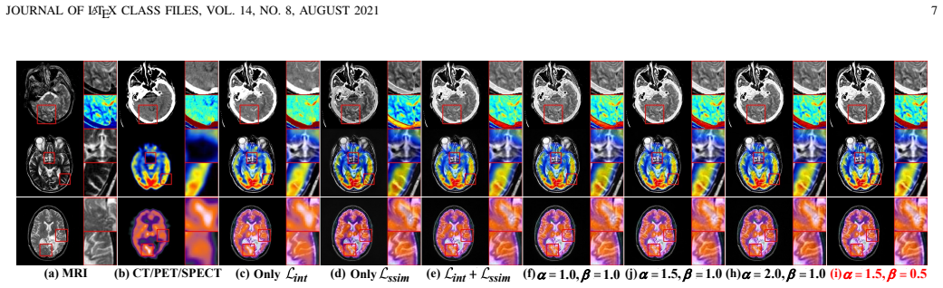
- [Section IV-A/B (Tables I and II)] The hyperparameter analysis in Section IV-B selects the intensity/SSIM weights (α=1.5, β=0.5) and the time-step combination (t1=5, t2=10, t3=20) by averaging metrics over the three test sets (Tables I and II), and then Tables V and VII report final comparisons computed on the same test sets. Because the test sets were used to choose the configuration, the reported margins over the nine baselines may reflect selection on the evaluation target rather than a genuine advantage. A held-out validation split or nested cross-validation is required before the state-of-the-art claim can be assessed.
- [Section IV-B (Table III)] The ablation labeled "w/o Dif." removes the diffusion process in Stage I and simultaneously removes the multi-time-step noisy inputs in Stage II ("the fusion module of different noise conditions was removed"). This conflates the effect of diffusion-based training with the effect of the multi-time-step input strategy. To attribute the improvement to diffusion-process training, the paper should compare against a control in which a conventionally trained UNet is fed the same multi-time-step noisy inputs, or in which the diffusion-trained UNet is used with a single time step.
- [Section IV-C (Tables V and VII)] The central comparison rests on point estimates over a single train/test split with only 90 training pairs and 50 test pairs per modality. Several winning margins are small (e.g., Table V: Proposed SCD 1.508 vs Cloud 1.378 and VIFF 0.600 vs 0.570; Table VII: Proposed Q_W 0.925 vs SHIP 0.926, where SHIP is slightly higher). No error bars, confidence intervals, or significance tests are reported, so the "outperforming" claim is not yet robust. At minimum, the authors should report results over multiple seeds or provide statistical tests.
- [Section III-C and IV-A] The forward diffusion process is defined with a total of T steps and a variance schedule α_t (Eqs. (1)-(2)), and Stage II uses t=5, 10, 20, but the paper never reports the value of T or the specific schedule used. Because the diffusion-trained UNet is the foundation of the feature extractor, the absence of these values prevents reproduction of Stage I and should be corrected.
minor comments (6)
- [Section III-E] The section heading "Loss fuction" should read "Loss function."
- [Section IV-A and throughout] The acronym "SOAT" appears where "SOTA" (state of the art) is intended; please correct it throughout, including "nine SOAT methods" in the experimental sections and figure captions.
- [Section IV-A and Section IV-C] The comparative method is introduced as "TL-SR" in Section IV-A but is referred to as "TLSR" in later sections and tables; please unify the naming.
- [Section IV-B (paragraph on the diffusion process)] The sentence "Most of the evaluation metrics across all test sets in Table V were also noticeably reduced" appears to refer to Table III, not Table V; please correct the cross-reference.
- [Section IV-B (Table IV discussion)] The statement "removing any module results in a significant performance drop" is not fully supported by Table IV: for MRI-CT, "w/o MSFF" yields higher AG (9.237 vs 9.000) and higher Q_AB/F (0.586 vs 0.571), and "w/o AMFF" yields higher Q_AB/F (0.588 vs 0.571). Please qualify this claim or explain how these increases are consistent with the stated conclusion.
- [Equation (4) and color-space descriptions] The notation IY_{B,t} introduces a superscript Y that is not defined when I_B is CT (grayscale). Clarify how the YCbCr luminance channel is used for CT as well as for PET/SPECT inputs.
Circularity Check
Final comparison rests on hyperparameters selected on the same test sets; the SOTA claim is partly a selection artifact.
-
fitted input called prediction
[Section IV-A (Implementation details) and Section IV-B (Tables I and II); final comparisons in Section IV-C (Tables V and VII)]
"The three test sets each contained 50 pairs of images for MRI-CT, MRI-PET, and MRI-SPECT. ... Table I: Objective evaluation for different values of α and β (averaged over three test sets). ... After considering the visual effects and quantitative analysis, we choose α=1.5 and β=0.5. ... Table II: Objective evaluation for different time-step combinations (averaged over three test sets). ... the combination of (5, 10, 20) yielding the best results."
The final SOTA claims (Tables V/VII, Figs. 7/10) are computed on the same 50-pair test sets whose metrics were used to select the configuration: α, β, and t1=5,t2=10,t3=20 were chosen because they gave the best/red values in Tables I and II, which are explicitly 'averaged over three test sets.' Thus the later 'prediction' that the chosen configuration outperforms baselines on those test sets is not an independent evaluation; it is the output of a selection procedure that tuned the model on the evaluation target. A held-out validation split or nested cross-validation is needed before the comparison can be read as evidence for the claimed advantage.
full rationale
The core derivation is not self-definitional: the fusion result is produced by a network trained with explicit intensity, gradient, and SSIM losses, and no predicted variable is defined as a fitted parameter. The diffusion feature extractor and attention modules are empirical design choices supported by ablations. Self-citations (LRFNet, Cloud, DSAGAN, MMIF-INet, DFENet) are baselines or related work and are not load-bearing for the derivation; no uniqueness theorem or ansatz is smuggled via citation. The main circular component is the test-set hyperparameter selection: Tables I and II are used to fix α, β, and the time steps, and the same test sets then produce the final comparison tables. This makes the reported advantage partially reflect selection on the evaluation target rather than an independent prediction. A separate validation split would remove this circular component; with the current protocol the SOTA claim is not fully independently assessed.
Assumptions & free parameters
free parameters (3)
- alpha (intensity loss weight) =
1.5
- beta (SSIM loss weight) =
0.5
- Diffusion time steps for fusion inputs =
5, 10, 20
assumptions (3)
- standard math The Gaussian diffusion forward process (Eqs. 1-2) and the UNet reverse process (Eq. 3) are valid for medical images.
- ad hoc to paper Diffusion features extracted at time steps 5, 10, and 20 capture complementary detailed information useful for fusion.
- domain assumption The ideal fused image's intensity and gradient equal the per-pixel maximum of the source images (Eqs. 15 and 18).
Cite this review
Pith. "Pith review of DM-FNet: Unified multimodal medical image fusion via diffusion process-trained encoder-decoder." pith.science (2026). https://pith.science/paper/3CMJQX5K
@misc{pith2026250615218,
  author       = {Pith},
  title        = {Pith review of: DM-FNet: Unified multimodal medical image fusion via diffusion process-trained encoder-decoder},
  year         = {2026},
  howpublished = {\url{https://pith.science/paper/3CMJQX5K}},
  note         = {Machine review of arXiv:2506.15218}
}
read the original abstract
Multimodal medical image fusion (MMIF) extracts the most meaningful information from multiple source images, enabling a more comprehensive and accurate diagnosis. Achieving high-quality fusion results requires a careful balance of brightness, color, contrast, and detail; this ensures that the fused images effectively display relevant anatomical structures and reflect the functional status of the tissues. However, existing MMIF methods have limited capacity to capture detailed features during conventional training and suffer from insufficient cross-modal feature interaction, leading to suboptimal fused image quality. To address these issues, this study proposes a two-stage diffusion model-based fusion network (DM-FNet) to achieve unified MMIF. In Stage I, a diffusion process trains UNet for image reconstruction. UNet captures detailed information through progressive denoising and represents multilevel data, providing a rich set of feature representations for the subsequent fusion network. In Stage II, noisy images at various steps are input into the fusion network to enhance the model's feature recognition capability. Three key fusion modules are also integrated to process medical images from different modalities adaptively. Ultimately, the robust network structure and a hybrid loss function are integrated to harmonize the fused image's brightness, color, contrast, and detail, enhancing its quality and information density. The experimental results across various medical image types demonstrate that the proposed method performs exceptionally well regarding objective evaluation metrics. The fused image preserves appropriate brightness, a comprehensive distribution of radioactive tracers, rich textures, and clear edges. The code is available at https://github.com/HeDan-11/DM-FNet.
Figures


Figures from the paper (8 more)


Reference graph
Works this paper leans on
-
[1]
Wei Tang, Fazhi He, Y . Liu, and Yansong Duan. Matr: Multimodal medical image fusion via multiscale adaptive transformer.IEEE Trans- actions on Image Processing, 31:5134–5149, 2022
work page 2022
-
[2]
Dan He, Weisheng Li, Guofen Wang, Yuping Huang, and Shiqiang Liu. Lrfnet: A real-time medical image fusion method guided by detail information.Computers in biology and medicine, 173:108381, 2024. JOURNAL OF LATEX CLASS FILES, VOL. 14, NO. 8, AUGUST 2021 13
work page 2024
-
[3]
Cigdem Serifoglu Yilmaz, V olkan Murat Yılmaz, and Oguz G ¨ung¨or. On the use of the sos metaheuristic algorithm in hybrid image fusion methods to achieve optimum spectral fidelity.International Journal of Remote Sensing, 41:3993 – 4021, 2020
work page 2020
-
[4]
Joint image fusion and denoising via three-layer decomposition and sparse representation
Xiaosong Li, Fuqiang Zhou, and Haishu Tan. Joint image fusion and denoising via three-layer decomposition and sparse representation. Knowl. Based Syst., 224:107087, 2021
work page 2021
-
[5]
Shaozhuang Ye, Tuo Wang, Mingyue Ding, and Xuming Zhang. F-darts: Foveated differentiable architecture search based multimodal medical image fusion.IEEE Transactions on Medical Imaging, 42:3348–3361, 2023
work page 2023
-
[6]
Xiaoqing Luo, Yuanhao Gao, Anqi Wang, Zhancheng Zhang, and Xiaojun Wu. Ifsepr: A general framework for image fusion based on separate representation learning.IEEE Transactions on Multimedia, 25:608–623, 2023
work page 2023
-
[7]
Guofen Wang, Weisheng Li, Xinbo Gao, Bin Xiao, and Jiao Du. Multimodal medical image fusion based on multichannel coupled neural p systems and max-cloud models in spectral total variation domain. Neurocomputing, 480:61–75, 2022
work page 2022
-
[8]
Gaurav Bhatnagar, Q. M. Jonathan Wu, and Zheng Liu. Directive contrast based multimodal medical image fusion in nsct domain.IEEE Transactions on Multimedia, 15(5):1014–1024, 2013
work page 2013
Show all 69 references
-
[9]
A novel approach based on grasshopper optimization algorithm for medical image fusion.Expert Syst
Phu-Hung Dinh. A novel approach based on grasshopper optimization algorithm for medical image fusion.Expert Syst. Appl., 171:114576, 2021
2021
-
[10]
A new approach to medical image fusion based on the improved extended difference-of-gaussians combined with the coati optimization algorithm.Biomed
Thi-Hong-Ha Le, Phu-Hung Dinh, Van-Hieu Vu, and Long Giang Nguyen. A new approach to medical image fusion based on the improved extended difference-of-gaussians combined with the coati optimization algorithm.Biomed. Signal Process. Control., 93:106175, 2024
2024
-
[11]
Densefuse: A fusion approach to infrared and visible images.IEEE TIP, 28:2614–2623, 2018
Hui Li and Xiaojun Wu. Densefuse: A fusion approach to infrared and visible images.IEEE TIP, 28:2614–2623, 2018
2018
-
[12]
Ifcnn: A general image fusion framework based on convolutional neural network.Inf
Yu Zhang, Yu Liu, Peng Sun, Han Yan, Xiaolin Zhao, and Li Zhang. Ifcnn: A general image fusion framework based on convolutional neural network.Inf. Fusion, 54:99–118, 2020
2020
-
[13]
Dfenet: A dual-branch feature enhanced network integrating transform- ers and convolutional feature learning for multimodal medical image fusion.Biomed
Weisheng Li, Yin Zhang, Guofen Wang, Yuping Huang, and Ruyue Li. Dfenet: A dual-branch feature enhanced network integrating transform- ers and convolutional feature learning for multimodal medical image fusion.Biomed. Signal Process. Control., 80:104402, 2023
2023
-
[14]
Fu, Baiqing He, Jie Yang, Jianpeng Liu, Aijia Ouyang, and Ya Wang
J. Fu, Baiqing He, Jie Yang, Jianpeng Liu, Aijia Ouyang, and Ya Wang. Cdrnet: Cascaded dense residual network for grayscale and pseudocolor medical image fusion.Computer methods and programs in biomedicine, 234:107506, 2023
2023
-
[15]
Multi-level difference information replenishment for medical image fusion.Applied Intelligence, 53:4579–4591, 2022
Luping Chen, Xue Wang, Yan Zhu, and Rencan Nie. Multi-level difference information replenishment for medical image fusion.Applied Intelligence, 53:4579–4591, 2022
2022
-
[16]
Mactfusion: Lightweight cross transformer for adaptive multimodal medical image fusion.IEEE journal of biomedical and health informatics, PP, 2024
Xinyu Xie, Xiaozhi Zhang, Xinglong Tang, Jiaxi Zhao, Dongping Xiong, Lijun Ouyang, Bin Yang, Hong Zhou, Bingo Wing-Kuen Ling, and Kok-Lay Teo. Mactfusion: Lightweight cross transformer for adaptive multimodal medical image fusion.IEEE journal of biomedical and health informati...
2024
-
[17]
Liu, Yansong Duan, and Tongzhen Si
Wei Tang, Fazhi He, Y . Liu, Yansong Duan, and Tongzhen Si. Datfuse: Infrared and visible image fusion via dual attention transformer.IEEE Transactions on Circuits and Systems for Video Technology, 33:3159– 3172, 2023
2023
-
[18]
Wei Tang, Fazhi He, and Y . Liu. Ydtr: Infrared and visible image fusion via y-shape dynamic transformer.IEEE Transactions on Multimedia, 25:5413–5428, 2023
2023
-
[19]
Xinyu Xie, Xiaozhi Zhang, Shengcheng Ye, Dongping Xiong, Lijun Ouyang, Bin Yang, Hong Zhou, and Yaping Wan. Mrscfusion: Joint residual swin transformer and multiscale cnn for unsupervised multi- modal medical image fusion.IEEE Transactions on Instrumentation and Measurement, 7...
2023
-
[20]
Fusionmamba: Dynamic feature enhancement for multimodal image fusion with mamba.ArXiv, abs/2404.09498, 2024
Xinyu Xie, Yawen Cui, Chio in Ieong, Tao Tan, Xiaozhi Zhang, Xubin Zheng, and Zitong Yu. Fusionmamba: Dynamic feature enhancement for multimodal image fusion with mamba.ArXiv, abs/2404.09498, 2024
2024 arXiv
-
[21]
Dsagan: A generative adversarial network based on dual-stream attention mechanism for anatomical and functional image fusion.Inf
Jun Fu, Weisheng Li, Jiao Du, and Liming Xu. Dsagan: A generative adversarial network based on dual-stream attention mechanism for anatomical and functional image fusion.Inf. Sci., 576:484–506, 2021
2021
-
[22]
Ddcgan: A dual-discriminator conditional generative adversarial network for multi-resolution image fusion.IEEE Transactions on Image Processing, 29:4980–4995, 2020
Jiayi Ma, Han Xu, Junjun Jiang, Xiaoguang Mei, and Xiao-Ping Zhang. Ddcgan: A dual-discriminator conditional generative adversarial network for multi-resolution image fusion.IEEE Transactions on Image Processing, 29:4980–4995, 2020
2020
-
[23]
Unified gradient- and intensity-discriminator generative adversarial net- work for image fusion.Inf
Huabing Zhou, Jilei Hou, Yanduo Zhang, Jiayi Ma, and Haibin Ling. Unified gradient- and intensity-discriminator generative adversarial net- work for image fusion.Inf. Fusion, 88:184–201, 2022
2022
-
[24]
Glioma segmentation-oriented multi-modal mr image fusion with adversarial learning.IEEE CAA J
Yu Liu, Yu Shi, Fuhao Mu, Juan Cheng, and Xun Chen. Glioma segmentation-oriented multi-modal mr image fusion with adversarial learning.IEEE CAA J. Autom. Sinica, 9:1528–1531, 2022
2022
-
[25]
Coconet: Coupled contrastive learning network with multi-level feature ensemble for multi-modality image fusion.IJCV ., 132:1748– 1775, 2022
Jinyuan Liu, Runji Lin, Guanyao Wu, Risheng Liu, Zhongxuan Luo, and Xin Fan. Coconet: Coupled contrastive learning network with multi-level feature ensemble for multi-modality image fusion.IJCV ., 132:1748– 1775, 2022
2022
-
[26]
Self-supervised fusion for multi-modal medical images via contrastive auto-encoding and convolutional information exchange.IEEE Computational Intelligence Magazine, 18:68–80, 2023
Ying Zhang, Ren qi Nie, Jinde Cao, and Chaozhen Ma. Self-supervised fusion for multi-modal medical images via contrastive auto-encoding and convolutional information exchange.IEEE Computational Intelligence Magazine, 18:68–80, 2023
2023
-
[27]
Zixiang Zhao, Hao Bai, Jiangshe Zhang, Yulun Zhang, Shuang Xu, Zudi Lin, Radu Timofte, and Luc Van Gool. Cddfuse: Correlation-driven dual- branch feature decomposition for multi-modality image fusion.2023 IEEE/CVF Conference on Computer Vision and Pattern Recognition (CVPR), p...
2023
-
[28]
A general paradigm with detail-preserving conditional invertible network for image fusion.International Journal of Computer Vision, 132(4):1029–1054, 2024
Wu Wang, Liang-Jian Deng, Ran Ran, and Gemine Vivone. A general paradigm with detail-preserving conditional invertible network for image fusion.International Journal of Computer Vision, 132(4):1029–1054, 2024
2024
-
[29]
Mmif-inet: Multimodal medical image fusion by invertible network.Inf
Dan He, Weisheng Li, Guofen Wang, Yuping Huang, and Shiqiang Liu. Mmif-inet: Multimodal medical image fusion by invertible network.Inf. Fusion, 114:102666, 2024
2024
-
[30]
An efficient approach to medical image fusion based on optimization and transfer learning with vgg19.Biomed
Oanh Cuong Do, Chi Mai Luong, Phu-Hung Dinh, and Giang Son Tran. An efficient approach to medical image fusion based on optimization and transfer learning with vgg19.Biomed. Signal Process. Control., 87:105370, 2024
2024
-
[31]
Mif-btf-mrn: Medical image fusion based on the bilateral texture filter and transfer learning with the resnet-101 network
Phu-Hung Dinh. Mif-btf-mrn: Medical image fusion based on the bilateral texture filter and transfer learning with the resnet-101 network. Biomed. Signal Process. Control., 100:106976, 2025
2025
-
[32]
Task-customized mixture of adapters for general image fusion.2024 IEEE/CVF Con- ference on Computer Vision and Pattern Recognition (CVPR), pages 7099–7108, 2024
Pengfei Zhu, Yang Sun, Bing Cao, and Qinghua Hu. Task-customized mixture of adapters for general image fusion.2024 IEEE/CVF Con- ference on Computer Vision and Pattern Recognition (CVPR), pages 7099–7108, 2024
2024
-
[33]
Zhang, Ren qi Nie, Jinde Cao, Chaozhen Ma, and Chengchao Wang
Y . Zhang, Ren qi Nie, Jinde Cao, Chaozhen Ma, and Chengchao Wang. Ss-ssan: a self-supervised subspace attentional network for multi-modal medical image fusion.Artificial Intelligence Review, 56:421 – 443, 2023
2023
-
[34]
Emfusion: An unsupervised enhanced medical image fusion network.Inf
Han Xu and Jiayi Ma. Emfusion: An unsupervised enhanced medical image fusion network.Inf. Fusion, 76:177–186, 2021
2021
-
[35]
Gesenet: A general semantic-guided network with couple mask ensemble for medical image fusion.IEEE transactions on neural networks and learning systems, PP, 2023
Jiawei Li, Jinyuan Liu, Shihua Zhou, Qian Zhang, and Nikola Kirilov Kasabov. Gesenet: A general semantic-guided network with couple mask ensemble for medical image fusion.IEEE transactions on neural networks and learning systems, PP, 2023
2023
-
[36]
Sdnet: A versatile squeeze-and-decomposition network for real-time image fusion.International Journal of Computer Vision, 129:2761 – 2785, 2021
Hao Zhang and Jiayi Ma. Sdnet: A versatile squeeze-and-decomposition network for real-time image fusion.International Journal of Computer Vision, 129:2761 – 2785, 2021
2021
-
[37]
Zhang, Deyu Meng, Radu Timofte, and Luc Van Gool
Zixiang Zhao, Hao Bai, Yuanzhi Zhu, Jiangshe Zhang, Shuang Xu, Yulun Zhang, K. Zhang, Deyu Meng, Radu Timofte, and Luc Van Gool. Ddfm: Denoising diffusion model for multi-modality image fusion.2023 IEEE/CVF International Conference on Computer Vision (ICCV), pages 8048–8059, 2023
2023
-
[38]
Fusiondiff: Multi-focus image fusion using denoising diffusion probabilistic models.Expert Syst
Mining Li, Ronghao Pei, Tianyou Zheng, Yang Zhang, and Weiwei Fu. Fusiondiff: Multi-focus image fusion using denoising diffusion probabilistic models.Expert Syst. Appl., 238:121664, 2023
2023
-
[39]
Dif-fusion: Toward high color fidelity in infrared and visible image fusion with diffusion models.IEEE Transactions on Image Processing, 32:5705– 5720, 2023
Jun Yue, Leyuan Fang, Shaobo Xia, Yue Deng, and Jiayi Ma. Dif-fusion: Toward high color fidelity in infrared and visible image fusion with diffusion models.IEEE Transactions on Image Processing, 32:5705– 5720, 2023
2023
-
[40]
Swinfusion: Cross-domain long-range learning for general image fusion via swin transformer.IEEE/CAA Journal of Automatica Sinica, 9:1200–1217, 2022
Jiayi Ma, Linfeng Tang, Fan Fan, Jun Huang, Xiaoguang Mei, and Yong Ma. Swinfusion: Cross-domain long-range learning for general image fusion via swin transformer.IEEE/CAA Journal of Automatica Sinica, 9:1200–1217, 2022
2022
-
[41]
Jinyu Wen, Fei wei Qin, Jiao Du, Mei Fang, Xinhua Wei, C. L. Philip Chen, and Ping Li. Msgfusion: Medical semantic guided two-branch network for multimodal brain image fusion.IEEE Transactions on Multimedia, 26:944–957, 2024
2024
-
[42]
Ct and mri medical image fusion using noise-removal and contrast enhancement scheme with convolutional neural network.Entropy, 24, 2022
Jameel Ahmed Bhutto, Lianfang Tian, Qiliang Du, Zheng-Chun Sun, Yu Lubin, and Muhammad Faizan Tahir. Ct and mri medical image fusion using noise-removal and contrast enhancement scheme with convolutional neural network.Entropy, 24, 2022
2022
-
[43]
Fatfusion: A functional–anatomical transformer for medical image fusion.Information Processing & Management, 61(4):103687, 2024
Wei Tang and Fazhi He. Fatfusion: A functional–anatomical transformer for medical image fusion.Information Processing & Management, 61(4):103687, 2024
2024
-
[44]
Jane Wang, and Xun Chen
Yu Liu, Chen Yu, Juan Cheng, Z. Jane Wang, and Xun Chen. Mm-net: A mixformer-based multi-scale network for anatomical and functional image fusion.IEEE Transactions on Image Processing, 33:2197–2212, 2024
2024
-
[45]
An improved hybrid JOURNAL OF LATEX CLASS FILES, VOL
Yanyu Liu, Yongsheng Zang, Dongming Zhou, Jinde Cao, Rencan Nie, Ruichao Hou, Zhaisheng Ding, and Jiatian Mei. An improved hybrid JOURNAL OF LATEX CLASS FILES, VOL. 14, NO. 8, AUGUST 2021 14 network with a transformer module for medical image fusion.IEEE Journal of Biomedical ...
2021
-
[46]
Jonathan Ho, Ajay Jain, and P. Abbeel. Denoising diffusion probabilistic models.ArXiv, abs/2006.11239, 2020
2006 arXiv
-
[47]
Diffusion- based network for unsupervised landmark detection.Knowl
Tao Wu, Kai Wang, Chuanming Tang, and Jianlin Zhang. Diffusion- based network for unsupervised landmark detection.Knowl. Based Syst., 292:111627, 2024
2024
-
[48]
Corrdiff: Corrective diffusion model for accurate mri brain tumor segmentation.IEEE Journal of Biomedical and Health Informatics, 28:1587–1598, 2024
Wenqing Li, Wenhui Huang, and Yuanjie Zheng. Corrdiff: Corrective diffusion model for accurate mri brain tumor segmentation.IEEE Journal of Biomedical and Health Informatics, 28:1587–1598, 2024
2024
-
[49]
Mikolaj Czerkawski and Christos Tachtatzis. Exploring the capability of text-to-image diffusion models with structural edge guidance for multispectral satellite image inpainting.IEEE Geoscience and Remote Sensing Letters, 21:1–5, 2023
2023
-
[50]
Yang, and Chaoyue Wang
Zuopeng Yang, Tianshu Chu, Xin Lin, Erdun Gao, Daqing Liu, J. Yang, and Chaoyue Wang. Eliminating contextual prior bias for semantic image editing via dual-cycle diffusion.IEEE Transactions on Circuits and Systems for Video Technology, 34:1316–1320, 2023
2023
-
[51]
Acdmsr: Accelerated conditional diffusion models for single image super-resolution.IEEE Transactions on Broadcasting, 70:492–504, 2023
Axi Niu, Trung Xuan Pham, Kang Zhang, Jinqiu Sun, Yu Zhu, Qingsen Yan, Inso Kweon, and Yanning Zhang. Acdmsr: Accelerated conditional diffusion models for single image super-resolution.IEEE Transactions on Broadcasting, 70:492–504, 2023
2023
-
[52]
Hanqun Cao, Cheng Tan, Zhangyang Gao, Yilun Xu, Guangyong Chen, Pheng-Ann Heng, and Stan Z. Li. A survey on generative diffusion models.IEEE Transactions on Knowledge and Data Engineering, 36:2814–2830, 2022
2022
-
[53]
Fleet, and Mohammad Norouzi
Chitwan Saharia, Jonathan Ho, William Chan, Tim Salimans, David J. Fleet, and Mohammad Norouzi. Image super-resolution via iterative refinement.IEEE Transactions on Pattern Analysis and Machine Intelligence, 45:4713–4726, 2021
2021
-
[54]
Dea-net: Single image dehazing based on detail-enhanced convolution and content-guided attention.IEEE Transactions on Image Processing, 33:1002–1015, 2023
Zixuan Chen, Zewei He, and Zhe ming Lu. Dea-net: Single image dehazing based on detail-enhanced convolution and content-guided attention.IEEE Transactions on Image Processing, 33:1002–1015, 2023
2023
-
[55]
M4fnet: Multimodal medical image fusion network via multi-receptive-field and multi-scale feature integration.Computers in biology and medicine, 159:106923, 2023
Zhaisheng Ding, Haiyan Li, Yi Guo, Dongming Zhou, Yanyu Liu, and Shidong Xie. M4fnet: Multimodal medical image fusion network via multi-receptive-field and multi-scale feature integration.Computers in biology and medicine, 159:106923, 2023
2023
-
[56]
Eskicioglu and Paul S
Ahmet M. Eskicioglu and Paul S. Fisher. Image quality measures and their performance.IEEE Trans. Commun., 43:2959–2965, 1995
1995
-
[57]
In-fibre bragg grating sensors.Measurement Science and Technology, 8:355 – 375, 1997
Yun jiang Rao. In-fibre bragg grating sensors.Measurement Science and Technology, 8:355 – 375, 1997
1997
-
[58]
Detail preserved fusion of visible and infrared images using regional saliency extraction and multi-scale image decomposition.Optics Com- munications, 341:199–209, 2015
Guangmang Cui, Huajun Feng, Zhi hai Xu, Qi Li, and Yue ting Chen. Detail preserved fusion of visible and infrared images using regional saliency extraction and multi-scale image decomposition.Optics Com- munications, 341:199–209, 2015
2015
-
[59]
Gemma Piella and Henk J. A. M. Heijmans. A new quality metric for image fusion.Proceedings 2003 International Conference on Image Processing (Cat. No.03CH37429), 3:III–173, 2003
2003
-
[60]
A new image quality metric for image fusion: The sum of the correlations of differences.Aeu- international Journal of Electronics and Communications, 69:1890– 1896, 2015
Veysel Aslantas ¸ and Emre Bendes. A new image quality metric for image fusion: The sum of the correlations of differences.Aeu- international Journal of Electronics and Communications, 69:1890– 1896, 2015
2015
-
[61]
A new image fusion performance metric based on visual information fidelity.Inf
Yu Han, Yunze Cai, Yin Cao, and Xiaoming Xu. A new image fusion performance metric based on visual information fidelity.Inf. Fusion, 14:127–135, 2013
2013
-
[62]
Xydeas and Vladimir S
Costas S. Xydeas and Vladimir S. Petrovic. Objective image fusion performance measure.Electronics Letters, 36:308–309, 2000
2000
-
[63]
Simoncelli, and Alan Conrad Bovik
Zhou Wang, Eero P. Simoncelli, and Alan Conrad Bovik. Multiscale structural similarity for image quality assessment.The Thrity-Seventh Asilomar Conference on Signals, Systems & Computers, 2003, 2:1398– 1402 V ol.2, 2003
2003
-
[64]
A non- reference image fusion metric based on mutual information of image features.Comput
Mohammad Haghighat, Ali Aghagolzadeh, and Hadi Seyedarabi. A non- reference image fusion metric based on mutual information of image features.Comput. Electr. Eng., 37:744–756, 2011
2011
-
[65]
Lrrnet: A novel representation learning guided fusion network for infrared and visible images.IEEE Trans
Hui Li, Tianyang Xu, Xiaojun Wu, Jiwen Lu, and Josef Kittler. Lrrnet: A novel representation learning guided fusion network for infrared and visible images.IEEE Trans. Pattern Anal. Mach. Intell, 45:11040– 11052, 2023
2023
-
[66]
Multi-interactive feature learning and a full-time multi-modality benchmark for image fusion and segmentation
Jinyuan Liu, Zhu Liu, Guanyao Wu, Long Ma, Risheng Liu, Wei Zhong, Zhongxuan Luo, and Xin-Yue Fan. Multi-interactive feature learning and a full-time multi-modality benchmark for image fusion and segmentation. InICCV, pages 8081–8090, 2023
2023
-
[67]
Correlation-guided discriminative cross-modality features network for infrared and visible image fusion.IEEE Transactions on Instrumentation and Measurement, 2023
Zhao Cai, Yong Ma, Jun Huang, Xiaoguang Mei, and Fan Fan. Correlation-guided discriminative cross-modality features network for infrared and visible image fusion.IEEE Transactions on Instrumentation and Measurement, 2023
2023
-
[68]
Probing synergistic high-order interaction in infrared and visible image fusion
Naishan Zheng, Man Zhou, Jie Huang, Junming Hou, Haoying Li, Yuan Xu, and Feng Zhao. Probing synergistic high-order interaction in infrared and visible image fusion. InCVPR, pages 26374–26385. IEEE, 2024
2024
-
[69]
Mmdrfuse: Distilled mini-model with dynamic refresh for multi- modality image fusion
Yanglin Deng, Tianyang Xu, Chunyang Cheng, Xiao-Jun Wu, and Josef Kittler. Mmdrfuse: Distilled mini-model with dynamic refresh for multi- modality image fusion. InACM MM, 2024. Dan Hereceived the B.S. degree from Hengyang Normal University, China, in 2020 and the M.S. degree f...
2024
Reviewed August 15, 2026 · model on record in the stance chip above.
Discussion (0). Continue with ORCID to comment.