REVIEW 3 major objections 5 minor 45 references
Deformable Medical Image Registration with Effective Anatomical Structure Representation and Divide-and-Conquer Network
T0 review · 3 major / 5 minor · reviewed 2026-08-06 · deepseek-v4-flash
Pith's one-line read ROI-based network beats VoxelMorph by up to 13% Dice.
desk verdict Promising ROI-based registration architecture, but the headline gains are inflated by selecting the GMM component count on the test set; the core idea still deserves a careful revision and a serious referee. read the letter →
The pith
A machine-rendered reading of the paper's core claim, the machinery that carries it, and where it could break.
The reading
What carries the argument
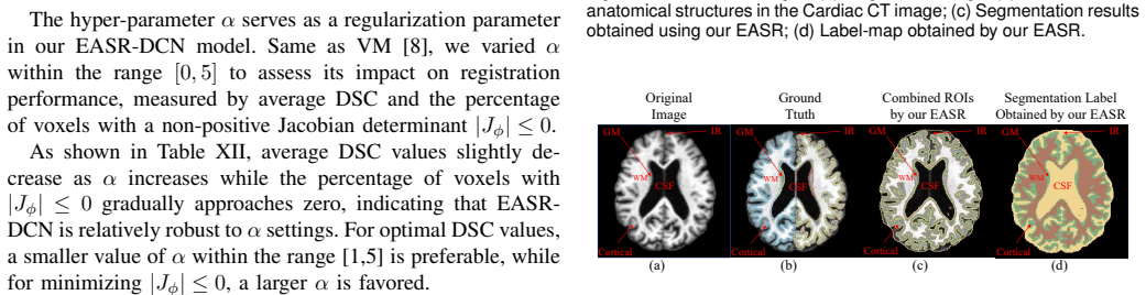
The load-bearing mechanism is the pairing of a Gaussian Mixture Model (GMM) ROI extraction with a multichannel Divide-and-Conquer Network (DCN). The GMM, fit jointly on the min-max normalized intensity arrays of the moving and fixed images by expectation-maximization, assigns each voxel to the Gaussian component with the highest responsibility, producing k paired masks (for brain MRI: cortex plus gray matter, white matter, and CSF plus irrelevant regions) that are applied to the original images to obtain k ROI pairs. The DCN feeds each ROI pair into its own encoder so each region learns its own feature alignment, and a shared decoder merges the encoded features through skip connections to emit one displacement vector field. Training minimizes normalized cross-correlation between the warped and fixed images plus a diffusion regularizer on the field, and the diffeomorphic variant derives the field from a stationary velocity field integrated by scaling-and-squaring.
What would settle it
Register a pair of synthetic or phantom images with known ground-truth deformation while gradually broadening the intensity distributions of two tissues until they overlap; if EASR-DCN's accuracy does not drop as overlap increases, or if swapping the ROI masks between moving and fixed images leaves the result unchanged, then ROI correspondence is not what drives the reported gains.
Extended reading notes
Core claim
The central claim is that explicit attention to anatomical regions—without any segmentation labels—is what limits current unsupervised registration, and that a divide-and-conquer architecture is the right way to exploit that structure. EASR-DCN represents both images by k effective ROIs obtained from a joint GMM fit on voxel intensities, feeds corresponding ROI pairs into k independent encoder streams so that each region's deformation is estimated without interference from other regions, and lets a shared decoder integrate the per-ROI feature alignments into one DVF. The DVF warps the moving image through a spatial transformer, and a diffeomorphic variant instead integrates a stationary velocity field with scaling-and-squaring. On OASIS brain MRI, cardiac MRI, hippocampus MRI, and cardiac CT, the method reports the highest Dice scores among the ten unsupervised baselines and is competitive with or better than the top weakly supervised methods, while producing fewer fold artifacts as measured by a lower percentage of non-positive Jacobian determinants.
Load-bearing premise
The load-bearing premise is that voxel intensities separate into the same meaningful regions in both images, so the ROI masks from the GMM correspond to the same anatomical structures across each image pair; if tissue intensities overlap or vary across subjects, the per-ROI alignment loses its anatomical meaning.
Editorial extensions
If this is right
- Unsupervised registration can close most of the gap to weakly supervised methods: EASR-DCN reports higher Dice than the best weakly supervised comparison on hippocampus MRI without needing any labels.
- Per-ROI independent alignment reduces deformation artifacts: EASR-DCN lowers the percentage of non-positive Jacobian determinants relative to the leading unsupervised baselines on all four datasets.
- The ROI decomposition transfers from MR to CT: on cardiac CT, EASR-DCN reports 85.72% Dice versus 83.96% for the second-best unsupervised method.
- The optimal ROI count depends on the anatomy and modality (k=3 for brain and hippocampus MRI, k=4 for cardiac MRI), so a k-selection step is part of applying the method to a new dataset.
Reading between the lines
- The central mechanism predicts that the method's advantage over whole-image registration grows with the number of distinct tissue compartments and shrinks as intensity distributions overlap; this could be tested by synthesizing images with controlled intensity overlap and measuring Dice as a function of overlap.
- A fully automatic version of the method is within reach: since registration accuracy peaks where GMM segmentation stays accurate, one could select k by maximizing per-ROI segmentation Dice on a small holdout set instead of searching on the test set, which the paper currently does.
- The divide-and-conquer design could be combined with any correspondence source, not just intensity: replacing the GMM masks with feature-based or shape-based regions might extend the same architecture to multimodal registration, a case the authors identify as a limitation.
Signed reviews
Editorial analysis
A structured set of objections, weighed in public.
Referee Report
Summary. The paper proposes EASR-DCN, an unsupervised deformable registration method that first represents a moving/fixed image pair by k intensity-based ROIs obtained from a Gaussian mixture model, then feeds corresponding ROI pairs into separate encoders of a divide-and-conquer network and integrates the resulting feature alignments into a single DVF. A diffeomorphic variant, EASR-DCN-diff, uses a stationary velocity field with scaling-and-squaring. The method is evaluated on OASIS brain MRI, hippocampus MRI, cardiac MRI, and cardiac CT against a large set of unsupervised and weakly-supervised baselines, with DSC, percentage of non-positive Jacobian determinant, and 95% Hausdorff distance as metrics, plus ablations, computational-cost comparisons, and a Learn2Reg OASIS evaluation. The central claim is that EASR-DCN substantially outperforms VoxelMorph and all unsupervised baselines and is competitive with weakly-supervised methods, while remaining unsupervised at training time.
Significance. If the reported results survive an unbiased hyperparameter-selection protocol, the paper makes a practically useful contribution: it demonstrates that intensity-based ROI decomposition combined with per-ROI independent encoding can improve registration accuracy and reduce DVF folding without any label supervision. The evaluation is unusually extensive for a registration paper: four datasets, more than twenty baselines, standard metrics with standard deviations, per-structure boxplots, DVF visualizations, and an external Learn2Reg leaderboard comparison. The promise to release code is another strength. However, the headline quantitative claims are currently tied to hyperparameters whose reported values appear to have been selected on the same test labels used for evaluation, which materially weakens the evidence for superiority over the baselines.
major comments (3)
- [Section IV.H, Table V] The number of GMM components k is selected using test-set performance. Table V reports DSC for k=1..5 on the three MRI datasets, and the entries that match the final EASR-DCN rows in Tables I and II (OASIS k=3: 89.20; Hippocampus k=3: 80.04; Cardiac k=4: 88.70) are exactly the best values in each column. Since Tables I and II are the test-set comparisons against all baselines, the headline 'superior to all unsupervised methods' claim is the result of choosing k from the same labels used to compute Dice. The problem is consequential: at k=3, Cardiac MRI DSC is 84.51, which is below CorrMLP's 85.91, and only the test-selected k=4 yields 88.70. The three-phase adaptive strategy described later in Section IV.H is presented as a future selection rule and was not used to produce Tables I and II. The authors should re-run the evaluation with k chosen by validation-only (e.g., the 5% holdout described in Phase 3) or report a nested cross-validation estimate that accounts for selection bias, and state explicitly which k values were chosen without access to test labels.
- [Section III.B, Eq. (3)] The joint GMM is the foundation of the claimed anatomical correspondence, but Eq. (3) min-max normalizes the moving and fixed images separately before concatenating them. This destroys the comparability of intensity values across the two images: a Gaussian component index k in the moving image and the same index in the fixed image are not guaranteed to represent the same anatomical structure. The paired-ROI assumption in Section III.C and the visualization in Fig. 3 therefore rest on an unverified premise. The paper's own Discussion acknowledges overlapping Hounsfield units in CT and intensity inhomogeneities in MR as limitations. To make the central claim load-bearing, the authors should either justify why separate normalization preserves cross-image component correspondence, modify the normalization so both images are mapped with the same reference statistics, or add an experiment that directly evaluates the anatomical consistency of corresponding ROI pairs (e.g., Dice between the GMM ROI in the fixed image and the warped GMM ROI from the moving image).
- [Section IV.N, Table XII] The smoothness weight alpha is also effectively selected on the test set. Table XII reports DSC and |J|<=0 for alpha in [0,5] on the three MRI datasets, and the final choices stated in the text (alpha=3 for OASIS Brain and Cardiac, alpha=1 for Hippocampus) coincide with the test DSC values used in Tables I and II (e.g., OASIS alpha=3 gives 89.20; Cardiac alpha=3 gives 88.70; Hippocampus alpha=1 gives 80.04). This introduces a second source of optimistic bias in the reported improvements over baselines. The authors should specify a single validation-based protocol for both alpha and k, report the values chosen on validation, and give the resulting test performance; the current presentation does not allow a reader to separate genuine method performance from hyperparameter search on the evaluation set.
minor comments (5)
- [Section IV.E] The text states that EASR-DCN reduces HdDist95 by 0.230 on Hippocampus MRI and 0.018 on Cardiac MRI, but Table I gives 0.018 for Hippocampus (2.415 to 2.397) and 0.230 for Cardiac (3.827 to 3.597); the two datasets are swapped.
- [Table X] The ablation table labels SAMReg as [28], but reference [28] is Kirillov et al., Segment Anything; the correct citation is [27] (Huang et al., One registration is worth two segmentations), which is also the citation used in the main text of Section IV.J.
- [Table VIII] The sentence 'When k=1... registration achieves perfect alignment with ground truth' is written about the GMM segmentation evaluation, not registration; it should say segmentation, not registration, to avoid confusion with Tables I and V.
- [Section III.D and Fig. 2] The diffeomorphic variant is described only by the scaling-and-squaring integration of a velocity field; the paper does not state how the network produces the velocity field instead of the DVF, nor whether the loss or architecture changes for EASR-DCN-diff. This is needed for reproducibility of the reported EASR-DCN-diff results.
- [Throughout] There are several typos that should be corrected: 'Netwok' in the Section III.C heading, 'Fxied imageMoving image' in the Fig. 9 caption, 'Ttuth' in the Fig. 14 and Fig. 15 captions, and 'ssume' in Section II.B.
Circularity Check
Headline cardiac MRI improvement is partly fitted: k is selected on the same test folds used to report the final DSC.
-
fitted input called prediction
[Section IV.H ('Analysis of k-value'), Tables V and I/II]
"Through systematic evaluation across three datasets (Table V), we incrementally varied k from 1 to 5, observing that performance initially improved and then declined as k exceeded the optimal value corresponding to the actual ROI counts. The results revealed that k=3 is optimal for OASIS Brain MRI and Hippocampus MRI. Cardiac MRI achieved peak performance at k=4, with higher values (k= 5) degrading the results in all cases. These findings informed our final parameter selection: k= 3 for the brain / hippocampus and k= 4 for the cardiac."
Table V reports DSC on the same test folds that later appear as the final EASR-DCN rows in Tables I and II (e.g., Cardiac MRI: k=3 gives 84.51 and k=4 gives 88.70; Table I reports 88.70). Section IV.H then uses these test-set DSCs to choose k. The headline improvement over VoxelMorph on Cardiac MRI (13.01%) is therefore the maximally selected test score rather than an independent estimate; at k=3 the method would rank below CorrMLP (85.91). The selection criterion and the reported outcome are the same measurement, so the claimed superiority on that dataset is fitted to the test labels rather than predicted.
full rationale
The only circular step is the test-set selection of the GMM component count k in Section IV.H: the k-values used for the final Tables I and II are chosen from Table V, which computes DSC on the same test folds, making the cardiac MRI headline the best of five test-set scores. This is a genuine case of a fitted parameter (k) being presented as the basis for a result whose evaluation metric is the same as the fitting criterion. I do not find other circularity: the EASR GMM ROI construction (Eqs. 2-10) is independent of the evaluation labels, the DCN is trained with unsupervised NCC/diffusion losses (Eqs. 13-15), and the Learn2Reg 2021 Task 3 results in Table IV are organizer-scored on a held-out test set, which independently corroborates the brain MRI claim. No load-bearing argument rests on a self-citation by the authors; citations to their own work (e.g., [18]) are only baselines. The Discussion's admission of intensity-inhomogeneity and overlapping-HU limitations is an accepted assumption, not a circular step. Because the central claim still has independent evidence for brain/CT and the circularity is confined to one headline comparison, the overall score is moderate rather than maximal.
Assumptions & free parameters
free parameters (2)
- k (number of GMM components) =
3 for brain MRI, 3 for hippocampus MRI, 4 for cardiac MRI, 3 for cardiac CT
- alpha (smoothness regularization weight) =
3 for OASIS Brain and Cardiac, 1 for Hippocampus
assumptions (3)
- domain assumption A Gaussian mixture model with K components adequately models the joint voxel intensity distribution of moving and fixed images.
- domain assumption The cluster labels obtained from the joint GMM establish valid anatomical correspondences between moving and fixed images, so pairing ROI_m^i with ROI_f^i is meaningful.
- standard math Standard facts of Gaussian mixture estimation with EM and the scaling-and-squaring integration of a stationary velocity field.
Cite this review
Pith. "Pith review of Deformable Medical Image Registration with Effective Anatomical Structure Representation and Divide-and-Conquer Network." pith.science (2026). https://pith.science/paper/YGTZ5OTH
@misc{pith2026250619222,
  author       = {Pith},
  title        = {Pith review of: Deformable Medical Image Registration with Effective Anatomical Structure Representation and Divide-and-Conquer Network},
  year         = {2026},
  howpublished = {\url{https://pith.science/paper/YGTZ5OTH}},
  note         = {Machine review of arXiv:2506.19222}
}
read the original abstract
Effective representation of Regions of Interest (ROI) and independent alignment of these ROIs can significantly enhance the performance of deformable medical image registration (DMIR). However, current learning-based DMIR methods have limitations. Unsupervised techniques disregard ROI representation and proceed directly with aligning pairs of images, while weakly-supervised methods heavily depend on label constraints to facilitate registration. To address these issues, we introduce a novel ROI-based registration approach named EASR-DCN. Our method represents medical images through effective ROIs and achieves independent alignment of these ROIs without requiring labels. Specifically, we first used a Gaussian mixture model for intensity analysis to represent images using multiple effective ROIs with distinct intensities. Furthermore, we propose a novel Divide-and-Conquer Network (DCN) to process these ROIs through separate channels to learn feature alignments for each ROI. The resultant correspondences are seamlessly integrated to generate a comprehensive displacement vector field. Extensive experiments were performed on three MRI and one CT datasets to showcase the superior accuracy and deformation reduction efficacy of our EASR-DCN. Compared to VoxelMorph, our EASR-DCN achieved improvements of 10.31\% in the Dice score for brain MRI, 13.01\% for cardiac MRI, and 5.75\% for hippocampus MRI, highlighting its promising potential for clinical applications. The code for this work will be released upon acceptance of the paper.
Figures


Figures from the paper (10 more)


Reference graph
Works this paper leans on
-
[27]
One registration is worth two segmentations,
S. Huang, T. Xu, Z. Shen, S. U. Saeed, W. Yan, D. Barratt, and Y . Hu, “One registration is worth two segmentations,” inInt. Conf. Med. Image Comput. Comput.-Assisted Intervention, pp. 665–675, Springer, 2024
work page 2024
-
[28]
A. Kirillov, E. Mintun, N. Ravi, H. Mao, C. Rolland, L. Gustafson, T. Xiao, S. Whitehead, A. C. Berg, W.-Y . Lo,et al., “Segment anything,” inProc. IEEE Int. Conf. Comput. Vis., pp. 4015–4026, 2023
work page 2023
-
[1]
Self-distilled hierarchical network for unsupervised deformable image registration,
S. Zhou, B. Hu, Z. Xiong, and F. Wu, “Self-distilled hierarchical network for unsupervised deformable image registration,”IEEE Transactions on Medical Imaging, vol. 42, no. 8, pp. 2162–2175, 2023
work page 2023
-
[2]
Symreg-gan: Symmetric image registration with generative adversarial networks,
Y . Zheng, X. Sui, Y . Jiang, T. Che, S. Zhang, J. Yang, and H. Li, “Symreg-gan: Symmetric image registration with generative adversarial networks,”IEEE Trans. Pattern Anal. Mach. Intell., 2021. MAet al.: DMIR WITH EFFECTIVE ANATOMICAL STRUCTURE REPRESENTATION AND DIVIDE-AND-CONQUER NETWORK 13
work page 2021
-
[3]
Shared multi-view data representation for multi-domain event detection,
Z. Yang, Q. Li, W. Liu, and J. Lv, “Shared multi-view data representation for multi-domain event detection,”IEEE Trans. Pattern Anal. Mach. Intell., vol. 42, no. 5, pp. 1243–1256, 2019
work page 2019
-
[4]
J.-Q. Zheng, Z. Wang, B. Huang, N. H. Lim, and B. W. Papie˙z, “Residual aligner-based network (ran): Motion-separable structure for coarse-to- fine discontinuous deformable registration,”Med. Image Anal., vol. 91, p. 103038, 2024
work page 2024
-
[5]
B. Kim, Y . Zhuang, T. S. Mathai, and R. M. Summers, “Otmorph: Unsupervised multi-domain abdominal medical image registration using neural optimal transport,”IEEE Trans. Med. Imaging, pp. 1–1, 2024
work page 2024
-
[6]
B. B. Avants, C. L. Epstein, M. Grossman, and J. C. Gee, “Symmetric diffeomorphic image registration with cross-correlation: evaluating au- tomated labeling of elderly and neurodegenerative brain,”Med. Image Anal., vol. 12, no. 1, pp. 26–41, 2008
work page 2008
Show all 45 references
-
[7]
Point set registration with mixture framework and variational inference,
X. Ma, S. Xu, J. Zhou, Q. Yang, Y . Yang, K. Yang, and S. H. Ong, “Point set registration with mixture framework and variational inference,”Pattern Recognit., vol. 104, p. 107345, 2020
2020
-
[8]
V oxelmorph: a learning framework for deformable medical image registration,
G. Balakrishnan, A. Zhao, M. R. Sabuncu, J. Guttag, and A. V . Dalca, “V oxelmorph: a learning framework for deformable medical image registration,”IEEE Trans. Med. Imaging, vol. 38, no. 8, pp. 1788–1800, 2019
2019
-
[9]
Rsegnet: a joint learning framework for deformable registration and segmentation,
L. Qiu and H. Ren, “Rsegnet: a joint learning framework for deformable registration and segmentation,”IEEE Trans. Autom. Sci. Eng., vol. 19, no. 3, pp. 2499–2513, 2021
2021
-
[10]
Weakly-supervised learning of multi-modal features for regularised iterative descent in 3d image registration,
M. Blendowski, L. Hansen, and M. P. Heinrich, “Weakly-supervised learning of multi-modal features for regularised iterative descent in 3d image registration,”Med. Image Anal., vol. 67, p. 101822, 2021
2021
-
[11]
Groupmorph: medical image registration via grouping network with contextual fusion,
Z. Tan, L. Zhang, Y . Lv, Y . Ma, and H. Lu, “Groupmorph: medical image registration via grouping network with contextual fusion,”IEEE Trans. Med. Imaging, 2024
2024
-
[12]
Bi-level learning of task-specific decoders for joint registration and one-shot medical image segmentation,
X. Fan, X. Wang, J. Gao, J. Wang, Z. Luo, and R. Liu, “Bi-level learning of task-specific decoders for joint registration and one-shot medical image segmentation,” inProc. IEEE Conf. Comput. Vis. Pattern Recognit., pp. 11726–11735, 2024
2024
-
[13]
Anatom- ically constrained and attention-guided deep feature fusion for joint segmentation and deformable medical image registration,
H. G. Khor, G. Ning, Y . Sun, X. Lu, X. Zhang, and H. Liao, “Anatom- ically constrained and attention-guided deep feature fusion for joint segmentation and deformable medical image registration,”Med. Image Anal., vol. 88, p. 102811, 2023
2023
-
[14]
Pc-reg: A pyramidal prediction– correction approach for large deformation image registration,
W. Yin, J.-J. Sonke, and E. Gavves, “Pc-reg: A pyramidal prediction– correction approach for large deformation image registration,”Med. Image Anal., vol. 90, p. 102978, 2023
2023
-
[15]
Progressively coupling network for brain mri registration in few-shot situation,
Z. Tan, H. Zhang, F. Tian, L. Zhang, W. Sun, and H. Lu, “Progressively coupling network for brain mri registration in few-shot situation,” in Proc. Int. Conf. Med. Image Comput. Comput.-Assisted Intervention, pp. 623–633, Springer, 2023
2023
-
[16]
Weakly-supervised biomechanically-constrained ct/mri registration of the spine,
B. Jian, M. F. Azampour, F. De Benetti, J. Oberreuter, C. Bukas, A. S. Gersing, S. C. Foreman, A.-S. Dietrich, J. Rischewski, J. S. Kirschke,et al., “Weakly-supervised biomechanically-constrained ct/mri registration of the spine,” inProc. Int. Conf. Med. Image Comput. Comput.-...
2022
-
[17]
Few-shot learning for deformable medical image registration with perception-correspondence decoupling and reverse teaching,
Y . He, T. Li, R. Ge, J. Yang, Y . Kong, J. Zhu, H. Shu, G. Yang, and S. Li, “Few-shot learning for deformable medical image registration with perception-correspondence decoupling and reverse teaching,”IEEE J. Biomed. Health. Inf., vol. 26, no. 3, pp. 1177–1187, 2022
2022
-
[18]
Deformable medical image registration with global–local transformation network and region simi- larity constraint,
X. Ma, H. Cui, S. Li, Y . Yang, and Y . Xia, “Deformable medical image registration with global–local transformation network and region simi- larity constraint,”Comput. Med. Imaging Graph., vol. 108, p. 102263, 2023
2023
-
[19]
Recursive decomposition network for deformable image registration,
B. Hu, S. Zhou, Z. Xiong, and F. Wu, “Recursive decomposition network for deformable image registration,”IEEE Journal of Biomedical and Health Informatics, vol. 26, no. 10, pp. 5130–5141, 2022
2022
-
[20]
Non-iterative coarse-to-fine registration based on single-pass deep cumulative learning,
M. Meng, L. Bi, D. Feng, and J. Kim, “Non-iterative coarse-to-fine registration based on single-pass deep cumulative learning,” inProc. Int. Conf. Med. Image Comput. Comput.-Assisted Intervention, pp. 88–97, Springer, 2022
2022
-
[21]
Spatial transformer networks,
M. Jaderberg, K. Simonyan, A. Zisserman,et al., “Spatial transformer networks,”Advances in neural information processing systems, vol. 28, pp. 2017–2025, 2015
2017
-
[22]
Transmorph: Transformer for unsupervised medical image registration,
J. Chen, E. C. Frey, Y . He, W. P. Segars, Y . Li, and Y . Du, “Transmorph: Transformer for unsupervised medical image registration,”Med. Image Anal., vol. 82, p. 102615, 2022
2022
-
[23]
Non-iterative coarse- to-fine transformer networks for joint affine and deformable image registration,
M. Meng, L. Bi, M. Fulham, D. Feng, and J. Kim, “Non-iterative coarse- to-fine transformer networks for joint affine and deformable image registration,” inProc. Int. Conf. Med. Image Comput. Comput.-Assisted Intervention, pp. 750–760, Springer, 2023
2023
-
[24]
Modet: Learning deformable image reg- istration via motion decomposition transformer,
H. Wang, D. Ni, and Y . Wang, “Modet: Learning deformable image reg- istration via motion decomposition transformer,” inProc. Int. Conf. Med. Image Comput. Comput.-Assisted Intervention, pp. 740–749, Springer, 2023
2023
-
[25]
Xmorpher: Full transformer for deformable medical image registration via cross attention,
J. Shi, Y . He, Y . Kong, J.-L. Coatrieux, H. Shu, G. Yang, and S. Li, “Xmorpher: Full transformer for deformable medical image registration via cross attention,” inInt. Conf. Med. Image Comput. Comput.-Assisted Intervention, pp. 217–226, Springer, 2022
2022
-
[26]
Deformer: Towards displacement field learning for unsupervised med- ical image registration,
J. Chen, D. Lu, Y . Zhang, D. Wei, M. Ning, X. Shi, Z. Xu, and Y . Zheng, “Deformer: Towards displacement field learning for unsupervised med- ical image registration,” inInt. Conf. Med. Image Comput. Comput.- Assisted Intervention, pp. 141–151, Springer, 2022
2022
-
[29]
Maximum likelihood from incomplete data via the em algorithm,
A. P. Dempster, N. M. Laird, and D. B. Rubin, “Maximum likelihood from incomplete data via the em algorithm,”Journal of the royal statistical society. Series B (methodological), pp. 1–38, 1977
1977
-
[30]
A log- euclidean framework for statistics on diffeomorphisms,
V . Arsigny, O. Commowick, X. Pennec, and N. Ayache, “A log- euclidean framework for statistics on diffeomorphisms,” inMedical Image Computing and Computer-Assisted Intervention–MICCAI 2006: 9th International Conference, Copenhagen, Denmark, October 1-6,
2006
-
[31]
Fast symmetric diffeomorphic image registra- tion with convolutional neural networks,
T. C. Mok and A. Chung, “Fast symmetric diffeomorphic image registra- tion with convolutional neural networks,” inProc. IEEE Conf. Comput. Vis. Pattern Recognit., pp. 4644–4653, 2020
2020
-
[32]
Large deformation diffeomorphic image registration with laplacian pyramid networks,
T. C. Mok and A. Chung, “Large deformation diffeomorphic image registration with laplacian pyramid networks,” inInt. Conf. Med. Image Comput. Comput.-Assisted Intervention, pp. 211–221, Springer, 2020
2020
-
[33]
Hy- permorph: Amortized hyperparameter learning for image registration,
A. Hoopes, M. Hoffmann, B. Fischl, J. Guttag, and A. V . Dalca, “Hy- permorph: Amortized hyperparameter learning for image registration,” inInformation Processing in Medical Imaging: 27th International Con- ference, IPMI 2021, Virtual Event, June 28–June 30, 2021, Proceedings ...
2021
-
[34]
Correlation-aware coarse-to-fine mlps for deformable medical image registration,
M. Meng, D. Feng, L. Bi, and J. Kim, “Correlation-aware coarse-to-fine mlps for deformable medical image registration,” inProc. IEEE Conf. Comput. Vis. Pattern Recognit., pp. 9645–9654, 2024
2024
-
[35]
A large annotated medical image dataset for the development and evaluation of segmentation algorithms,
A. L. Simpson, M. Antonelli, S. Bakas, M. Bilello, K. Farahani, B. Van Ginneken, A. Kopp-Schneider, B. A. Landman, G. Litjens, B. Menze,et al., “A large annotated medical image dataset for the development and evaluation of segmentation algorithms,”arXiv preprint arXiv:1902.09063, 2019
1902 arXiv
-
[36]
Learn2reg: comprehensive multi-task medical image registration challenge, dataset and evaluation in the era of deep learning,
A. Hering, L. Hansen, T. C. Mok, A. C. Chung, H. Siebert, S. H ¨ager, A. Lange, S. Kuckertz, S. Heldmann, W. Shao,et al., “Learn2reg: comprehensive multi-task medical image registration challenge, dataset and evaluation in the era of deep learning,”IEEE Trans. Med. Imaging, vo...
2022
-
[37]
Multi-scale patch and multi-modality atlases for whole heart segmentation of mri,
X. Zhuang and J. Shen, “Multi-scale patch and multi-modality atlases for whole heart segmentation of mri,”Med. Image Anal., vol. 31, pp. 77–87, 2016
2016
-
[38]
Advanced normalization tools (ants),
B. B. Avants, N. Tustison, G. Song,et al., “Advanced normalization tools (ants),”Insight j, vol. 2, no. 365, pp. 1–35, 2009
2009
-
[39]
Automatic differentiation in pytorch,
A. Paszke, S. Gross, S. Chintala, G. Chanan, E. Yang, Z. DeVito, Z. Lin, A. Desmaison, L. Antiga, and A. Lerer, “Automatic differentiation in pytorch,” inNIPS-W, 2017
2017
-
[40]
Measures of the amount of ecologic association between species,
L. R. Dice, “Measures of the amount of ecologic association between species,”Ecology, vol. 26, no. 3, pp. 297–302, 1945
1945
-
[41]
Comparing images using the hausdorff distance,
D. P. Huttenlocher, G. A. Klanderman, and W. J. Rucklidge, “Comparing images using the hausdorff distance,”IEEE Trans. Pattern Anal. Mach. Intell., vol. 15, no. 9, pp. 850–863, 1993
1993
-
[42]
Joint progressive and coarse-to-fine registration of brain mri via deformation field integration and non-rigid feature fusion,
J. Lv, Z. Wang, H. Shi, H. Zhang, S. Wang, Y . Wang, and Q. Li, “Joint progressive and coarse-to-fine registration of brain mri via deformation field integration and non-rigid feature fusion,”IEEE Transactions on Medical Imaging, vol. 41, no. 10, pp. 2788–2802, 2022
2022
-
[43]
Fast 3d registration with accurate optimisation and little learning for learn2reg 2021,
H. Siebert, L. Hansen, and M. P. Heinrich, “Fast 3d registration with accurate optimisation and little learning for learn2reg 2021,” in International Conference on Medical Image Computing and Computer- Assisted Intervention, pp. 174–179, Springer, 2021
2021
-
[44]
Conditional deformable image registra- tion with convolutional neural network,
T. C. Mok and A. C. Chung, “Conditional deformable image registra- tion with convolutional neural network,” inMedical Image Computing and Computer Assisted Intervention–MICCAI 2021: 24th International Conference, Strasbourg, France, September 27–October 1, 2021, Pro- ceedings,...
2021
-
[2006]
924–931, Springer, 2006
Proceedings, Part I 9, pp. 924–931, Springer, 2006
2006
Reviewed August 6, 2026 · model on record in the stance chip above.
Discussion (0). Continue with ORCID to comment.