REVIEW 3 major objections 5 minor 1 cited by
Nonlinear reversal of photo-excitation on the attosecond time scale improves ultrafast x-ray diffraction images
T0 review · 3 major / 5 minor · reviewed 2026-08-06 · deepseek-v4-flash
Pith's one-line read Intense sub-femtosecond X-ray pulses yield brighter diffraction with less electronic damage than 15-femtosecond pulses.
desk verdict Plausible new sub-fs x-ray diffraction effect, but the central comparison is undermined by a 1.8x fluence mismatch and no fluence-matched subsample. read the letter →
The pith
A machine-rendered reading of the paper's core claim, the machinery that carries it, and where it could break.
The reading
What carries the argument
The central mechanism is stimulated x-ray emission (the $\Gamma_{SE}$ term in the transition-rate equation of the Monte Carlo/MD model), which returns excited electrons from transiently resonant ionic states to the K-shell during the pulse. Because the sub-fs pulse duration is shorter than the few-femtosecond Auger-decay and nanoplasma-formation timescales, a population of ions with strongly enhanced elastic scattering cross sections (up to $10^4$–$10^6$ times the neutral value if the resonances stay narrow) is kept alive long enough to scatter many incident photons. The paper's simulations show that including stimulated emission is what lifts the sub-fs scattering cross section above the 15-fs one and keeps it rising with fluence.
What would settle it
Record diffraction and coincident ion spectra for the same type of Ne nanoparticles with sub-fs and 15-fs pulses while matching the per-particle fluence shot by shot (e.g., by selecting events with equal incident pulse energy and focal overlap), and check whether the sub-fs advantage in scattered photons per unit absorbed dose persists; a direct spectral search for stimulated-emission photons at the transient resonance during the pulse would also test the mechanism.
Extended reading notes
Core claim
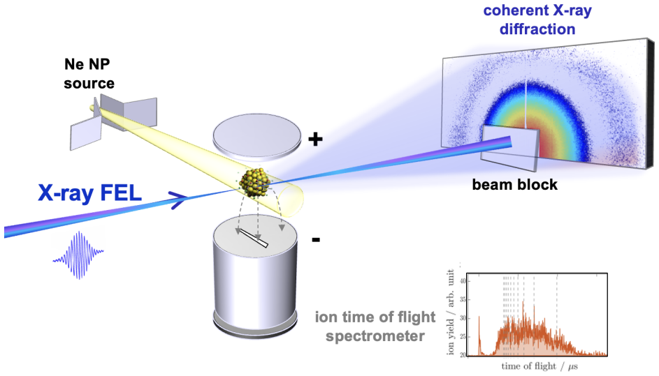
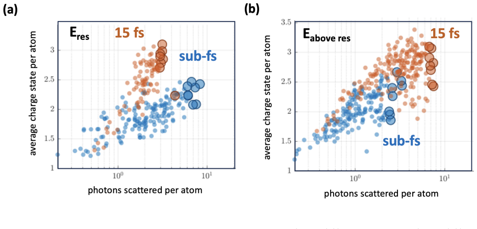
In the experiment, individual Ne nanoparticles of 60–100 nm diameter were hit by single X-ray pulses from the LCLS, either sub-femtosecond (XLEAP, ~0.3 fs) or ~15 fs, with photon energy scanned from 815 eV to 1000 eV across the Ne K-edge at 870 eV. The authors find that near the edge (866 eV) the sub-fs pulses yield diffraction images with more scattered photons per atom and a lower average charge state (around Ne2+–Ne4+) than the 15-fs pulses, which leave charge states above Ne6+; far above the edge (956 eV) the scattering advantage disappears while the charge-state reduction remains. The theoretical modeling attributes the brightening to stimulated emission: intense transient resonances (for instance 1s–3p transitions of ionized Ne) are cycled back to the ground state by the field, so that a small fraction of ions (less than 10–15 percent) scatter many photons each, amplifying the total cross section by up to two orders of magnitude in the fluence-scaling simulation. The same mechanism, the authors argue, means that the diffraction signal increasingly comes from bound electrons that mirror the pristine ionic structure, making sub-fs pulses a path to diffraction-before-destruction imaging with sub-nanometer resolution.
Load-bearing premise
The central comparison assumes that the sub-fs and 15-fs pulses delivered the same x-ray fluence to each nanoparticle, so the observed brighter diffraction and lower charge state are caused by the shorter pulse duration rather than by a difference in photon dose.
Editorial extensions
If this is right
- Sub-fs exposures shift the diffraction signal from delocalized nanoplasma electrons toward bound electrons, so the images more faithfully mirror the pristine ionic structure of the sample.
- Because the scattering cross section continues to rise with pulse energy when stimulated emission is included, the usual electronic-bleaching limit on X-ray imaging intensity can be pushed upward by orders of magnitude.
- The same mechanism should apply to other light elements with short Auger lifetimes, such as oxygen and nitrogen, and with hard X-rays it could extend sub-nanometer-resolution imaging to heavier elements.
- The result reframes electronic damage as partly reversible: instead of only outrunning damage, the pulse can undo some photo-excitation through stimulated emission during the exposure.
- Fully coherent sub-fs pulses open the prospect of coherent control of the X-ray-matter interaction through Rabi oscillations, as predicted for Ne+ and demonstrated in the XUV.
Reading between the lines
- A decisive test of the mechanism would be to match per-particle fluence exactly between the two pulse durations; the paper's reported average pulse energies (62 µJ sub-fs vs 114 µJ for 15-fs attenuated) mean a dose effect has not been fully excluded.
- If stimulated emission is the operative channel, its spectral signature (a directional, phase-matched emission at the resonance frequency) should be detectable in the forward-scattered or side-scattered spectrum during the sub-fs pulse, which would distinguish it from mere transient-resonance fluorescence.
- The recycling picture suggests that the enhancement should peak at the resonance frequency and disappear with detuning, mirroring the observed loss of the scattering advantage at 956 eV; a systematic scan of the detuning dependence would test the mechanism's resonance character.
- Because the paper's simulations treat the field classically through Einstein coefficients, a fully quantum treatment of the bound-bound transition might reveal Rabi-cycling signatures at the highest intensities, extending the mechanism toward coherent control.
Editorial analysis
A structured set of objections, weighed in public.
Referee Report
Summary. The manuscript reports a single-particle X-ray diffraction and ion-spectroscopy study of individual Ne nanoparticles exposed to ~0.3 fs XLEAP pulses and ~15 fs SASE pulses tuned near the Ne K-edge. The authors observe that near resonance (866 eV) the sub-fs pulses yield brighter diffraction patterns together with lower average charge states than the 15 fs pulses, while above the resonance (956 eV) the diffraction is dimmer for sub-fs pulses. They interpret the near-resonance behavior as a combination of outrunning Auger-Meitner decay and nanoplasma formation and of stimulated emission, which recycles excited electrons back into the K-shell and prolongs transient resonances. A Monte Carlo/MD simulation using Hartree-Fock-Slater cross sections and Einstein coefficients, with stimulated emission toggled on and off, is presented in support of this interpretation. The paper concludes that intense sub-fs X-ray pulses can partially reverse electronic damage and improve diffraction-before-destruction imaging.
Significance. If the central comparison survives a properly fluence-matched analysis, the result would be an important advance: it would challenge the usual picture that electronic bleaching sets an unavoidable dose limit and would identify stimulated emission as a controllable mechanism for increasing scattering while reducing absorbed dose. The experiment has notable strengths: diffraction and ion spectra are recorded shot-to-shot from the same individual nanoparticles, the data set is large, and the simulation is a forward calculation using fixed atomic data rather than a fit to the observed trends. The on/off comparison of stimulated emission in the model and the predicted fluence scaling in Supplements Figure 6 provide falsifiable tests. The main obstacle is that the experimental comparison is currently confounded by unequal incident fluence between the two pulse modes, so the significance cannot be fully assessed until that issue is resolved.
major comments (3)
- [Measurement; Supplements, Table I and Experimental Details] The main text states that 'The x-ray fluence was similar for both pulse durations (see Supplements)' and later refers to 'a similar photon number' for the two pulse types. Supplements Table I lists mean on-target pulse energies of (62 ± 26) µJ for XLEAP sub-fs pulses and (114 ± 20) µJ for SASE attenuated 10–15 fs pulses, and the Experimental Details state that both modes are focused to the same ~1.2 µm FWHM. The incident fluence for the 15 fs pulses is therefore about 1.8 times larger than for the sub-fs pulses, not similar. Since both axes of Figure 3 (scattered photons per atom and average charge state Q) increase with incident fluence, the reported curve separation could be a dose effect rather than a pulse-duration benefit. I request a fluence-matched analysis, for example by restricting both data sets to overlapping on-target pulse-energy bins or by normalizing each event to its measured per-shot fluence, with identical selection criteria applied to both pulse modes.
- [Measurement, Figure 3; Supplements, Data Processing and Figure 9] The selection of the brightest shots, including the top-5% cross-section values and the eight-brightest images, is based on scattered signal, while the pulse-energy histograms in Supplements Figure 9 have different shapes for the two modes, with a high-energy tail in the sub-fs distribution. Unless the comparison is restricted to events with comparable incident pulse energy, the highlighted subsets may sample different parts of the focal fluence distribution. In addition, the absolute scattering cross sections used for the top-5% estimate depend on the assumed beam diameter d_FWHM = 1.17 µm, which is itself calibrated from the sub-fs data below the absorption edge; the uncertainty in this normalization should be propagated into the comparison and reported.
- [Modeling; Supplements, Theory] The simulation is the main evidence for the stimulated-emission mechanism, but the Supplement acknowledges two discrepancies with the 15 fs data set: a predicted enhancement below the Ne edge that is absent in the experiment and a mismatch in the photon energy of the maximum scattering. These discrepancies are not quantified in the main text, and the statement that the simulation 'overall agrees' with the data is stronger than this acknowledgement supports. Since the 15 fs reference is essential to the central comparison, the authors should show the simulated and experimental cross sections at the specific energies of Figure 3, quantify how the stimulated-emission contribution changes when the model is adjusted to remove the discrepancies, and report the sensitivity of the conclusion to the ad hoc phase-shift explanation offered in the Supplement.
minor comments (5)
- [Figure 3 caption and reference list] The Figure 3 caption contains the typos 'depicated' and 'reschosonance' ('depicted' and 'resonance'), and the reference list contains several misspelled names (e.g., 'Kräsignssig', 'Erke'); a careful proofread is needed.
- [Supplements, Data Processing, Eq. (20)] Equation (20) and the surrounding text use 'Iinc' together with 'total pulse power' without specifying whether Iinc is peak intensity, time-averaged intensity, or fluence; please define the quantity and state how the pulse duration enters the normalization of the extracted cross section.
- [Figure 3] The central claim of diverging and converging slopes in Figure 3 is based on data with large shot-to-shot scatter, but no confidence intervals or regression uncertainties are reported; please provide them, for example from binned medians or a weighted fit.
- [Supplements, Theory] The sentence stating that the simulation 'overall agrees' with the experimental data should be softened to reflect the two acknowledged discrepancies, which are described qualitatively but not quantified.
- [Outlook] The statement that sub-fs pulses are 'fully temporal coherent' is too strong for near-Fourier-limited XLEAP pulses; please qualify the degree of coherence supported by the pulse characterization.
Circularity Check
No significant circularity: the experimental observation and the stimulated-emission simulation are independent, and the only calibration constant is applied symmetrically.
full rationale
The paper's central contrast—sub-fs pulses yield brighter diffraction and lower average charge than 15 fs pulses—is an experimental measurement reduced from raw diffraction and ion time-of-flight data; it is not derived from the model. The simulation (Eq. 1) is a forward Monte Carlo calculation with Hartree-Fock-Slater cross sections and Einstein-coefficient rates; stimulated emission is toggled on/off as a controlled test rather than fitted to reproduce the observed enhancement. The only reported fit is the beam diameter d_FWHM = 1.17 µm, calibrated on sub-fs data below the Ne K-edge against literature scattering values; because the same diameter is used for both pulse durations, this absolute normalization cannot by construction create the pulse-duration-dependent difference in slope seen in Figure 3. Citations to prior simulation work [16,24,38,43,44] are methodological and not invoked as an external uniqueness proof. The fluence-match assertion ("The x-ray fluence was similar for both pulse durations") is potentially contradicted by Supplements Table I (62 ± 26 µJ vs 114 ± 20 µJ), but an unequal-fluence confound is an experimental validity threat, not a circular derivation; it does not make the prediction equal to its input. The paper even acknowledges discrepancies between simulation and 15 fs data, indicating the model is not tautologically matched to experiment.
Assumptions & free parameters
free parameters (1)
- FEL beam diameter d_FWHM used for fluence normalization =
1.17 µm FWHM
assumptions (4)
- domain assumption Simulated transition rates (photoionization, Auger, fluorescence, resonant excitation, impact ionization, recombination, stimulated emission) from the Hartree-Fock-Slater model are accurate for Ne ions in the FEL field.
- domain assumption Delocalized electrons are uniformly distributed inside the cluster for the form factor calculation (Eq. 5 in Supplements Theory).
- domain assumption The average charge state Q extracted from ion time-of-flight spectra is a faithful relative measure of total absorbed energy per atom.
- standard math Standard quantum electrodynamics and atomic physics (Einstein coefficients, Thomson scattering, atomic form factors) apply at these intensities.
Cite this review
Pith. "Pith review of Nonlinear reversal of photo-excitation on the attosecond time scale improves ultrafast x-ray diffraction images." pith.science (2026). https://pith.science/paper/SG45BYY3
@misc{pith2026250619394,
  author       = {Pith},
  title        = {Pith review of: Nonlinear reversal of photo-excitation on the attosecond time scale improves ultrafast x-ray diffraction images},
  year         = {2026},
  howpublished = {\url{https://pith.science/paper/SG45BYY3}},
  note         = {Machine review of arXiv:2506.19394}
}
read the original abstract
The advent of isolated and intense sub-femtosecond X-ray pulses enables tracking of quantummechanical motion of electrons in molecules and solids. The combination of X-ray spectroscopy and diffraction imaging is a powerful approach to visualize non-equilibrium dynamics in systems beyond few atoms. However, extreme x-ray intensities introduce significant electronic damage, limiting material contrast and spatial resolution. Here we show that newly available intense subfemtosecond (sub-fs) x-ray FEL pulses can outrun most ionization cascades and partially reverse x-ray damage through stimulated x-ray emission in the vicinity of a resonance. In our experiment, we compared thousands of coherent x-ray diffraction patterns and simultaneously recorded ion spectra from individual Ne nanoparticles injected into the FEL focus. Our experimental results and theoretical modeling reveal that x-ray diffraction increases and the average charge state decreases in particles exposed to sub-fs pulses compared to those illuminated with 15-femtosecond pulses. Sub-fs exposures outrun most Auger decays and impact ionization processes, and enhance nonlinear effects such as stimulated emission, which cycle bound electrons between different states. These findings demonstrate that intense sub-fs x-ray FEL pulses are transformative for advancing high-resolution imaging and spectroscopy in chemical and material sciences, and open the possibilities of coherent control of the interaction between x-rays and complex specimen beyond few atoms.
Figures

Figures from the paper (6 more)


Forward citations
Cited by 1 Pith paper
-
Dichography: Two-frame Ultrafast Imaging from a Single Diffraction Pattern
Dichography retrieves two separate images from a single diffraction pattern that holds two superimposed scattering signals, demonstrated with two-color X-ray pulses on helium nanodroplets.
Reference graph
Works this paper leans on
- [1]
-
[2]
M. M. Seibert, T. Ekeberg, F. R. N. C. Maia, M. Svenda, J. Andreasson, O. Jönsson, D. Odić, B. Iwan, A. Rocker, D. Westphal, M. Hantke, D. P. DePonte, A. Barty, J. Schulz, 14 L.Gumprecht, N.Coppola, A.Aquila, M.Liang, T.A.White, A.Martin, C.Caleman, S.Stern, C. Abergel, V. Seltzer, J.-M. Claverie, C. Bostedt, J. D. Bozek, S. Boutet, A. A. Miahnahri, M. Me...
work page 2011
-
[3]
A. Barty, C. Caleman, A. Aquila, N. Timneanu, L. Lomb, T. A. White, J. Andreasson, D. Arn- lund, S. Bajt, T. R. M. Barends, M. Barthelmess, M. J. Bogan, C. Bostedt, J. D. Bozek, R. Coffee, N. Coppola, J. Davidsson, D. P. DePonte, R. B. Doak, T. Ekeberg, V. Elser, S. W. Epp, B. Erk, H. Fleckenstein, L. Foucar, P. Fromme, H. Graafsma, L. Gumprecht, J. Hajdu...
work page 2012
- [4]
- [5]
-
[6]
N. D. Loh, C. Y. Hampton, A. V. Martin, D. Starodub, R. G. Sierra, A. Barty, A. Aquila, J. Schulz, L. Lomb, J. Steinbrener,et al., Fractal morphology, imaging and mass spectrometry 15 of single aerosol particles in flight, Nature486, 513 (2012)
work page 2012
-
[7]
E. A. Schriber, D. W. Paley, R. Bolotovsky, D. J. Rosenberg, R. G. Sierra, A. Aquila, D. Mendez, F. Poitevin, J. P. Blaschke, A. Bhowmick,et al., Chemical crystallography by serial femtosecond x-ray diffraction, Nature601, 360 (2022)
work page 2022
-
[8]
C. Bostedt, E. Eremina, D. Rupp, M. Adolph, H. Thomas, M. Hoener, A. R. B. de Cas- tro, J. Tiggesbäumker, K.-H. Meiwes-Broer, T. Laarmann, H. Wabnitz, G. Porro, V. Paton, L. Preis, R. Schneider, R. Treusch, D. Twyford, E. Jamieson, R. Hüttermann, S. Pruvost, J. Kohn, and K. Ziegler, Ultrafast X-Ray Scattering of Xenon Nanoparticles: Imaging Tran- sient St...
work page 2012
Show all 58 references
-
[9]
Gorkhover, M
T. Gorkhover, M. Adolph, D. Rupp, S. Schorb, S. W. Epp, B. Erk, L. Foucar, R. Hartmann, N. Kimmel, K. U. Kühnel, D. Rolles, B. Rudek, A. Rudenko, R. Andritschke, A. Aquila, J. D. Bozek, N. Coppola, T. Erke, F. Filsinger, H. Gorke, H. Graafsma, L. Gumprecht, G. Hauser, S. Herrm...
2012
-
[10]
D. Rupp, L. Flückiger, M. Adolph, A. Colombo, T. Gorkhover, M. Harmand, M. Krikunova, J. P. Müller, T. Oelze, Y. Ovcharenko, M. Richter, M. Sauppe, S. Schorb, R. Treusch, D. Wolter, C. Bostedt, and T. Möller, Imaging plasma formation in isolated nanoparticles with ultrafast re...
2020 doi
-
[11]
L. F. Gomez, K. R. Ferguson, J. P. Cryan, C. Bacellar, R. M. P. Tanyag, C. Jones, S. Schorb, D. Anielski, A. Belkacem, C. Bernando, et al., Shapes and vorticities of superfluid helium nanodroplets, Science345, 906 (2014)
2014
-
[12]
Gorkhover, S
T. Gorkhover, S. Schorb, R. Coffee, M. Adolph, L. Foucar, D. Rupp, A. Aquila, J. D. Bozek, S. W. Epp, B. Erk, L. Gumprecht, L. Holmegaard, A. Hartmann, R. Hartmann, G. Hauser, P. Holl, A. Hömke, P. Johnsson, N. Kimmel, K. U. Kühnel, M. Messerschmidt, C. Reich, A.Rouzée, B.Rude...
2016
-
[13]
Y. Ihm, D. H. Cho, D. Sung, D. Nam, C. Jung, T. Sato, S. Kim, J. Park, S. Kim, M. Gallagher- Jones, et al., Direct observation of picosecond melting and disintegration of metallic nanopar- ticles, Nature communications10, 1 (2019)
2019
-
[14]
K. R. Ferguson, M. Bucher, T. Gorkhover, S. Boutet, H. Fukuzawa, J. E. Koglin, Y. Kumagai, A. Lutman, A. Marinelli, M. Messerschmidt,et al., Transient lattice contraction in the solid- to-plasma transition, Science advances2, e1500837 (2016)
2016
-
[15]
Peltz, J
C. Peltz, J. A. Powell, P. Rupp, A. Summers, T. Gorkhover, M. Gallei, I. Halfpap, E. Anton- sson, B. Langer, C. Trallero-Herrero,et al., Few-femtosecond resolved imaging of laser-driven nanoplasma expansion, New Journal of Physics24, 043024 (2022)
2022
-
[16]
P. J. Ho and C. Knight, Large-scale atomistic calculations of clusters in intense x-ray pulses, Journal of Physics B: Atomic, Molecular and Optical Physics50, 104003 (2017)
2017
-
[17]
S.-K. Son, L. Young, and R. Santra, Impact of hollow-atom formation on coherent x-ray scattering at high intensity, Physical Review A—Atomic, Molecular, and Optical Physics83, 033402 (2011)
2011
-
[18]
Schropp and C
A. Schropp and C. G. Schroer, Dose requirements for resolving a given feature in an object by coherent x-ray diffraction imaging, New Journal of Physics12, 035016 (2010)
2010
-
[19]
Aquila, A
A. Aquila, A. Barty, C. Bostedt, S. Boutet, G. Carini, P. Drell, S. Doniach, K. Downing, T. Earnest, H. Elmlund,et al., The LINAC coherent light source single particle imaging road map, Structural Dynamics2, 041701 (2015)
2015
-
[20]
Young, E
L. Young, E. P. Kanter, B. Kräsignssig, Y. Li, A. M. March, S. T. Pratt, R. Santra, S. H. Southworth, N. Rohringer, L. F. Dimauro, G. Doumy, C. A. Roedig, N. Berrah, L. Fang, M. Hoener, P. H. Bucksbaum, J. P. Cryan, S. Ghimire, J. M. Glownia, D. A. Reis, J. D. Bozek, C. Bosted...
2010
-
[21]
Jurek and G
Z. Jurek and G. Faigel, The effect of tamper layer on the explosion dynamics of atom clusters, The European Physical Journal D50, 35 (2008)
2008
-
[22]
U.Lorenz, N.Kabachnik, E.Weckert,andI.Vartanyants,Impactofultrafastelectronicdamage in single-particle x-ray imaging experiments, Physical Review E—Statistical, Nonlinear, and Soft Matter Physics86, 051911 (2012). 17
2012
-
[23]
Kroll, C
T. Kroll, C. Weninger, R. Alonso-Mori, D. Sokaras, D. Zhu, L. Mercadier, V. P. Majety, A. Marinelli, A. Lutman, M. W. Guetg, et al., Stimulated x-ray emission spectroscopy in transition metal complexes, Physical review letters120, 133203 (2018)
2018
-
[24]
P. J. Ho, B. J. Daurer, M. F. Hantke, J. Bielecki, A. Al Haddad, M. Bucher, G. Doumy, K. R. Ferguson, L. Flückiger, T. Gorkhover,et al., The role of transient resonances for ultra-fast imaging of single sucrose nanoclusters, Nature communications11, 167 (2020)
2020
-
[25]
Rohringer, D
N. Rohringer, D. Ryan, R. A. London, M. Purvis, F. Albert, J. Dunn, J. D. Bozek, C. Bostedt, A. Graf, R. Hill,et al., Atomic inner-shell x-ray laser at 1.46 nanometres pumped by an x-ray free-electron laser, Nature481, 488 (2012)
2012
-
[26]
Eichmann, H
U. Eichmann, H. Rottke, S. Meise, J.-E. Rubensson, J. Söderström, M. Agåker, C. Såthe, M.Meyer, T.Baumann, R.Boll, et al.,Photon-recoilimaging: Expandingtheviewofnonlinear x-ray physics, Science369, 1630 (2020)
2020
-
[27]
Duris, S
J. Duris, S. Li, T. Driver, E. G. Champenois, J. P. MacArthur, A. A. Lutman, Z. Zhang, P. Rosenberger, J. W. Aldrich, R. Coffee, G. Coslovich, F.-J. Decker, J. M. Glownia, G. Hart- mann, W. Helml, A. Kamalov, J. Knurr, J. Krzywinski, M.-F. Lin, J. P. Marangos, M. Nantel, A. Na...
2020 arXiv
-
[28]
Barnard, N
S.Li, T.Driver, P.Rosenberger, E.G.Champenois, J.Duris, A.Al-Haddad, V.Averbukh, J.C. Barnard, N. Berrah, C. Bostedt,et al., Attosecond coherent electron motion in auger-meitner decay, Science375, 285 (2022)
2022
-
[29]
Franz, S
P. Franz, S. Li, T. Driver, R. R. Robles, D. Cesar, E. Isele, Z. Guo, J. Wang, J. P. Duris, K. Larsen, et al., Terawatt-scale attosecond x-ray pulses from a cascaded superradiant free- electron laser, Nature Photonics , 1 (2024)
2024
-
[30]
Driver, M
T. Driver, M. Mountney, J. Wang, L. Ortmann, A. Al-Haddad, N. Berrah, C. Bostedt, E. G. Champenois, L. F. DiMauro, J. Duris,et al., Attosecond delays in x-ray molecular ionization, Nature 632, 762 (2024)
2024
-
[31]
Rohringer and R
N. Rohringer and R. Santra, Resonant auger effect at high x-ray intensity, Physical Review A—Atomic, Molecular, and Optical Physics77, 053404 (2008). 18
2008
-
[32]
Schulz, A
H.Wabnitz, L.Bittner, A.R.B.deCastro, R.Döhrmann, P.Gürtler, T.Laarmann, W.Laasch, J. Schulz, A. Swiderski, K. von Haeften, T. Möller, B. Faatz, A. Fateev, J. Feldhaus, C. Gerth, U. Hahn, E. Saldin, E. Schneidmiller, K. Sytchev, K. Tiedtke, R. Treusch, and M. Yurkov, Multiple ...
2002
-
[33]
P. Emma, R. Akre, J. Arthur, R. Bionta, C. Bostedt, J. Bozek, A. Brachmann, P. Bucksbaum, R. Coffee, F.-J. Decker, Y. Ding, D. Dowell, S. Edstrom, A. Fisher, J. Frisch, S. Gilevich, J. Hastings, G. Hays, P. Hering, Z. Huang, R. Iverson, H. Loos, M. Messerschmidt, A. Miah- nahr...
2010
-
[34]
Osipov, C
T. Osipov, C. Bostedt, J.-C. Castagna, K. R. Ferguson, M. Bucher, S. C. Montero, M. L. Swiggers, R. Obaid, D. Rolles, A. Rudenko,et al., The lamp instrument at the linac coherent light source free-electron laser, Review of Scientific Instruments89 (2018)
2018
-
[35]
Walter, T
P. Walter, T. Osipov, M.-F. Lin, J. Cryan, T. Driver, A. Kamalov, A. Marinelli, J. Robinson, M. H. Seaberg, T. J. Wolf,et al., The time-resolved atomic, molecular and optical science instrument at the linac coherent light source, Journal of synchrotron radiation29, 957 (2022)
2022
-
[36]
Dragone, P
A. Dragone, P. Caragiulo, B. Markovic, R. Herbst, K. Nishimura, B. Reese, S. Herrmann, P. Hart, G. Blaj, J. Segal, A. Tomada, J. Hasi, G. Carini, C. Kenney, and G. Haller, epix: A class of front-end asics for second generation lcls integrating hybrid pixel detectors, in2013 IE...
2013
-
[37]
Guinier, G
A. Guinier, G. Fournet, C. B. Walker, and K. L. Yudowitch,Small-angle Scattering of X-rays, Structure of matter series (Wiley, New York, London, 1955)
1955
-
[38]
Kuschel, P
S. Kuschel, P. J. Ho, A. Al Haddad, F. F. Zimmermann, L. Flueckiger, M. R. Ware, J. Duris, J. P. MacArthur, A. Lutman, M.-F. Lin,et al., Non-linear enhancement of ultrafast x-ray diffraction through transient resonances, Nature Communications16, 847 (2025)
2025
-
[39]
Arbeiter and T
M. Arbeiter and T. Fennel, Rare-gas clusters in intense vuv, xuv and soft x-ray pulses: signa- tures of the transition from nanoplasma-driven cluster expansion to coulomb explosion in ion and electron spectra, New Journal of Physics13, 053022 (2011). 19
2011
-
[40]
E.P.Kanter, B.Krässig, Y.Li, A.M.March, P.Ho, N.Rohringer, R.Santra, S.H.Southworth, L. F. Dimauro, G. Doumy, C. A. Roedig, N. Berrah, L. Fang, M. Hoener, P. H. Bucksbaum, S. Ghimire, D. A. Reis, J. D. Bozek, C. Bostedt, and M. Messerschmidt, Unveiling and driving hidden reson...
2011
-
[41]
Rudek, S.-K
B. Rudek, S.-K. Son, L. Foucar, S. W. Epp, B. Erk, R. Hartmann, M. Adolph, R. An- dritschke, A. Aquila, N. Berrah, C. Bostedt, J. Bozek, N. Coppola, F. Filsinger, H. Gorke, T. Gorkhover, H. Graafsma, L. Gumprecht, A. Hartmann, G. Hauser, S. Herrmann, H. Hirse- mann, P. Holl, A...
2012
-
[42]
Rörig, S.-K
A. Rörig, S.-K. Son, T. Mazza, P. Schmidt, T. M. Baumann, B. Erk, M. Ilchen, J. Laksman, V. Music, S. Pathak,et al., Multiple-core-hole resonance spectroscopy with ultraintense x-ray pulses, Nature Communications14, 5738 (2023)
2023
-
[43]
P. J. Ho, C. Knight, M. Tegze, G. Faigel, C. Bostedt, and L. Young, Atomistic three- dimensional coherent x-ray imaging of nonbiological systems, Physical Review A94, 063823 (2016)
2016
-
[44]
P. J. Ho, C. Bostedt, S. Schorb, and L. Young, Theoretical tracking of resonance-enhanced multiple ionization pathways in x-ray free-electron laser pulses, Phys. Rev. Lett.113, 253001 (2014)
2014
-
[45]
Nandi, E
S. Nandi, E. Olofsson, M. Bertolino, S. Carlström, F. Zapata, D. Busto, C. Callegari, M. Di Fraia, P. Eng-Johnsson, R. Feifel, G. Gallician, M. Gisselbrecht, S. Maclot, L. Neoričić, J. Peschel, O. Plekan, K. C. Prince, R. J. Squibb, S. Zhong, P. V. Demekhin, M. Meyer, C. Miron...
2022
-
[46]
Richter, U
F. Richter, U. Saalmann, E. Allaria, M. Wollenhaupt, B. Ardini, A. Brynes, C. Callegari, G.Cerullo, M.Danailov, A.Demidovich, K.Dulitz, R.Feifel, M.D.Fraia, S.D.Ganeshamandi- 20 ram, L. Giannessi, N. Gölz, S. Hartweg, B. von Issendorff, T. Laarmann, F. Landmesser, Y. Li, M. Ma...
2024
-
[47]
Attwood and A
D. Attwood and A. Sakdinawat,X-Rays and Extreme Ultraviolet Radiation(Cambridge Uni- versity Press, 2016)
2016
-
[48]
B. L. Henke and J. W. DuMond, Submicroscopic structure determination by long wavelength x-ray diffraction, Journal of Applied Physics26, 903 (1955)
1955
-
[49]
J. Kirz, C. Jacobsen, and M. Howells, Soft x-ray microscopes and their biological applications, Quarterly reviews of biophysics28, 33 (1995)
1995
-
[50]
The Even-Lavie valve as a source for high intensity supersonic beam
U. Even, “The Even-Lavie valve as a source for high intensity supersonic beam”, EPJ Tech- niques and Instrumentation2, 17 (2015)
2015
-
[51]
Langbehn, Imaging the shapes and dynamics of superfluid helium nanodroplets, Doctoral thesis, Technische Universität Berlin (2021)
B. Langbehn, Imaging the shapes and dynamics of superfluid helium nanodroplets, Doctoral thesis, Technische Universität Berlin (2021)
2021
-
[52]
Langbehn, K
B. Langbehn, K. Sander, Y. Ovcharenko, C. Peltz, A. Clark, M. Coreno, R. Cucini, M. Drabbels, P. Finetti, M. Di Fraia, L. Giannessi, C. Grazioli, D. Iablonskyi, A. C. LaForge, T. Nishiyama, V. Oliver Álvarez de Lara, P. Piseri, O. Plekan, K. Ueda, J. Zimmermann, K. C. Prince, ...
2018
-
[53]
Dragone, P
A. Dragone, P. Caragiulo, B. Markovic, R. Herbst, B. Reese, S. C. Herrmann, P. A. Hart, J. Segal, G. A. Carini, C. J. Kenney, and G. Haller, ePix: a class of architectures for second generation LCLS cameras, Journal of Physics: Conference Series493, 012012 (2014)
2014
-
[54]
K. A. Larsen, K. Borne, R. Obaid, A. Kamalov, Y. Liu, X. Cheng, J. James, T. Driver, K. Li, Y. Liu, A. Sakdinawat, C. David, T. J. A. Wolf, J. P. Cryan, P. Walter, and M.-F. Lin, Compact single-shot soft X-ray photon spectrometer for free-electron laser diagnostics, Optics Exp...
2023
-
[55]
M. L. Klein and J. A. Venables, Rare gas solids, Academic Press London2 (1997)
1997
-
[56]
Walter, A
P. Walter, A. Yamada, W. Zhang, I. Mattioli, T. Kawaguchi, M. Yabashi, H. Mimura, K. Tsuboi, H. Tanaka, Y. Katsumura,et al., Effects of multiple ionization in the interaction 21 of intense x rays with a Xe cluster, J. Synchrotron Rad.6, 1 (2022)
2022
-
[57]
K. R. Ferguson,Crystal structure determinations of xenon nanoparticles and X-ray induced transient lattice contraction in the solid-to-plasma transition, Ph.D. thesis, Stanford University (2016)
2016
-
[58]
T. M. Inc., MATLAB version: 23.2.0 (R2023b) (2023). 22 SUPPLEMENTS Theory We employed Monte-Carlo/Molecular-Dynamics (MC/MD) calculations to simulate the scattering cross sections of the Ne clusters [24,38,43] to model the full electron and nuclear dynamics in an atomistic man...
2023
Reviewed August 6, 2026 · model on record in the stance chip above.
Discussion (0). Continue with ORCID to comment.