REVIEW 4 major objections 5 minor 85 references
Dichography: Two-frame Ultrafast Imaging from a Single Diffraction Pattern
T0 review · 4 major / 5 minor · reviewed 2026-08-05 · deepseek-v4-flash
Pith's one-line read Dichography retrieves two time-delayed images of a sample from a single diffraction pattern in which their X-ray scattering signals overlap.
desk verdict A genuinely new algorithmic separation of mixed diffraction patterns, well validated on double-hits; the two-color nanodroplet survival claim is plausible but underdetermined by the shared-sphere constraint and sparse data. read the letter →
The pith
A machine-rendered reading of the paper's core claim, the machinery that carries it, and where it could break.
The reading
What carries the argument
The load-bearing mechanism is the dichographic intensity projector. At each detector pixel the two current field estimates provide amplitudes M_A, M_B and phases phi_A, phi_B; the amplitude pair is treated as a two-dimensional vector and renormalized so that its length equals the square root of the measured intensity while its direction—the relative-amplitude phase phi_M—is preserved. This one operation couples two otherwise independent iterative phase-retrieval loops, so conventional HIO/ER-type algorithms can be adapted almost directly. For the nanodroplet data, a Droplet-CDI constraint fixes both frames to the same pre-fitted spherical helium envelope, leaving only the inner xenon density
What would settle it
On simulated two-color data where the helium envelope expands or distorts by 750 fs while the xenon doping is identical, run Dichography with the fixed spherical-envelope constraint: if the reconstructions still show two spherical envelopes with matching xenon features, the constraint alone can manufacture the paper's main evidence. Experimentally, the same two-color nanodroplet patterns could be reconstructed with and without the shared-sphere constraint; if the matching xenon structures only appear when the shared envelope is forced, the survival claim fails.
Extended reading notes
Core claim
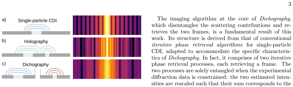
The central claim is that when the recorded intensity obeys I(q) = |psi_A(q)|^2 + |psi_B(q)|^2—two independent scattered fields whose intensities add without an interference cross-term—both fields, and therefore both sample densities rho_A and rho_B, can be retrieved from the single mixed diffraction pattern. The retrieval runs two otherwise separate phase-retrieval reconstructions that are coupled only when the data are enforced: at each pixel the two field amplitudes are rescaled together so that the sum of their squared magnitudes matches the measurement, while all phases are kept. Applied to two-color X-ray data from xenon-doped helium nanodroplets, the reconstructions show a 1.0 keV fra
Load-bearing premise
The load-bearing premise is that the recorded pattern is exactly the incoherent sum of two independent scattered fields; for the nanodroplet result it is also assumed that both frames share the same pre-fitted spherical helium envelope, so a droplet that deformed within 750 fs would still be reconstructed as spherical and could produce apparent agreement between the frames.
Editorial extensions
If this is right
- Two-frame structural movies of isolated nanoparticles at terahertz-scale time separations become possible from a single detector exposure using existing two-color X-ray pulse modes.
- Any experiment whose detector signal is an incoherent sum—two particles in the focus, overlapping broadband or polychromatic scattering, or two exposures on one frame—becomes a candidate for the same algorithmic separation.
- The nanodroplet results imply that within 20 nm, the xenon dopant distribution is unchanged 750 fs after the first pulse, so the early damage from the pump pulse does not yet rearrange the heavy-atom skeleton.
- A moderate increase in two-color pulse brightness or the addition of per-photon energy information should move Dichography from a few optimal patterns to routine reconstructions.
- Combined with an optical-laser prepulse or attosecond two-color pulses, Dichography would allow direct imaging of shock-wave expansion, disintegration, and electronic-excitation dynamics in nanomatter.
Reading between the lines
- A decisive test the paper does not perform: run Dichography on simulated two-color data in which the helium envelope is allowed to expand or distort between the two frames. Because the shared-sphere DCDI constraint would force both frames to be spherical anyway, this would reveal whether apparent frame consistency can be manufactured by the prior.
- The different pixel scales of the two frames (5.57 nm versus 4.64 nm per pixel) give a built-in ghost detector: a genuine physical feature must occupy different pixel counts in the two reconstructions, while a ghost appears at the wrong scale. This could be formalized as a quantitative consistency score.
- The paper's uniqueness observation—knowing one frame correctly reduces the other to ordinary CDI—suggests a path to a full proof that the dichographic problem has no spurious solutions, which would put the method on the same footing as conventional phase retrieval.
- The vector-rescaling projector generalizes naturally to N incoherently summed fields by renormalizing an N-dimensional amplitude vector at each pixel, so the same machinery may extend to three-frame or spectrally multiplexed imaging.
Editorial analysis
A structured set of objections, weighed in public.
Referee Report
Summary. The paper introduces Dichography, an algorithmic extension of single-particle CDI that separates a single recorded diffraction pattern I(q) into two incoherent intensity contributions |ψ_A|^2 + |ψ_B|^2 (Eq. 1) and reconstructs the two corresponding densities ρ_A and ρ_B. The method is demonstrated on two experimental datasets: (i) two-color XFEL data from Xe-doped superfluid He nanodroplets at the European XFEL (1.0 keV and 1.2 keV pulses separated by 750 fs), where the spherical He envelope is imposed via DCDI and only the Xe doping is retrieved; and (ii) SwissFEL double-hit data in which two Ag nanocubes/nanoparticles are illuminated by the same pulse. Successful Ag reconstructions show separated four-fold-symmetric diffraction patterns with ghosting roughly two orders of magnitude below the true signal. The two-color reconstructions show similar Xe features in both frames, which the authors interpret as evidence that no significant structural damage occurs within 750 fs. Appendices describe the iterative projector (Appendix A), oversampling and uniqueness (Appendix B), the Memetic Phase Retrieval adaptation 'Equinox' (Appendix C), experimental details (Appendix D), and DCDI implementation (Appendix E).
Significance. If the central claims hold, Dichography offers a genuinely new capability: two time-delayed structural snapshots from a single detector readout, avoiding spectral filtering or beam splitting. The paper has concrete strengths: the double-hit results are visually compelling and are supported by an open-source implementation (Equinox) and public data; the disentangled nanocube patterns show the expected four-fold symmetry; and the paper is candid about the missing uniqueness proof and about the role of DCDI. The significance of the ultrafast-movie claim, however, rests on the two-color nanodroplet reconstructions, which are fewer in number and depend on a pre-imposed spherical helium envelope. Because that dependency is not stress-tested in the main text, the physical conclusion should be treated as promising but not fully established.
major comments (4)
- [Sec. III.B and Appendix E] The survival inference at 750 fs is partly circular with respect to the DCDI constraint. Both frames are reconstructed inside the same pre-fitted spherical helium envelope; the algorithm cannot represent deformation, expansion, or mass loss, so it would output spherical-envelope frames even if the droplet had changed. The cited support is a companion paper (Ref. [35]) and an ion-displacement estimate of ≤18.6 nm, which is below but close to the 19–20 nm resolution and accounts only for ionic Coulomb motion. A null control is needed: run the same pipeline on simulated two-color data with a deliberately deformed droplet (or wrong fixed radius) and show that retrieved Xe features do not artificially reproduce the pump-frame structure. Without this, the abstract's 'provides evidence' claim goes beyond what the reconstruction alone establishes.
- [Sec. III.A vs. III.B, Figs. 2–4] The double-hit validation does not transfer quantitatively to the two-color regime. Double-hits have disjoint supports (>4 µm separation), same wavelength, high photon counts, and no shared envelope; two-color data have overlapping supports, 1–10 photons/pixel (Fig. 4 color bars), unbalanced brightness (64% and 3×), and enforced spherical envelopes. The Fig. 2 ghosting estimate (≈10^-2) therefore does not calibrate the low-count, overlapping-support geometry of Fig. 4. The main text should include a two-color ghosting benchmark with the DCDI constraint, beyond the reference to Supplemental Sec. S1.
- [Appendix B1, Eq. (B4)] Appendix B1 explicitly states that uniqueness is unproven. Eq. (B4) is conditional: if one frame is already correct, the other is unique; it does not establish uniqueness of the pair from I(q) in Eq. (1). The inherent ambiguity I = J + (I−J) means the method's success in the two-color regime is an empirical claim. The authors should provide a formal uniqueness/stability result or a quantitative ground-truth test using the actual DCDI constraint and noise level.
- [Sec. III.B, Fig. 7 and Appendix E] The pixel-scaling argument excluding ghosting is suggestive but not quantitative. Different pixel sizes (5.57 nm vs 4.64 nm, Eq. (E1)) make a true structure span different pixel counts, but a ghost generated in the target frame could be rescaled by the target frame's constraints. A numerical test is needed: inject a strong feature in one frame's Fourier data and check whether it leaks into the other at the wrong scale.
minor comments (5)
- [Introduction] Typo: 'snapshtos' should be 'snapshots'.
- [Sec. III.B] The phrase 'controllable time delays delay' contains a duplicated word; 'delay' appears twice.
- [Sec. III.A] The sentence 'The number of photons scattered by the first frame in Fig. 2c' is confusing: Fig. 2c is a density map, not a scattering frame. The intended reference appears to be Fig. 3c or a disentangled pattern.
- [Appendix B] The oversampling count O_d' > 4 (Eq. B2) treats the two supports' areas independently, but in the two-color case the two frames share the same compact support region; the practical oversampling is smaller than in the double-hit case, and the text should state this explicitly.
- [References] The Supplemental Material placeholder 'URL_will_be_provided_by_the_publisher' should be resolved; the companion paper Ref. [35] should be explicitly identified as a companion preprint.
Circularity Check
Nanodroplet survival claim rests on a self-cited fitting assumption (D constant over 750 fs); the Dichography method itself is independently validated by double-hit nanocube benchmarks.
-
self citation load bearing
[Section III B and Appendix E (DCDI prerequisites; Ref. [35])]
"The applicability of the DCDI method to the two-color pump-probe data relies on two fundamental prerequisites: the ability to determine the droplet size a priori, and the structural integrity of the droplet, which must be preserved over the 750 fs time delay between the two XFEL pulses. Both of these conditions have been verified and demonstrated in Ref. [35]... In this procedure, the droplet size D is assumed constant between the two scattering events."
The load-bearing premise for the two-color nanodroplet reconstruction is that the helium droplet remains a hard sphere of constant size over the 750 fs delay. The paper cites the same-author companion paper (Ref. [35]) as having verified this premise, but that verification consists of fitting the two-color radial profile as the incoherent sum of two Mie intensities while assuming D is constant between the two pulses. A fit made under the constancy assumption cannot independently establish constancy; it can only report compatibility. The present paper then imposes that fitted spherical envelope as a fixed DCDI constraint on both frames, so the late frame is forced to share the same spherical support. Consequently the 'no structural damage' inference for the nanodroplet branch is conditional
full rationale
The core Dichography method is not circular: the split of I into |psi_A|^2 + |psi_B|^2 is enforced by the iterative intensity projector (Eq. A1) and is independently benchmarked on experimental double-hit data against known silver nanocube morphologies and on simulated data. The two-color xenon-doped droplet branch, however, imports a load-bearing premise from Ref. [35], a companion paper with overlapping authors: droplet integrity over 750 fs. That companion paper's fit assumes a single constant droplet size D between the two pulses, so treating it as an independent verification of droplet integrity is a mild circular step. The paper itself also concedes that Dichography uniqueness is unproven (Appendix B 1), which is a limitation rather than a circularity. Overall, the central imaging claim retains independent content, but the nanodroplet survival claim is partially anchored in a self-cited fitting assumption, giving a score of 4.
Assumptions & free parameters
free parameters (3)
- Helium droplet radius R (used value 350 nm) =
R = 350 nm (diameter ~700 nm)
- Relative two-pulse brightness (1.0 keV vs 1.2 keV) =
1.2 keV frame 64% brighter than 1.0 keV (Fig. 4a); ~3x brighter (Fig. 4d)
- Signal-to-noise cutoff for resolution =
theta_max ~ 1.5 deg, giving ~19-20 nm
assumptions (6)
- standard math Far-field kinematic scattering: the scattered field is the Fourier transform of the sample density, rho proportional to F^{-1}[psi]
- domain assumption Mutual incoherence of the two scattered fields, so that I = |psi_A|^2 + |psi_B|^2 without a cross term (Eq. 1)
- domain assumption Support constraint and sufficient oversampling: each frame is compact with zero scattering outside, and O'_d > 4 is sufficient (Eqs. B1-B3)
- ad hoc to paper Uniqueness of the dichographic solution (the iterative algorithm converges to the true pair of frames)
- domain assumption The helium droplet is a pristine hard sphere of the Mie-fitted size for both pulses, valid as a DCDI constraint at 750 fs delay
- domain assumption Charging-model bound: maximum ion displacement in 750 fs is 18.6 nm, below the ~19 nm resolution
Cite this review
Pith. "Pith review of Dichography: Two-frame Ultrafast Imaging from a Single Diffraction Pattern." pith.science (2026). https://pith.science/paper/RZ643YPH
@misc{pith2026250820153,
  author       = {Pith},
  title        = {Pith review of: Dichography: Two-frame Ultrafast Imaging from a Single Diffraction Pattern},
  year         = {2026},
  howpublished = {\url{https://pith.science/paper/RZ643YPH}},
  note         = {Machine review of arXiv:2508.20153}
}
read the original abstract
We experimentally demonstrate that pairs of time-delayed ultrabright and ultrashort X-ray pulses of two different colors, delivered by modern X-ray Free Electron Lasers, can provide two time-delayed snapshots of a sample. We introduce Dichography, a method that algorithmically separates the diffraction signals overlapping on the detector and independently retrieves the two images of the specimen. We employ Dichography to reconstruct two views of individual xenon-doped helium nanodroplets with 20 nm spatial resolution. The consistency of structures observed in both images at delays up to 750 fs provides evidence that, under these illumination conditions, significant structural damage only occurs at longer timescales. We further validate the method by imaging pairs of silver nanoparticles intercepted by the same light pulse. Dichography enables a new class of experiments across physics, chemistry, and materials science, making a significant step toward the original promise of X-ray free-electron lasers to capture ultrafast movies of nanomatter.
Figures


Figures from the paper (5 more)


Reference graph
Works this paper leans on
-
[35]
K. R. Ferguson, M. Bucher, T. Gorkhover, S. Boutet, H. Fukuzawa, J. E. Koglin, Y. Kumagai, A. Lutman, A. Marinelli, M. Messerschmidt, K. Nagaya, J. Turner, K. Ueda, G. J. Williams, P. H. Bucksbaum, and C. Bost- edt, Transient lattice contraction in the solid-to-plasma transition, Science Advances 2, e1500837 (2016)
work page 2016
-
[1]
Iterative phase retrieval Coherent Diffraction Imaging (CDI) is an indirect imaging method that requires extensive numerical anal- ysis to obtain the sample image from the recorded data. The scattered field ψ encodes the complete information about the spatial density of the sample, ρ, as they are mathematically linked by a Fourier Transform (FT) op- erati...
-
[2]
Constraining experimental data In Dichography, the experimentally acquired informa- tion on the sum of the two scattering signals has to be constrained, i.e., propagated to the two frames of the re- construction, to make the two estimates of the field, ψA and ψB, satisfy the condition in Eq. (A1). An intuitive description of the operation is reported in F...
-
[3]
equations
Real-space constraint The application of the real-space constraint, i.e., the support function, is performed independently on the two frames, as if they were two separate reconstruction proce- dures using iterative algorithms for conventional single- particle CDI. This means that each of the two densities, ρA and ρB, is constrained to its own independent ...
-
[4]
They are designed to minimize the difference between the mea- sured experimental data and the Fourier intensities of the reconstructed sample image
Uniqueness of the solution All imaging algorithms for conventional CDI are, in practice, optimization algorithms [7, 32, 40]. They are designed to minimize the difference between the mea- sured experimental data and the Fourier intensities of the reconstructed sample image. Thus, the phase re- trieval problem is an optimization problem. The recon- structi...
-
[5]
Silver nanoparticles at SwissFEL The two-color experiment was performed at the Mal- oja endstation of SwissFEL [68, 69]. Wet-chemically grown silver nanoparticles were injected into the exper- imental chamber, kept under vacuum conditions, using an electrospray and a set of aerodynamic lenses [70, 71]. The electrospray disperses the liquid containing the ...
-
[6]
The XFEL was operated at a repetition rate of 10 Hz with an elec- tron energy of 16 GeV
Two-color experiment at the European XFEL The experiment was performed at the Nano-sized Quantum Systems (NQS) endstation of the Small Quan- tum Systems (SQS) instrument [73–75], located on the SASE3 branch of the European XFEL [76]. The XFEL was operated at a repetition rate of 10 Hz with an elec- tron energy of 16 GeV. Two consecutive sets of XFEL undul...
-
[7]
Z. Guo, T. Driver, S. Beauvarlet, D. Cesar, J. Duris, P. L. Franz, O. Alexander, D. Bohler, C. Bostedt, V. Aver- bukh, et al. , Experimental demonstration of attosecond pump–probe spectroscopy with an x-ray free-electron laser, Nature Photonics 18, 691 (2024)
work page 2024
Show all 85 references
-
[8]
Franz, S
P. Franz, S. Li, T. Driver, R. R. Robles, D. Cesar, E. Isele, Z. Guo, J. Wang, J. P. Duris, K. Larsen, et al., Terawatt- scale attosecond x-ray pulses from a cascaded superradi- ant free-electron laser, Nature Photonics 18, 698 (2024)
2024
-
[9]
J. Yan, W. Qin, Y. Chen, W. Decking, P. Dijkstal, M. Guetg, I. Inoue, N. Kujala, S. Liu, T. Long, et al. , Terawatt-attosecond hard x-ray free-electron laser at high repetition rate, Nature Photonics 18, 1293 (2024)
2024
-
[10]
P. K. Maroju, C. Grazioli, M. Di Fraia, M. Moioli, D. Er- tel, H. Ahmadi, O. Plekan, P. Finetti, E. Allaria, L. Gi- annessi, et al. , Complex attosecond waveform synthesis at fel fermi, Applied Sciences 11, 9791 (2021)
2021
-
[11]
Duris, S
J. Duris, S. Li, T. Driver, E. G. Champenois, J. P. MacArthur, A. A. Lutman, Z. Zhang, P. Rosenberger, J. W. Aldrich, R. Coffee, et al., Tunable isolated attosec- ond x-ray pulses with gigawatt peak power from a free- electron laser, Nature Photonics 14, 30 (2020)
2020
-
[12]
R. A. Kirian and H. N. Chapman, Imaging of objects by coherent diffraction of x-ray free-electron laser pulses, in Synchrotron Light Sources and Free-Electron Lasers (Springer International Publishing, 2016) pp. 1135–1195
2016
-
[13]
Colombo and D
A. Colombo and D. Rupp, Imaging clusters and their dynamics with single-shot coherent diffraction (2023)
2023
-
[14]
H. N. Chapman, A. Barty, M. J. Bogan, S. Boutet, M. Frank, S. P. Hau-Riege, S. Marchesini, B. W. Woods, 16 S. Bajt, W. H. Benner, et al. , Femtosecond diffrac- tive imaging with a soft-x-ray free-electron laser, Nature Physics 2, 839 (2006)
2006
-
[15]
Kimura, Y
T. Kimura, Y. Joti, A. Shibuya, C. Song, S. Kim, K. Tono, M. Yabashi, M. Tamakoshi, T. Moriya, T. Os- hima, et al. , Imaging live cell in micro-liquid enclosure by x-ray laser diffraction, Nat. Commun. 5, 3052 (2014)
2014
-
[16]
M. M. Seibert, T. Ekeberg, F. R. N. C. Maia, M. Svenda, J. Andreasson, O. J¨ onsson, D. Odi´ c, B. Iwan, A. Rocker, D. Westphal, M. Hantke, D. P. DePonte, A. Barty, J. Schulz, L. Gumprecht, N. Coppola, A. Aquila, M. Liang, T. A. White, A. Martin, C. Caleman, S. Stern, C. Aberg...
2011
-
[17]
Ekeberg, D
T. Ekeberg, D. Assalauova, J. Bielecki, R. Boll, B. J. Daurer, L. A. Eichacker, L. E. Franken, D. E. Galli, L. Gelisio, L. Gumprecht, et al. , Observation of a sin- gle protein by ultrafast x-ray diffraction, Light: Science & Applications 13, 15 (2024)
2024
-
[18]
Colombo, S
A. Colombo, S. Dold, P. Kolb, N. Bernhardt, P. Behrens, J. Correa, S. D¨ usterer, B. Erk, L. Hecht, A. Heil- rath, R. Irsig, N. Iwe, J. Jordan, B. Kruse, B. Lang- behn, B. Manschwetus, F. Martinez, K.-H. Meiwes- Broer, K. Oldenburg, C. Passow, C. Peltz, M. Sauppe, F. Seel, R. ...
2023
-
[19]
Langbehn, K
B. Langbehn, K. Sander, Y. Ovcharenko, C. Peltz, A. Clark, M. Coreno, R. Cucini, M. Drabbels, P. Finetti, M. Di Fraia, L. Giannessi, C. Grazioli, D. Iablonskyi, A. C. LaForge, T. Nishiyama, V. Oliver ´Alvarez de Lara, P. Piseri, O. Plekan, K. Ueda, J. Zimmermann, K. C. Prince,...
2018
-
[20]
Gorkhover, S
T. Gorkhover, S. Schorb, R. N. Coffee, M. Adolph, L. Foucar, D. Rupp, A. Aquila, J. D. Bozek, S. W. Epp, B. Erk, L. Gumprecht, L. Holmegaard, A. Hart- mann, R. Hartmann, G. Hauser, P. Holl, A. H¨ omke, P. Johnsson, N. Kimmel, K. U. K¨ uhnel, M. Messer- schmidt, C. Reich, A. Ro...
2016
-
[21]
Fl¨ uckiger, D
L. Fl¨ uckiger, D. Rupp, M. Adolph, T. Gorkhover, M. Krikunova, M. M¨ uller, T. Oelze, Y. Ovcharenko, M. Sauppe, S. Schorb, C. Bostedt, S. D¨ usterer, M. Har- mand, H. Redlin, R. Treusch, and T. M¨ oller, Time- resolved x-ray imaging of a laser-induced nanoplasma and its neutr...
2016
-
[22]
Langbehn, Y
B. Langbehn, Y. Ovcharenko, A. Clark, M. Coreno, R. Cucini, A. Demidovich, M. Drabbels, P. Finetti, M. D. Fraia, L. Giannessi, C. Grazioli, D. Iablonskyi, A. C. LaForge, T. Nishiyama, V. O. ´A. de Lara, C. Peltz, P. Piseri, O. Plekan, K. Sander, K. Ueda, T. Fennel, K. C. Princ...
2022
-
[23]
Bacellar, A
C. Bacellar, A. S. Chatterley, F. Lackner, C. D. Pem- maraju, R. M. P. Tanyag, D. Verma, C. Bernando, S. M. O. O’Connell, M. Bucher, K. R. Ferguson, T. Gorkhover, R. N. Coffee, G. Coslovich, D. Ray, T. Os- ipov, D. M. Neumark, C. Bostedt, A. F. Vilesov, and O. Gessner, Anisotr...
2022
-
[24]
S. Dold, T. Reichenbach, A. Colombo, J. Jordan, I. Barke, P. Behrens, N. Bernhardt, J. Correa, S. D¨ usterer, B. Erk, T. Fennel, L. Hecht, A. Heilrath, R. Irsig, N. Iwe, P. Kolb, B. Kruse, B. Langbehn, B. Man- schwetus, P. Marienhagen, F. Martinez, K.-H. Meiwes- Broer, K. Olde...
2025
-
[25]
Serkez, W
S. Serkez, W. Decking, L. Froehlich, N. Gerasimova, J. Gr¨ unert, M. Guetg, M. Huttula, S. Karabekyan, A. Koch, V. Kocharyan, et al. , Opportunities for two- color experiments in the soft x-ray regime at the euro- pean xfel, Applied Sciences 10, 2728 (2020)
2020
-
[26]
Lutman, R
A. Lutman, R. Coffee, Y. Ding, Z. Huang, J. Krzywin- ski, T. Maxwell, M. Messerschmidt, and H.-D. Nuhn, Ex- perimental demonstration of femtosecond two-color x-ray free-electron lasers, Physical Review Letters 110, 134801 (2013)
2013
-
[27]
Marinelli, D
A. Marinelli, D. Ratner, A. Lutman, J. Turner, J. Welch, F.-J. Decker, H. Loos, C. Behrens, S. Gilevich, A. Mi- ahnahri, S. Vetter, T. Maxwell, Y. Ding, R. Coffee, S. Wakatsuki, and Z. Huang, High-intensity double- pulse X-ray free-electron laser, Nature Communications 6, 6369 (2015)
2015
-
[28]
Allaria, D
E. Allaria, D. Castronovo, P. Cinquegrana, P. Craievich, M. Dal Forno, M. Danailov, G. D’Auria, A. Demidovich, G. De Ninno, S. Di Mitri, et al. , Two-stage seeded soft- x-ray free-electron laser, Nature Photonics 7, 913 (2013)
2013
-
[29]
T. Hara, Y. Inubushi, T. Katayama, T. Sato, H. Tanaka, T. Tanaka, T. Togashi, K. Togawa, K. Tono, M. Yabashi, et al., Two-colour hard x-ray free-electron laser with wide tunability, Nature communications 4, 2919 (2013)
2013
-
[30]
E. Prat, P. Dijkstal, E. Ferrari, R. Ganter, P. Jurani´ c, A. Malyzhenkov, S. Reiche, T. Schietinger, G. Wang, A. A. Haddad, et al. , Widely tunable two-color x-ray free-electron laser pulses, Physical Review Research 4, 17 L022025 (2022)
2022
-
[31]
Str¨ uder, S
L. Str¨ uder, S. Epp, D. Rolles, R. Hartmann, P. Holl, G. Lutz, H. Soltau, R. Eckart, C. Reich, K. Heinzinger, C. Thamm, A. Rudenko, F. Krasniqi, K.-U. K¨ uhnel, C. Bauer, C.-D. Schr¨ oter, R. Moshammer, S. Techert, D. Miessner, M. Porro, O. H¨ alker, N. Meidinger, N. Kim- mel...
2010
-
[32]
Kuster, K
M. Kuster, K. Ahmed, K.-E. Ballak, C. Danilevski, M. Ekmedˇ zi´ c, B. Fernandes, P. Gessler, R. Hartmann, S. Hauf, P. Holl, M. Meyer, J. Monta˜ no, A. M¨ unnich, Y. Ovcharenko, N. Rennhack, T. R¨ uter, D. Rupp, D. Schlosser, K. Setoodehnia, R. Schmitt, L. Str¨ uder, R. M. P. T...
2021
-
[33]
Hinger, A
V. Hinger, A. al Haddad, R. Barten, A. Bergamaschi, M. Br¨ uckner, M. Carulla, S. Chiriotti-Alvarez, R. Di- napoli, S. Ebner, E. Fr¨ ojdh, et al. , Advancing the jungfrau detector toward low-energy x-ray applications, Journal of Instrumentation 17 (09), C09027
-
[34]
Marchesini, H
S. Marchesini, H. He, H. N. Chapman, S. P. Hau-Riege, A. Noy, M. R. Howells, U. Weierstall, and J. C. H. Spence, X-ray image reconstruction from a diffraction pattern alone, Phys. Rev. B 68, 140101(R) (2003)
2003
-
[36]
Hecht, Zweifarben-Streubildaufnahme von Helium- Nanotr¨ opfchen: Planung, Durchf¨ uhrung und erste Ergeb- nisse, Master’s thesis, Technische Universit¨ at Berlin, Berlin (2018)
L. Hecht, Zweifarben-Streubildaufnahme von Helium- Nanotr¨ opfchen: Planung, Durchf¨ uhrung und erste Ergeb- nisse, Master’s thesis, Technische Universit¨ at Berlin, Berlin (2018)
2018
-
[37]
Sauppe, T
M. Sauppe, T. Bischoff, C. Bomme, C. Bostedt, A. Colombo, B. Erk, T. Feigl, L. Fl¨ uckiger, T. Gorkhover, A. Heilrath, K. Kolatzki, Y. Kumagai, B. Langbehn, J. P. M¨ oller, C. Passow, D. Ramm, D. Rolles, D. Rompo- tis, J. Sch¨ afer-Zimmermann, B. Senfftleben, R. Treusch, A. Ul...
2024
-
[38]
Marchesini, Invited article: A unified evaluation of iterative projection algorithms for phase retrieval, Rev
S. Marchesini, Invited article: A unified evaluation of iterative projection algorithms for phase retrieval, Rev. Sci. Instrum. 78, 011301 (2007)
2007
-
[39]
Nolting, C
F. Nolting, C. Bostedt, T. Schietinger, and H. Braun, The swiss light source and swissfel at the paul scherrer institute, The European Physical Journal Plus 138, 126 (2023)
2023
-
[40]
See Supplemental Material at URL_will_be_provided_ by_the_publisher
-
[41]
Hecht, Y
L. Hecht, Y. Ovcharenko, A. Ø. Lægdsmand, B. Bas- tian, T. M. Baumann, A. Colombo, S. De, A. D. Fanis, S. Dold, T. Fennel, R. Hartmann, K. Ko- latzki, S. Krishnan, B. Kruse, A. C. Laforge, B. Lang- behn, S. Mandal, T. Mazza, C. Medina, C. Peltz, T. Pfeifer, B. Senfftleben, K. ...
2025 arXiv
-
[42]
R. M. P. Tanyag, C. Bernando, C. F. Jones, C. Bacel- lar, K. R. Ferguson, D. Anielski, R. Boll, S. Carron, J. P. Cryan, L. Englert,et al., Communication: X-ray co- herent diffractive imaging by immersion in nanodroplets, Structural Dynamics 2 (2015)
2015
-
[43]
Gessner and A
O. Gessner and A. F. Vilesov, Imaging quantum vortices in superfluid helium droplets, Annual review of physical chemistry 70, 173 (2019)
2019
-
[44]
Ulmer, A
A. Ulmer, A. Heilrath, B. Senfftleben, S. M. O’Connell- Lopez, B. Kruse, L. Seiffert, K. Kolatzki, B. Langbehn, A. Hoffmann, T. M. Baumann, et al., Generation of large vortex-free superfluid helium nanodroplets, Physical re- view letters 131, 076002 (2023)
2023
-
[45]
R. M. P. Tanyag, C. F. Jones, C. Bernando, S. M. O. O’Connell, D. Verma, and A. F. Vilesov, CHAPTER
-
[46]
Dulieu and A
Experiments with Large Superfluid Helium Nan- odroplets, in Theoretical and Computational Chemistry Series, edited by O. Dulieu and A. Osterwalder (Royal Society of Chemistry, Cambridge, 2017) pp. 389–443
2017
-
[47]
Colombo, M
A. Colombo, M. Sauppe, A. A. Haddad, K. Ayyer, M. Babayan, R. Boll, R. Dagar, S. Dold, T. Fen- nel, L. Hecht, G. Knopp, K. Kolatzki, B. Langbehn, F. R. N. C. Maia, A. Mall, P. Mazumder, T. Mazza, Y. Ovcharenko, I. C. Polat, D. Raiser, J. C. Sch¨ afer- Zimmermann, K. Schnorr, M...
2025
-
[48]
E. Prat, Z. Geng, C. Kittel, A. Malyzhenkov, F. Mar- cellini, S. Reiche, T. Schietinger, and P. Craievich, At- tosecond time-resolved measurements of electron and photon beams with a variable polarization x-band ra- diofrequency deflector at an x-ray free-electron laser, Ad- v...
2025
-
[49]
Kuschel, P
S. Kuschel, P. J. Ho, A. Al Haddad, F. F. Zimmermann, L. Flueckiger, M. R. Ware, J. Duris, J. P. MacArthur, A. Lutman, M.-F. Lin, X. Li, K. Nakahara, J. W. Aldrich, P. Walter, L. Young, C. Bostedt, A. Marinelli, and T. Gorkhover, Non-linear enhancement of ultrafast X-ray diffr...
2025 doi
-
[50]
Ulmer, P
A. Ulmer, P. J. Ho, B. Langbehn, S. Kuschel, L. Hecht, R. Obaid, S. Dold, T. Driver, J. Duris, M.-F. Lin, D. Cesar, P. Franz, Z. Guo, P. A. Hart, A. Kamalov, K. A. Larsen, X. Li, M. Meyer, K. Nakahara, R. G. Radloff, R. Robles, L. R¨ onnebeck, N. Sudar, A. M. Summers, L. Young...
-
[51]
J. C. Sch¨ afer-Zimmermann, T. von Scheven, K. Ko- latzki, B. Kruse, B. Langbehn, T. M¨ oller, N. Monserud, M. Sauppe, B. Sch¨ utte, B. Senfftleben, R. M. P. Tanyag, A. Ulmer, T. Fennel, M. J. J. Vrakking, A. Rouz´ ee, and D. Rupp, Laser-induced transient opacity in helium nan...
2025
-
[52]
Vicario, S
C. Vicario, S. Bettoni, A. Lutman, A. Dax, M. Huppert, and A. Trisorio, Two-color x-ray free-electron laser by photocathode laser emittance spoiler, Physical Review Accelerators and Beams 24, 060703 (2021)
2021
-
[53]
Marr and J
G. Marr and J. West, Absolute photoionization cross- section tables for helium, neon, argon, and krypton in the vuv spectral regions, Atomic Data and Nuclear Data Tables 18, 497 (1976)
1976
-
[54]
Allaria, L
E. Allaria, L. Badano, S. Bassanese, F. Capotondi, D. Castronovo, P. Cinquegrana, M. B. Danailov, G. D’Auria, A. Demidovich, R. De Monte, G. De Ninno, S. Di Mitri, B. Diviacco, W. M. Fawley, M. Ferianis, E. Ferrari, G. Gaio, D. Gauthier, L. Giannessi, F. Iaz- zourene, G. Kurdi...
2015
-
[55]
Rossbach, J
J. Rossbach, J. R. Schneider, and W. Wurth, 10 years of pioneering X-ray science at the Free-Electron Laser FLASH at DESY, Physics Reports 808, 1 (2019)
2019
-
[56]
B. L. Henke, E. M. Gullikson, and J. C. Davis, X-ray interactions: photoabsorption, scattering, transmission, and reflection at e= 50-30,000 ev, z= 1-92, Atomic data and nuclear data tables 54, 181 (1993)
1993
-
[57]
D. Rupp, L. Fl¨ uckiger, M. Adolph, A. Colombo, T. Gorkhover, M. Harmand, M. Krikunova, J. P. M¨ uller, T. Oelze, Y. Ovcharenko, et al. , Imaging plasma for- mation in isolated nanoparticles with ultrafast resonant scattering, Structural Dynamics 7, 034303 (2020)
2020
-
[58]
A. A. Lutman, J. P. MacArthur, M. Ilchen, A. O. Lindahl, J. Buck, R. N. Coffee, G. L. Dakovski, L. Dammann, Y. Ding, H. A. D¨ urr, et al. , Polarization control in an x-ray free-electron laser, Nature photonics 10, 468 (2016)
2016
-
[59]
Kittel, A
C. Kittel, A. Sarracini, S. Augustin, N. Yang, A. Al Had- dad, E. Ferrari, G. Knopp, J. Knurr, A. S. Morillo- Candas, I. Swiderska, et al., Demonstration of full polar- ization control of soft x-ray pulses with apple x undula- tors at swissfel using recoil ion momentum spectro...
2024
-
[60]
Allaria, B
E. Allaria, B. Diviacco, C. Callegari, P. Finetti, B. Mahieu, J. Viefhaus, M. Zangrando, G. De Ninno, G. Lambert, E. Ferrari, et al. , Control of the polar- ization of a vacuum-ultraviolet, high-gain, free-electron laser, Physical Review X 4, 041040 (2014)
2014
-
[61]
J. Yan, H. Hao, S. Huang, J. Li, V. N. Litvinenko, P. Liu, S. F. Mikhailov, V. G. Popov, G. Swift, N. A. Vinokurov, et al., Polarization control of a free-electron laser oscilla- tor using helical undulators of opposite helicities, Physi- cal Review Accelerators and Beams 23, ...
2020
-
[62]
Marotzke, D
S. Marotzke, D. Gupta, R.-P. Wang, M. Pavelka, S. Dziarzhytski, C. von Korff Schmising, S. Jana, N. Thielemann-K¨ uhn, T. Amrhein, M. Weinelt, et al. , First experiments with ultrashort, circularly polarized soft x-ray pulses at flash2, Structural Dynamics 12 (2025)
2025
-
[63]
St¨ ohr, X-ray magnetic circular dichroism spectroscopy of transition metal thin films, Journal of Electron Spec- troscopy and Related Phenomena 75, 253 (1995)
J. St¨ ohr, X-ray magnetic circular dichroism spectroscopy of transition metal thin films, Journal of Electron Spec- troscopy and Related Phenomena 75, 253 (1995)
1995
-
[64]
Cireasa, A
R. Cireasa, A. Boguslavskiy, B. Pons, M. Wong, D. Descamps, S. Petit, H. Ruf, N. Thir´ e, A. Ferr´ e, J. Suarez, et al. , Probing molecular chirality on a sub- femtosecond timescale, Nature Physics 11, 654 (2015)
2015
-
[65]
S. B. Wilkins, P. D. Spencer, P. D. Hatton, S. P. Collins, M. D. Roper, D. Prabhakaran, and A. T. Boothroyd, Direct observation of orbital ordering in la 0.5sr1.5mno4 using soft x-ray diffraction, Phys. Rev. Lett. 91, 167205 (2003)
2003
-
[66]
J. R. Fienup, Phase retrieval algorithms: a comparison, Appl. Opt. 21, 2758 (1982)
1982
-
[67]
Abbey, L
B. Abbey, L. W. Whitehead, H. M. Quiney, D. J. Vine, G. A. Cadenazzi, C. A. Henderson, K. A. Nugent, E. Bal- aur, C. T. Putkunz, A. G. Peele, et al., Lensless imaging using broadband x-ray sources, Nature Photonics 5, 420 (2011)
2011
-
[68]
Huijts, S
J. Huijts, S. Fernandez, D. Gauthier, M. Kholodtsova, A. Maghraoui, K. Medjoubi, A. Somogyi, W. Boutu, and H. Merdji, Broadband coherent diffractive imaging, Na- ture Photonics 14, 618 (2020)
2020
-
[69]
C. Chen, H. Gu, and S. Liu, Ultra-broadband diffractive imaging with unknown probe spectrum, Light: Science & Applications 13, 213 (2024)
2024
-
[70]
D. Rupp, N. Monserud, B. Langbehn, M. Sauppe, J. Zim- mermann, Y. Ovcharenko, T. M¨ oller, F. Frassetto, L. Po- letto, A. Trabattoni, et al. , Coherent diffractive imaging of single helium nanodroplets with a high harmonic gen- eration source, Nature communications 8, 493 (2017)
2017
-
[71]
J. Miao, D. Sayre, and H. N. Chapman, Phase retrieval from the magnitude of the fourier transforms of nonperi- odic objects, J. Opt. Soc. Am. A 15, 1662 (1998)
1998
-
[72]
Favre-Nicolin, F
V. Favre-Nicolin, F. Mastropietro, J. Eymery, D. Ca- macho, Y. Niquet, B. Borg, M. Messing, L.-E. Werners- son, R. Algra, E. Bakkers, et al. , Analysis of strain and stacking faults in single nanowires using bragg coherent diffraction imaging, New Journal of Physics 12, 035013 (2010)
2010
-
[73]
Guizar-Sicairos and P
M. Guizar-Sicairos and P. Thibault, Ptychography: A so- lution to the phase problem, Physics Today74, 42 (2021)
2021
-
[74]
Colombo, D
A. Colombo, D. E. Galli, L. De Caro, F. Scattarella, and E. Carlino, Facing the phase problem in coherent diffrac- tive imaging via memetic algorithms, Sci. Rep. 7, 42236 (2017)
2017
-
[75]
Z. Sun, A. Al Haddad, S. Augustin, G. Knopp, J. Knurr, K. Schnorr, and C. Bostedt, Ultrafast single-particle imaging with intense x-ray pulses, Chimia76, 529 (2022)
2022
-
[76]
PSI, Swissfel maloja experimental station (Aug 2025), https://www.psi.ch/en/swissfel/maloja
2025
-
[77]
Bielecki, M
J. Bielecki, M. F. Hantke, B. J. Daurer, H. K. Reddy, D. Hasse, D. S. Larsson, L. H. Gunn, M. Svenda, A. Munke, J. A. Sellberg, et al. , Electrospray sample in- jection for single-particle imaging with x-ray lasers, Sci- ence Advances 5, eaav8801 (2019)
2019
-
[78]
M. F. Hantke, J. Bielecki, O. Kulyk, D. Westphal, D. S. Larsson, M. Svenda, H. K. Reddy, R. A. Kirian, J. Andreasson, J. Hajdu, et al. , Rayleigh-scattering mi- croscopy for tracking and sizing nanoparticles in focused aerosol beams, IUCrJ 5, 673 (2018). 19
2018
-
[79]
Mozzanica, M
A. Mozzanica, M. Andr¨ a, R. Barten, A. Bergamaschi, S. Chiriotti, M. Br¨ uckner, R. Dinapoli, E. Fr¨ ojdh, D. Greiffenberg, F. Leonarski, et al. , The jungfrau de- tector for applications at synchrotron light sources and xfels, Synchrotron Radiation News 31, 16 (2018)
2018
-
[80]
SQS, Small Quantum Systems Instrument (Aug 2025), https://www.xfel.eu/facility/instruments/ sqs/index_eng.html
2025
-
[81]
Tschentscher, C
T. Tschentscher, C. Bressler, J. Gr¨ unert, A. Madsen, A. P. Mancuso, M. Meyer, A. Scherz, H. Sinn, and U. Za- strau, Photon beam transport and scientific instruments at the european xfel, Applied Sciences 7, 592 (2017)
2017
-
[82]
Mazza, T
T. Mazza, T. M. Baumann, R. Boll, A. De Fa- nis, P. Grychtol, M. Ilchen, J. Monta˜ no, V. Music, Y. Ovcharenko, N. Rennhack, D. E. Rivas, A. R¨ orig, P. Schmidt, S. Usenko, P. Zio lkowski, D. La Civita, M. Vannoni, H. Sinn, B. Keitel, E. Pl¨ onjes, U. F. Jastrow, A. Sorokin, K...
2023
-
[83]
Decking, S
W. Decking, S. Abeghyan, P. Abramian, A. Abramsky, A. Aguirre, C. Albrecht, P. Alou, M. Altarelli, P. Alt- mann, K. Amyan, et al. , A mhz-repetition-rate hard x- ray free-electron laser driven by a superconducting linear accelerator, Nat. Photonics 14, 391 (2020)
2020
-
[84]
Mie, Beitr¨ age zur Optik tr¨ uber Medien, speziell kolloidaler Metall¨ osungen, Annalen der Physik 10.1002/andp.19083300302 (1908)
G. Mie, Beitr¨ age zur Optik tr¨ uber Medien, speziell kolloidaler Metall¨ osungen, Annalen der Physik 10.1002/andp.19083300302 (1908)
1908 doi
-
[85]
Bostedt, H
C. Bostedt, H. Thomas, M. Hoener, T. M¨ oller, U. Saal- mann, I. Georgescu, C. Gnodtke, and J.-M. Rost, Fast electrons from multi-electron dynamics in xenon clus- ters induced by inner-shell ionization, New J. Phys. 12, 083004 (2010)
2010
Reviewed August 5, 2026 · model on record in the stance chip above.
Discussion (0). Continue with ORCID to comment.