REVIEW 5 major objections 6 minor 47 references
Phenylalanine modification in plasma-driven biocatalysis revealed by solvent accessibility and reactive dynamics in combination with protein mass spectrometry
T0 review · 5 major / 6 minor · reviewed 2026-08-06 · deepseek-v4-flash
Pith's one-line read A solvent-accessible-surface screen predicts which amino acid residues plasma species modify, and mass spectrometry confirms three phenylalanine oxidations on CviUPO.
desk verdict Useful screening workflow with an honest limitations section, but the central validation claim is not supported: the MS search was seeded by the prediction, and the residue-level top hits from SASA were not the experimentally modified sites. read the letter →
The pith
A machine-rendered reading of the paper's core claim, the machinery that carries it, and where it could break.
The reading
What carries the argument
The machinery is a two-stage computational pipeline. First, a SASA interaction analysis places a probe molecule at each solvent-accessible point about 1.4 Å from the protein surface, rotates it to the best orientation, and computes a local interaction energy using the ReaxFF reactive force field, a bond-order potential that allows bonds to break and form during the simulation; post-processing ranks residues by total and frequency-normalized (relative) interactions, using the thirty strongest energy points. This yields a three-dimensional interaction map for each plasma species. Second, reactive molecular dynamics validate the hot spots: short 75 fs simulations at the ten lowest-energy positions test whether a covalent bond forms, and longer high-concentration simulations with randomly placed species check whether the residue preferences survive diffusion and competition with solvent. The pipeline was run on three enzymes, CviUPO, AaeUPO, and GapA, and experimentally checked on CviUPO.
What would settle it
Plasma-treat CviUPO with and without a hydroxyl-radical scavenger and compare oxidation of Phe51, Phe67, and Phe208 by mass spectrometry; if oxidation persists when OH radicals are scavenged, the claim that OH drives these modifications is wrong. A complementary check would compute the OH–phenylalanine reaction energy with a high-level quantum-chemical method on a model compound and compare it with the ReaxFF value used in the screening.
Extended reading notes
Core claim
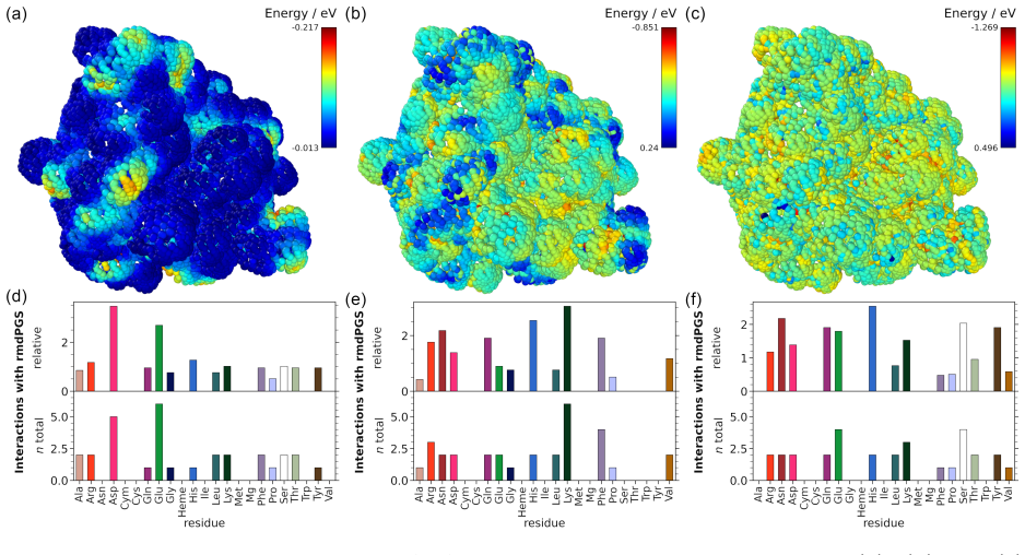
The central claim is that local interaction energies computed at solvent-accessible points on a protein surface identify the residues most likely to be chemically modified by reactive plasma species. The method places one model plasma species at a time on surface points 1.4 Å from the protein, relaxes its orientation, and computes a local interaction energy $E_{\mathrm{int}} = E_{\mathrm{SASA}} - E_{\mathrm{macro\,mol}} - E_{\mathrm{probe\,mol}}$ from the ReaxFF reactive force field. Hot spots cluster on specific residues; reactive molecular dynamics at the ten lowest-energy sites shows that H, O, and OH form bonds there, and high-concentration simulations reproduce the same residue preferences even with random starting positions. For CviUPO, the screen predicted strong interactions with surface-exposed phenylalanines, and dielectric-barrier-discharge plasma treatment followed by tryptic digestion and LC-MS/MS detected significant oxidation of Phe51, Phe67, and Phe208 in all replicates. The paper therefore claims that SASA-based screening can direct targeted mass spectrometry searches and, more generally, locate protein-surface regions of interest for small reactive molecules.
Load-bearing premise
The load-bearing premise is that the reactive force field faithfully represents the chemistry of plasma species such as hydroxyl radicals, and the paper's own supporting information concedes that distinguishing OH radicals from OH ions in ReaxFF 'remains highly uncertain'; if that chemistry is misrepresented, the predicted interaction profiles and modification sites may not reflect real plasma conditions.
Editorial extensions
If this is right
- If the method is right, any enzyme can be screened before plasma experiments to obtain a short list of residues to search for modification in MS data, reducing the search space for time-consuming proteomics.
- For CviUPO, the oxidation of Phe51 and Phe67, both near the substrate channel, flags surface-exposed aromatic residues as a potential cause of activity loss under plasma treatment.
- The solvent simulations say most reactive species react with water before reaching the enzyme; this directly supports immobilization or geometry changes that lengthen the diffusion path as protection strategies.
- At elevated temperatures, unfolding exposes buried residues and the heme cofactor to attack, so thermal control of the reaction environment should limit damage to catalytically essential regions.
- Because the interaction energy definition is not specific to plasma species, the same SASA screen could be used to locate binding or modification sites for other small reactive molecules.
Reading between the lines
- A testable extension beyond this paper: benchmark the SASA screen on enzymes with already published plasma- or oxidant-induced modification maps, asking whether the rank order of predicted hot spots matches experimentally observed modified residues.
- Editorial inference: the screen's residue preferences may be driven mostly by surface exposure and electrostatics rather than by accurate radical chemistry; comparing SASA hot spots against a surface-exposure-only ranking would show how much the reactive dynamics add.
- If Phe oxidation near the substrate channel indeed impairs catalysis, engineering solvent-exposed phenylalanines into less oxidizable residues could improve enzyme stability under plasma-driven conditions.
- The current MS search covered only a small panel of modifications, so the actual damage may be broader; a wider search including nitration and carbonyl formation could test whether other SASA-flagged residues, such as lysine and arginine, are modified under conditions that favor reactive nitrogen species.
Signed reviews
Editorial analysis
A structured set of objections, weighed in public.
Referee Report
Summary. The paper proposes a computational workflow to predict which amino-acid residues of enzymes are modified by plasma-generated species (PGS) such as H2O2, OH, O2, O, NO, and H. The workflow combines a SASA-based interaction analysis, in which a single probe species is placed at solvent-accessible surface points and ReaxFF interaction energies are computed (Eq. 1), with reactive molecular dynamics (ReaxFF with the Monti et al. force field) as a short-time validation of bond formation at predicted positions. The method is applied to three enzymes, with CviUPO as the main experimental subject. For CviUPO, the analysis predicts strong OH interactions with Lys, Phe, and Arg; a DBD plasma-treated sample is then analyzed by LC-MS/MS, and the authors report significant oxidation of Phe51, Phe67, Phe208, Met42, and Met220, claiming that the predicted modification of phenylalanine was experimentally confirmed. The Conclusion and the Supplementary Information explicitly acknowledge that the Phe prediction prompted the inclusion of Phe modifications in the MS database search and that ReaxFF cannot reliably distinguish OH radicals from OH ions.
Significance. If validated, the proposed screening workflow would be practically useful for the plasma-biocatalysis community as a cheap computational prescreen for ranking amino-acid residues on a protein surface with respect to reactivity toward plasma-generated species, and for directing time-consuming mass spectrometry searches. The manuscript has notable strengths: the SASA-analysis package is released on GitHub, the simulation and mass spectrometry data are provided, and the SI candidly documents the ReaxFF species limitation. However, the current evidence does not establish the central claim as stated. The MS validation is partly circular because the search space was seeded by the prediction; Phe oxidation is a known general plasma effect; the agreement is at the level of residue type rather than residue identity; and the force-field ambiguity acknowledged in the SI affects the entire interaction-energy ranking. The paper currently supports a screening heuristic with a plausible consistency check, not an independently confirmed predictive method.
major comments (5)
- [Conclusion] The Conclusion states that 'the Phe modification by plasma treatment has thus been brought into focus by the SASA analysis, which prompted the inclusion of Phe modifications in the MS database search.' Because the prediction seeded the database search, the subsequent detection of Phe oxidation is not an independent confirmation of the method; it is a consistency check whose search space was conditioned on the prediction. This circularity is compounded by the fact that Phe hydroxylation is a well-documented plasma-induced modification of amino acids in the very literature cited by the authors (refs. 43 and 44), so its observation would be expected even if the SASA screen carried no predictive information. The Abstract's claim that predictions were 'confirmed experimentally by mass spectrometry' should be weakened to a consistency statement, or the authors should provide a discriminating validation, e.g., a prospective or pre-registered MS search over a fixed modification list in which the Phe prediction was made before the search was executed.
- [Results, SASA interaction analysis (CviUPO); Supplementary Table 1] The experimental match is at the amino-acid-type level, not the residue level. The ten strongest OH interaction points at 300 K listed in Supplementary Table 1 map to Leu225, Asp91, Arg84, Val153, and other non-Phe residues, and neither the short reactive-MD validation nor the high-concentration simulations (Fig. 5) report bond formation at Phe51, Phe67, or Phe208; they report Phe only as a residue type. The SASA evidence for Phe comes from the frequency-normalized counts in Fig. 2(e), i.e., Phe is over-represented in the pool of strong-interaction points, not from residue-specific interaction energies at the three positions later observed by MS. The paper should explicitly state that the method predicts which amino-acid types are preferential targets and that residue-level correspondence between prediction and experiment is not currently demonstrated.
- [SI, 'Explanation to the species nature in ReaxFF'] The SI states that distinguishing OH radicals from OH ions in ReaxFF 'remains highly uncertain' and that 'any conclusions drawn from these simulations should be interpreted with caution,' and the main text acknowledges that the modeled 'rmdPGS ... are not real plasma-generated species.' Since the SASA interaction energies (Eq. 1) and all bond-formation statistics in the reactive MD are computed with this force field, the mechanistic attribution of Phe oxidation to OH and O in the Conclusion is not supported by a species-resolved model. This caveat should be moved into the main text and the mechanistic claims reframed as applying to the modeled species only. A concrete strengthening would be to benchmark the force field against an experimentally known relative reactivity order, e.g., the high susceptibility of Met and Cys reported in refs. 43-45, before using it to rationalize Phe-selective modification.
- [Results, Mass spectrometry of plasma-treated CviUPO] The screen produced a false negative for a substantial fraction of the experimentally observed modifications: the SASA analysis reports no interactions with Met or Cys because these residues are rare and buried, yet Met42 and Met220 are oxidized in all three replicates. The paper's explanation that buried residues become accessible post hoc does not rescue the predictive claim, since the same argument could apply to many buried residues and is not testable from the reported data. The authors should quantify the screen's miss rate (two of the five observed modified residues belong to a residue type the screen excluded) and discuss whether the Met oxidation indicates that the static SASA screen misses modifications that require protein dynamics, radical diffusion into the protein interior, or species not represented in the model. The weak experimental signal for the strongly predicted Lys/Arg modifications (detected only in some replicates and at low relative intensity) further limits what the current experiment can establish about the method's selectivity.
- [Table 1] The mass spectrometry table reports modified Phe51, Phe67, and Phe208, with Phe51 and Phe67 located in the same 33-residue tryptic peptide, but no site-localization evidence (e.g., fragment-ion coverage or a localization score) is provided, and no error bars or significance tests are given for the relative intensities. Because the headline claim is residue-specific oxidation, the authors should report the localization confidence for each modified site (e.g., the number of site-determining fragment ions) and descriptive statistics over the three replicates, and define the 'relative intensity > 100' acceptance criterion.
minor comments (6)
- [Figure 5] The figure caption is internally inconsistent: it is titled 'Bond analysis for high concentrations of hydrogen' while the surrounding text and the axis description refer to OH, and the caption itself mentions 'an additional OH atom' while the panels are described in terms of hydrogen. Please reconcile the caption with the plotted species.
- [References] References 39 and 43 appear to cite the same article by Takai et al. (2014) with different journal names (Journal of Physics D: Applied Physics vs. Plasma Sources Science and Technology); please deduplicate and correct.
- [Computational details] In Eqs. (2) and (3), the quantity 'Interactions Total Number' is not defined; please specify whether it is the total number of SASA points, the number of points above the top-30 cutoff, or the summed interaction counts over the analyzed set.
- [Computational details] The default cutoff of the thirty strongest interaction energies is described only as 'determined empirically' with no supporting data; a brief sensitivity check (e.g., top 10 vs. top 50) would help the reader judge the robustness of the residue rankings and is recommended given the load this parameter carries.
- [Table 1] The acceptance criterion 'relative intensity > 100' is undocumented; please state how the relative intensities were computed (e.g., normalization to peptide or protein abundance) and why the threshold of 100 was chosen.
- [Throughout] Several typos and LaTeX artifacts remain in the text, including 'occurring in textitCviUPO', the broken 'N V Ten' ensemble label, and the phrase 'are display in Figure 5'; a careful proofread is needed.
Circularity Check
Partial circularity: the MS 'confirmation' of Phe modification is not independent because the SASA prediction was used to seed the MS database search, although unanticipated Met oxidation and weak Lys/Arg signals prevent a fully closed loop.
-
self definitional
[Conclusion (last paragraph); compare 'Mass spectrometry of plasma-treated CviUPO' section]
"The Phe modification by plasma treatment has thus been brought into focus by the SASA analysis, which prompted the inclusion of Phe modifications in the MS database search."
The paper's central validation loop is: SASA analysis predicts strong Phe/Lys/Arg interactions, the MS search then includes Phe, Arg, and Lys modifications 'because of their predicted strong interaction with ROS such as OH,' and the same experiment finds Phe+O and is presented as confirmation. This is not an independent test: the modification being tested was added to the search because the prediction said to look for it, so the positive Phe hit is an expected consequence of the search design rather than evidence that the SASA screen adds predictive value.
full rationale
The main circularity is the seeded MS validation: the Phe hypothesis was generated by the SASA analysis and then the MS search was explicitly designed to include Phe modifications, so detecting Phe+O does not independently validate the method. This is a genuine partial circularity. However, the loop is not closed: Met oxidation was found although SASA predicted no Met/Cys interactions, and predicted Lys/Arg modifications were largely not observed, giving the experiment independent content. The simulation-internal checks (short ReaxFF MD at the ten lowest SASA-energy points, and high-concentration MD comparisons) are consistency checks using the same force field; they do not independently validate the SASA approximation but are not circular in the harmful sense. No load-bearing self-citation is present: the ReaxFF force field is external (Monti et al. 2013), and the authors explicitly note that they did not develop it. The SI caveat that ReaxFF 'lacks the precision required to unambiguously differentiate OH radicals from OH ions' and that conclusions 'should be interpreted with caution' weakens the computational prediction but is an honest limitation, not circularity. The known background that plasma readily oxidizes phenylalanine (refs. 43, 44) further weakens the evidential value of the Phe hit, but that is a correctness/novelty concern, not a circularity one. Overall score 4 reflects partial circularity while acknowledging independent elements in the experimental outcome.
Assumptions & free parameters
free parameters (1)
- SASA post-processing cutoff =
30 strongest interaction energies
assumptions (3)
- domain assumption ReaxFF with the Monti et al. 2013 biomolecule force field adequately represents the reactive chemistry of plasma-generated species.
- domain assumption SASA interaction energies computed in vacuum with a single static probe at solvent-accessible points correlate with covalent modification propensity in solution.
- domain assumption The targeted mass spectrometry search covers the modifications that matter for validation.
Cite this review
Pith. "Pith review of Phenylalanine modification in plasma-driven biocatalysis revealed by solvent accessibility and reactive dynamics in combination with protein mass spectrometry." pith.science (2026). https://pith.science/paper/T22UNPH7
@misc{pith2026250620205,
  author       = {Pith},
  title        = {Pith review of: Phenylalanine modification in plasma-driven biocatalysis revealed by solvent accessibility and reactive dynamics in combination with protein mass spectrometry},
  year         = {2026},
  howpublished = {\url{https://pith.science/paper/T22UNPH7}},
  note         = {Machine review of arXiv:2506.20205}
}
read the original abstract
Biocatalysis is an emerging field that provides an environmentally friendly alternative to conventional catalysis, but still it faces some challenges. One of the major difficulties for biocatalysts that require reactive species like H2O2 as co-substrates lies in the concentration of these reactive species. On the one hand, they are used as reactants, but on the other hand, they inactivate the enzymes at high concentrations. When utilizing non-thermal plasma to deliver H2O2 for biocatalysis, it is essential to understand the potential interactions between plasma-generated species (PGS) and enzymes. This is particularly important because, alongside \ch{H2O2}, other reactive species such as hydroxyl radicals, atomic oxygen, superoxide, and nitric oxide are also produced. The investigation of the localized reactivity of the solvent accessible surface area (SASA) of an enzyme, with certain species, is an important tool for predicting these interactions. In combination with reactive molecular dynamics (MD) simulations this enabled us to identify amino acid residues that are likely targets for modifications by the PGS. A subset of the theoretical predictions made in the present study were confirmed experimentally by mass spectrometry, underlining the utility of the SASA and MD based screening approach to direct time-consuming experiments and assist their interpretation.
Figures


Figures from the paper (20 more)


Reference graph
Works this paper leans on
-
[1]
Catalysis: Concepts and Green Applications, 2nd ed.; Wiley-VCH Verlag, 2017
Rothenberg, G. Catalysis: Concepts and Green Applications, 2nd ed.; Wiley-VCH Verlag, 2017
work page 2017
-
[2]
Berg, J. M.; Tymoczko, J. L.; Gatto, G. J.; Stryer, L. Biochemie; Springer Berlin Heidelberg, 2018
work page 2018
-
[3]
Bell, E. L.; Finnigan, W.; France, S. P.; Green, A. P.; Hayes, M. A.; Hepworth, L. J.; Lovelock, S. L.; Niikura, H.; Osuna, S.; Romero, E.; Ryan, K. S.; Turner, N. J.; Flitsch, S. L. Biocatalysis. Nature Reviews Methods Primers 2021, 1, 1--21
work page 2021
-
[4]
C.; Baldenius, K.; Bornscheuer, U
Wu, S.; Snajdrova, R.; Moore, J. C.; Baldenius, K.; Bornscheuer, U. T. Biocatalysis: Enzymatic Synthesis for Industrial Applications. Angewandte Chemie International Edition 2021, 60, 88--119
work page 2021
-
[5]
Vasudevan, P. T.; Weiland, R. H. Deactivation of catalase by hydrogen peroxide. Biotechnology and Bioengineering 1990, 36, 783--789
work page 1990
-
[6]
Suicide Inactivation of Peroxidases and the Challenge of Engineering More Robust Enzymes
Valderrama, B.; Ayala, M.; Vazquez-Duhalt, R. Suicide Inactivation of Peroxidases and the Challenge of Engineering More Robust Enzymes . Chemistry & Biology 2002, 9, 555--565
work page 2002
-
[7]
Karich, A.; Scheibner, K.; Ullrich, R.; Hofrichter, M. Exploring the catalase activity of unspecific peroxygenases and the mechanism of peroxide-dependent heme destruction. Journal of Molecular Catalysis B: Enzymatic 2016, 134, 238--246
work page 2016
-
[8]
Wapshott-Stehli, H. L.; Grunden, A. M. In situ H2O2 generation methods in the context of enzyme biocatalysis. Enzyme and Microbial Technology 2021, 145, 109744
work page 2021
Show all 47 references
-
[9]
F.; Hollmann, F.; Alcalde, M.; Kourist, R.; Bandow, J
Yayci, A.; Álvaro Gómez Baraibar; Krewing, M.; Fueyo, E. F.; Hollmann, F.; Alcalde, M.; Kourist, R.; Bandow, J. E. Plasma-Driven in Situ Production of Hydrogen Peroxide for Biocatalysis. ChemSusChem 2020, 13, 2072--2079
2020
-
[10]
Yayci, A.; Dirks, T.; Kogelheide, F.; Alcalde, M.; Hollmann, F.; Awakowicz, P.; Bandow, J. E. Microscale Atmospheric Pressure Plasma Jet as a Source for Plasma-Driven Biocatalysis. ChemCatChem 2020, 12, 5893--5897
2020
-
[11]
I.; Walsh, J
Dickenson, A.; Britun, N.; Nikiforov, A.; Leys, C.; Hasan, M. I.; Walsh, J. L. The generation and transport of reactive nitrogen species from a low temperature atmospheric pressure air plasma source. Physical Chemistry Chemical Physics 2018, 20, 28499--28510
2018
-
[12]
2022; https://zenodo.org/records/6637571
GROMACS 2022.2 Source code. 2022; https://zenodo.org/records/6637571
2022
-
[13]
J.; Murtola, T.; Schulz, R.; P \' a ll, S.; Smith, J
Abraham, M. J.; Murtola, T.; Schulz, R.; P \' a ll, S.; Smith, J. C.; Hess, B.; Lindahl, E. GROMACS : High performance molecular simulations through multi-level parallelism from laptops to supercomputers. SoftwareX 2015, 1-2, 19--25
2015
-
[14]
Berendsen, H. J. C.; van der Spoel, D.; van Drunen, R. GROMACS : A message-passing parallel molecular dynamics implementation. Computer Physics Communications 1995, 91, 43--56
1995
-
[15]
GROMACS 4: 0.167em Algorithms for Highly Efficient , Load - Balanced , and Scalable Molecular Simulation
Hess, B.; Kutzner, C.; van der Spoel, D.; Lindahl, E. GROMACS 4: 0.167em Algorithms for Highly Efficient , Load - Balanced , and Scalable Molecular Simulation . Journal of Chemical Theory and Computation 2008, 4, 435--447
2008
-
[16]
GROMACS 3.0: a package for molecular simulation and trajectory analysis
Lindahl, E.; Hess, B.; van der Spoel, D. GROMACS 3.0: a package for molecular simulation and trajectory analysis. Molecular modeling annual 2001, 7, 306--317
2001
-
[17]
J.; Kutzner, C.; Hess, B.; Lindahl, E
P \' a ll, S.; Abraham, M. J.; Kutzner, C.; Hess, B.; Lindahl, E. Tackling Exascale Software Challenges in Molecular Dynamics Simulations with GROMACS . Solving Software Challenges for Exascale . Cham, 2015; pp 3--27
2015
-
[18]
Heterogeneous parallelization and acceleration of molecular dynamics simulations in GROMACS
P \' a ll, S.; Zhmurov, A.; Bauer, P.; Abraham, M.; Lundborg, M.; Gray, A.; Hess, B.; Lindahl, E. Heterogeneous parallelization and acceleration of molecular dynamics simulations in GROMACS . The Journal of Chemical Physics 2020, 153, 134110
2020
-
[19]
R.; Smith, J
Pronk, S.; P \' a ll, S.; Schulz, R.; Larsson, P.; Bjelkmar, P.; Apostolov, R.; Shirts, M. R.; Smith, J. C.; Kasson, P. M.; van der Spoel, D.; Hess, B.; Lindahl, E. GROMACS 4.5: a high-throughput and highly parallel open source molecular simulation toolkit. Bioinformatics 2013...
2013
-
[20]
E.; Berendsen, H
Van Der Spoel, D.; Lindahl, E.; Hess, B.; Groenhof, G.; Mark, A. E.; Berendsen, H. J. C. GROMACS : Fast , flexible, and free. Journal of Computational Chemistry 2005, 26, 1701--1718
2005
-
[21]
A.; Armacost, K
Soteras Guti \' e rrez, I.; Lin, F.-Y.; Vanommeslaeghe, K.; Lemkul, J. A.; Armacost, K. A.; Brooks, C. L.; MacKerell, A. D. Parametrization of halogen bonds in the CHARMM general force field: Improved treatment of ligand--protein interactions. Bioorganic & Medicinal Chemistry ...
2016
-
[22]
Vanommeslaeghe, K.; Hatcher, E.; Acharya, C.; Kundu, S.; Zhong, S.; Shim, J.; Darian, E.; Guvench, O.; Lopes, P.; Vorobyov, I.; Mackerell Jr., A. D. CHARMM general force field: A force field for drug-like molecules compatible with the CHARMM all-atom additive biological force ...
2010
-
[23]
Vanommeslaeghe, K.; MacKerell, A. D. J. Automation of the CHARMM General Force Field ( CGenFF ) I : Bond Perception and Atom Typing . Journal of Chemical Information and Modeling 2012, 52, 3144--3154
2012
-
[24]
P.; MacKerell, A
Vanommeslaeghe, K.; Raman, E. P.; MacKerell, A. D. J. Automation of the CHARMM General Force Field ( CGenFF ) II : Assignment of Bonded Parameters and Partial Atomic Charges . Journal of Chemical Information and Modeling 2012, 52, 3155--3168
2012
-
[25]
Yu, W.; He, X.; Vanommeslaeghe, K.; MacKerell Jr., A. D. Extension of the CHARMM general force field to sulfonyl-containing compounds and its utility in biomolecular simulations. Journal of Computational Chemistry 2012, 33, 2451--2468
2012
-
[26]
From Proteins to Perturbed Hamiltonians: A Suite of Tutorials for the GROMACS -2018 Molecular Simulation Package
Lemkul, J. From Proteins to Perturbed Hamiltonians: A Suite of Tutorials for the GROMACS -2018 Molecular Simulation Package. Living Journal of Computational Molecular Science 2019, 1
2018
-
[27]
2023; https://zenodo.org/records/10806852
LAMMPS : Large -scale Atomic / Molecular Massively Parallel Simulator . 2023; https://zenodo.org/records/10806852
2023
-
[28]
M.; Fogarty, J
Aktulga, H. M.; Fogarty, J. C.; Pandit, S. A.; Grama, A. Y. Parallel reactive molecular dynamics: Numerical methods and algorithmic techniques. Parallel Computing 2012, 38, 245--259
2012
-
[29]
Fast Parallel Algorithms for Short - Range Molecular Dynamics
Plimpton, S. Fast Parallel Algorithms for Short - Range Molecular Dynamics . Journal of Computational Physics 1995, 117, 1--19
1995
-
[30]
P.; Aktulga, H
Thompson, A. P.; Aktulga, H. M.; Berger, R.; Bolintineanu, D. S.; Brown, W. M.; Crozier, P. S.; in 't Veld, P. J.; Kohlmeyer, A.; Moore, S. G.; Nguyen, T. D.; Shan, R.; Stevens, M. J.; Tranchida, J.; Trott, C.; Plimpton, S. J. LAMMPS - a flexible simulation tool for particle-b...
2022
-
[31]
Duin, A. C. V.; Dasgupta, S.; Lorant, F.; Goddard, W. A. ReaxFF: A Reactive Force Field for Hydrocarbons. Journal of Physical Chemistry A 2001, 105, 9396--9409
2001
-
[32]
L.; Shin, Y
Monti, S.; Corozzi, A.; Fristrup, P.; Joshi, K. L.; Shin, Y. K.; Oelschlaeger, P.; Duin, A. C. V.; Barone, V. Exploring the conformational and reactive dynamics of biomolecules in solution using an extended version of the glycine reactive force field. Physical Chemistry Chemic...
2013
-
[33]
VMD -- V isual M olecular D ynamics
Humphrey, W.; Dalke, A.; Schulten, K. VMD -- V isual M olecular D ynamics. Journal of Molecular Graphics 1996, 14, 33--38
1996
-
[34]
P.; Wright, W
Varshney, A.; Brooks, F. P.; Wright, W. V. Linearly Scalable Computation of Smooth Molecular Surfaces. IEEE Computer Graphics and Applications 1994, 14, 19--25
1994
-
[35]
Visualization and analysis of atomistic simulation data with OVITO –the Open Visualization Tool
Stukowski, A. Visualization and analysis of atomistic simulation data with OVITO –the Open Visualization Tool . Modelling and Simulation in Materials Science and Engineering 2009, 18, 015012
2009
-
[36]
Studier, F. W. Protein production by auto-induction in high-density shaking cultures. Protein Expression and Purification 2005, 41, 207--234
2005
-
[37]
Reaction of OH with phenylalanine in neutral aqueous solution
Solar, S. Reaction of OH with phenylalanine in neutral aqueous solution. Radiation Physics and Chemistry (1977) 1985, 26, 103--108
1977
-
[38]
Chemical modification of amino acids by atmospheric-pressure cold plasma in aqueous solution
Takai, E.; Kitamura, T.; Kuwabara, J.; Ikawa, S.; Yoshizawa, S.; Shiraki, K.; Kawasaki, H.; Arakawa, R.; Kitano, K. Chemical modification of amino acids by atmospheric-pressure cold plasma in aqueous solution. Journal of Physics D: Applied Physics 2014, 47, 285403
2014
-
[39]
Reactive molecular dynamics simulations on interaction mechanisms of cold atmospheric plasmas and peptides
Guo, J.-S.; Tian, S.-Q.; Zhang, Y.-T. Reactive molecular dynamics simulations on interaction mechanisms of cold atmospheric plasmas and peptides. Physics of Plasmas 2023, 30, 043512
2023
-
[40]
Chemical fingerprints of cold physical plasmas – an experimental and computational study using cysteine as tracer compound
Lackmann, J.-W.; Wende, K.; Verlackt, C.; Golda, J.; Volzke, J.; Kogelheide, F.; Held, J.; Bekeschus, S.; Bogaerts, A.; Schulz-von der Gathen, V.; Stapelmann, K. Chemical fingerprints of cold physical plasmas – an experimental and computational study using cysteine as tracer c...
2018
-
[41]
Gonz \'a lez-Benjumea, A.; Carro, J.; Renau-M \'i nguez, C.; Linde, D.; Fern \'a ndez-Fueyo, E.; Guti \'e rrez, A.; Mart \'i nez, A. T. Fatty acid epoxidation by Collariella virescens peroxygenase and heme-channel variants. Catalysis Science & Technology 2020, 10, 717--725
2020
-
[42]
Chemical modification of amino acids by atmospheric-pressure cold plasma in aqueous solution
Takai, E.; Kitamura, T.; Kuwabara, J.; Ikawa, S.; Yoshizawa, S.; Shiraki, K.; Kawasaki, H.; Arakawa, R.; Kitano, K. Chemical modification of amino acids by atmospheric-pressure cold plasma in aqueous solution. Plasma Sources Science and Technology 2014, 47, 285403
2014
-
[43]
E.-D.; Aadil, R
Liu, Z.-W.; Zhou, Y.-X.; Wang, F.; Tan, Y.-C.; Cheng, J.-H.; Bekhit, A. E.-D.; Aadil, R. M.; Liu, X.-B. Oxidation induced by dielectric barrier discharge (DBD) plasma treatment reduces IgG/IgE binding capacity and improves the functionality of glycinin. Food chemistry 2021, 36...
2021
-
[44]
Lackmann, J.-W.; Baldus, S.; Steinborn, E.; Edengeiser, E.; Kogelheide, F.; Langklotz, S.; Schneider, S.; Leichert, L. I. O.; Benedikt, J.; Awakowicz, P.; Bandow, J. E. A dielectric barrier discharge terminally inactivates RNase A by oxidizing sulfur-containing amino acids and...
2015
-
[45]
Yayci, A.; Dirks, T.; Kogelheide, F.; Alcalde, M.; Hollmann, F.; Awakowicz, P.; Bandow, J. E. Protection strategies for biocatalytic proteins under plasma treatment. Journal of Physics D: Applied Physics 2020, 54, 035204
2020
-
[46]
J.; Van Genechten, K.; Gasteiger, J
Mortier, W. J.; Van Genechten, K.; Gasteiger, J. Electronegativity equalization: application and parametrization. Journal of the American Chemical Society 1985, 107, 829--835
1985
-
[47]
Explanation to the species nature in ReaxFF
Senftle, T. P.; Hong, S.; Islam, M. M.; Kylasa, S. B.; Zheng, Y.; Shin, Y. K.; Junkermeier, C.; Engel-Herbert, R.; Janik, M. J.; Aktulga, H. M.; Verstraelen, T.; Grama, A.; Duin, A. C. V. The ReaxFF reactive force-field: development, applications and future directions. npj Com...
1977
Reviewed August 6, 2026 · model on record in the stance chip above.
Discussion (0). Continue with ORCID to comment.