REVIEW 3 major objections 6 minor 51 references
Similarity Memory Prior is All You Need for Medical Image Segmentation
T0 review · 3 major / 6 minor · reviewed 2026-08-06 · deepseek-v4-flash
Pith's one-line read Sim-MPNet claims that a prototype memory bank of category similarity priors improves medical image segmentation beyond state-of-the-art methods on four public datasets.
desk verdict Plausible memory-attention module, but the headline SOTA claim is confounded by a 256 vs 224 input resolution gap and missing error bars. read the letter →
The pith
A machine-rendered reading of the paper's core claim, the machinery that carries it, and where it could break.
The reading
What carries the argument
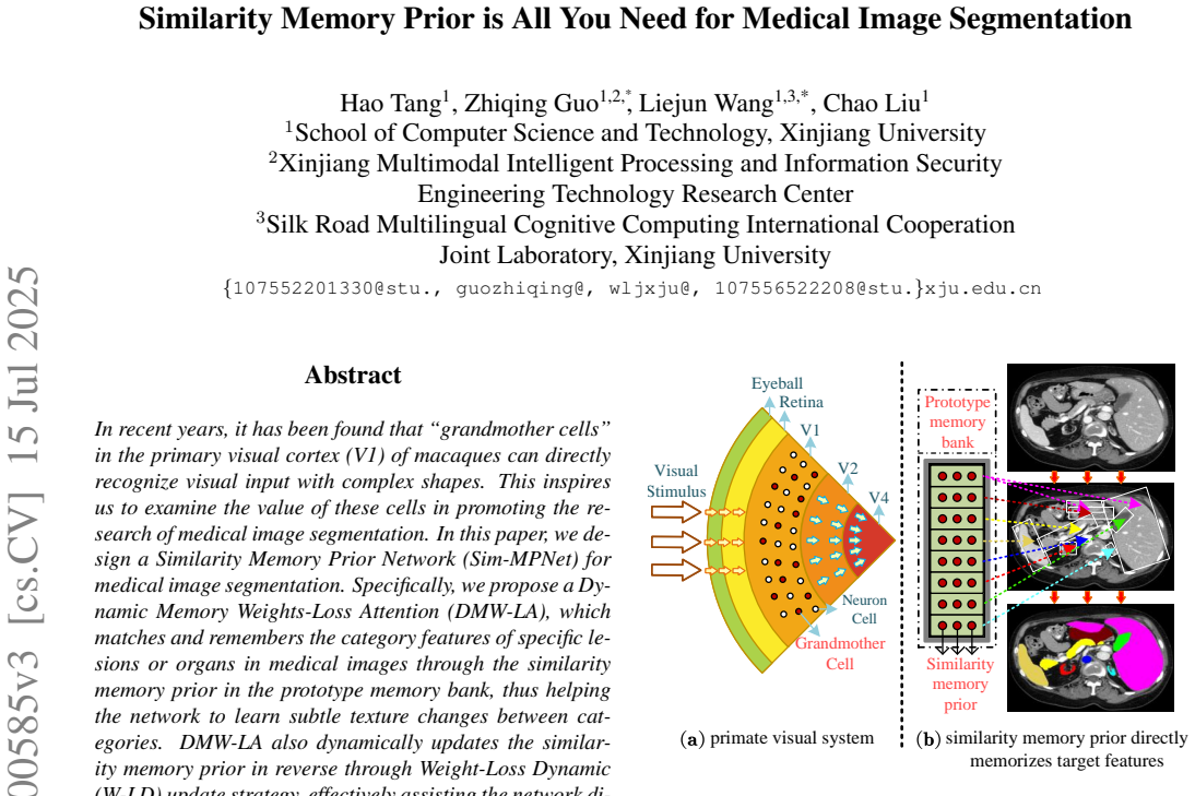
The load-bearing object is the prototype memory bank containing similarity memory priors, one cluster of prototype vectors per segmentation category, initialized by K-means on the global-interaction features. DMW-LA matches each query region to the nearest similarity core prior using cosine similarity, then runs self-attention with the matched prototype acting as both Key and Value, which is how the network "remembers" a category. The W-LD update strategy replaces the $K$ lowest-weight columns of a prototype with the $K$ highest-weight columns of matched current features, with $K$ modulated by the epoch-to-epoch loss difference, $K = (-\alpha(\mathrm{Loss}_\nabla - \mathrm{Loss}_\Delta)+\beta)\theta$, where $\alpha=\beta=0.5$ and $\frac{M}{4}\leq K\leq\frac{3M}{4}$. DS-GIM computes windowed cosine similarities to weight decay coefficients and then uses euclidean distances between spatial elements to split and re-attend to the similarity map, embedding distance information into the features.
What would settle it
Rerun Sim-MPNet and the strongest baselines on the same four datasets with both encoders at 224x224 and with, say, five training seeds per method; if the mean Dice gaps shrink to within run-to-run variation or reverse on any dataset, the claimed advantage is not established.
Extended reading notes
Core claim
The central claim is that explicitly memorizing category-level prototypes gives Sim-MPNet a direct route to the anatomical or pathological structures it must segment, and this route is what produces its accuracy gains. In the authors' telling, CNNs passively respond to local texture and ViTs model global texture statistics, but neither identifies the semantic object itself; the prototype memory bank plays the role of "grandmother cells" by responding selectively to specific organ or lesion categories. With the Weight-Loss Dynamic update rule, the bank replaces its least informative entries with the most informative current features, and the number of replaced entries is tied to whether the loss is falling. The Double-Similarity Global Internal Enhancement Module then refines the feature distribution internally, and a dual-encoder design with MaxViT supplies global context. Reported DSC values are 92.18 on ACDC, 83.12 on SegPC-2021, 90.85 on ISIC-2018, and 84.34 on Synapse, each above the compared baselines.
Load-bearing premise
The SOTA claim assumes the comparison is apples-to-apples: the same data splits, the same training budget, and no advantage from Sim-MPNet's second encoder running at 256x256 while the baselines use 224x224, with no variance or significance tests reported.
Editorial extensions
If this is right
- Prototype-memory category modeling can be inserted alongside CNNs and ViTs as a third modeling paradigm for medical images, since the dual-encoder ablation shows DMW-LA contributes more than the MaxViT encoder alone.
- The W-LD update rule lets the memory bank track training progress, which should keep prototypes informative in long training schedules; the paper reports no training collapse across 300 epochs.
- On small-organ and multi-cell datasets, the memory prior's direct category matching is claimed to reduce boundary errors, reflected in lower HD95 on ACDC, SegPC-2021, and ISIC-2018.
- Because only the number of K-means clusters changes between datasets, the method is claimed to generalize across MRI, microscopic, dermoscopic, and CT modalities without per-dataset hyperparameter tuning.
Reading between the lines
- If the memory-bank mechanism generalizes, a testable extension would be to replace unsupervised K-means initialization with class labels or weak supervision, which could sharpen the priors since categories are currently only implied by the number of clusters.
- The resolution asymmetry, Sim-MPNet's second encoder at 256x256 versus 224x224 for the baselines, is the most plausible alternative explanation for the reported gains, and a matched-resolution control experiment would settle whether the memory prior itself carries the improvement.
- The same similarity-memory design could be carried over to few-shot or continual medical segmentation, where a fixed category count is not available; the dynamic update rule would then need a mechanism to create and retire prototypes rather than only update a fixed set.
- The reported per-organ wins and losses, such as lower DSC on spleen, aorta, and liver than some baselines, suggest the method's benefit is category-dependent; if the memory prior is the cause, the largest gains should appear on categories with compact and repeatable appearances.
Editorial analysis
A structured set of objections, weighed in public.
Referee Report
Summary. The paper proposes Sim-MPNet, a dual-encoder medical image segmentation network. The first encoder is built from a new Dynamic Memory Weights-Loss Attention (DMW-LA) module that stores and updates a prototype memory bank of "similarity memory priors," inspired by grandmother cells in primate V1; the second encoder uses MaxViT. A Double-Similarity Global Internal Enhancement Module (DS-GIM) is also introduced. Experiments are reported on ACDC, SegPC-2021, ISIC-2018, and Synapse, and the paper claims that Sim-MPNet outperforms previous state-of-the-art methods on all four datasets.
Significance. If the claim is established, the memory-prior mechanism would be a useful new direction for category-aware medical image segmentation, and the paper has several strengths: it provides code, evaluates on four public datasets, reports both DSC and HD95, and includes ablations and visualizations. However, the central empirical claim is not yet supported because the comparison with baselines is confounded by input resolution and by the absence of variance or significance information. The ablations in Table 3 do not isolate the proposed module. The idea is interesting, but the evidence presented in the current manuscript is insufficient to justify the stated state-of-the-art conclusion.
major comments (3)
- [§4.2, Table 2 footnote] The central SOTA comparison is confounded by input resolution. Section 4.2 states that the two encoders use 224×224 and 256×256 inputs, and the Table 2 footnote confirms that Sim-MPNet's FLOPs are computed for a dual encoder at these two resolutions while all baselines use 224×224. The reported gains over the best baseline are between 0.70 and 2.40 DSC points, which are small enough that the higher input resolution of encoder 2 could explain part or all of the improvement. To support the claim that the similarity memory prior is the cause, please add matched-resolution baselines (for example, Sim-MPNet with both encoders at 224×224, or existing methods trained at 256×256) and show that the reported differences persist.
- [§4.3, Tables 1–2] No standard deviations, confidence intervals, or significance tests are reported for any of the four datasets. Margins such as 0.74 DSC on ISIC-2018 and 0.70 DSC on ACDC are within the range of run-to-run variability commonly observed in medical image segmentation. Please report results over at least three independent runs as mean±std, and include a paired significance test for the main comparisons and the ablations so that the reader can assess whether the improvements are reliable.
- [§4.4, Table 3] The ablation in Table 3 does not isolate the contribution of DMW-LA. The row "DMW-LA (Ours)" uses only encoder 1 at 224×224, the row "— MaxViT" uses only encoder 2 at 256×256, and the row "MaxViT MaxViT" uses both resolutions. Thus encoder type, input resolution, and parameter count are varied simultaneously, and the performance gap between the first two rows cannot be attributed to DMW-LA. Please add resolution-matched and, where possible, parameter-matched ablations.
minor comments (6)
- [Title] The running title in the full text contains a spacing error: "All Y ou Need" should read "All You Need".
- [§1] The Introduction cites "Figure 7(a)" and "Figure 7(b)" when referring to the primate visual system and the prototype memory bank, but Figure 7 appears only in the supplementary material. The first references should point to the main-text figure (Figure 1) or the figures should be renumbered.
- [§3.2] The set notation for the similarity memory prior and the similarity core prior is malformed (e.g., double braces and a stray "then ... o"); please rewrite these definitions in standard set notation.
- [§3.2, Eq. (5)] K is an integer number of columns, but the right-hand side of Eq. (5) is not guaranteed to be an integer; please specify the rounding rule and provide a justification or sensitivity analysis for the hand-set values α=0.5, β=0.5, and the bounds M/4 and 3M/4.
- [§3.3, Eq. (6)] The decay-coefficient formula in Eq. (6) is difficult to parse: the expression "2−2.5− 5·n/l" is ambiguous and the constants 0.25 and 2.5 are not motivated. Please clarify the notation and explain how these constants were chosen.
- [Table 2] The inference time column Ti is reported without standard deviations and without a description of the hardware/software measurement protocol beyond the GPU name; please add those details or remove the column.
Circularity Check
No significant circularity: the paper's performance claims are empirical comparisons on held-out test sets, and the memory-prior module is learned end-to-end rather than derived from the outcome it predicts.
full rationale
The paper's central claim is an empirical one: Sim-MPNet achieves higher DSC and lower HD95 than prior methods on four public datasets (Tables 1-2). No step in the method section derives a prediction from a parameter that was fitted to that same prediction. The prototype memory bank is initialized by K-means on the network's own features and then updated with the W-LD rule; this is a trainable component evaluated on held-out test data, so the test scores are not equal by construction to any training signal. The ablations in Tables 3-4 vary the presence of DMW-LA, DS-GIM, and W-LD and report independent test-set DSCs; they do not rename the fitted components as outcomes. The only self-citation, Ref. [42] (TransUNet++), is used solely to state that the ACDC train/test split is consistent with that work; the claim of better segmentation performance does not rest on that citation. The Table 2 footnote shows a resolution mismatch (Sim-MPNet's second encoder uses 256x256 while baselines use 224x224), and no variance or significance tests are reported; those are comparison confounds that bear on the strength of the SOTA claim, but they do not make the result circular, because the reported DSC values are not constructed from the inputs of the comparison. Overall, the empirical claim is self-contained and falsifiable, so the circularity score is 0.
Assumptions & free parameters
free parameters (6)
- alpha (W-LD decay factor) =
0.5
- beta (W-LD offset) =
0.5
- K bounds for memory update =
M/4 to 3M/4, initial M/2
- Loss weights =
0.7 Dice + 0.3 CE
- DS-GIM decay constants =
0.25, 2.5, 5 in Eq. (6)
- Encoder 2 input resolution =
256x256
assumptions (4)
- domain assumption Cosine similarity between query and prototype core prior correctly matches category features
- standard math K-means clustering provides a good initialization for the memory bank
- ad hoc to paper Replacing low-weight memory columns with high-weight input columns improves memory quality
- domain assumption The 'grandmother cell' analogy: V1 neurons directly recognize complex shapes, motivating prototype priors
invented entities (1)
-
similarity memory prior
Cite this review
Pith. "Pith review of Similarity Memory Prior is All You Need for Medical Image Segmentation." pith.science (2026). https://pith.science/paper/G443HPHJ
@misc{pith2026250700585,
  author       = {Pith},
  title        = {Pith review of: Similarity Memory Prior is All You Need for Medical Image Segmentation},
  year         = {2026},
  howpublished = {\url{https://pith.science/paper/G443HPHJ}},
  note         = {Machine review of arXiv:2507.00585}
}
read the original abstract
In recent years, it has been found that "grandmother cells" in the primary visual cortex (V1) of macaques can directly recognize visual input with complex shapes. This inspires us to examine the value of these cells in promoting the research of medical image segmentation. In this paper, we design a Similarity Memory Prior Network (Sim-MPNet) for medical image segmentation. Specifically, we propose a Dynamic Memory Weights-Loss Attention (DMW-LA), which matches and remembers the category features of specific lesions or organs in medical images through the similarity memory prior in the prototype memory bank, thus helping the network to learn subtle texture changes between categories. DMW-LA also dynamically updates the similarity memory prior in reverse through Weight-Loss Dynamic (W-LD) update strategy, effectively assisting the network directly extract category features. In addition, we propose the Double-Similarity Global Internal Enhancement Module (DS-GIM) to deeply explore the internal differences in the feature distribution of input data through cosine similarity and euclidean distance. Extensive experiments on four public datasets show that Sim-MPNet has better segmentation performance than other state-of-the-art methods. Our code is available on https://github.com/vpsg-research/Sim-MPNet.
Figures


Figures from the paper (4 more)


Reference graph
Works this paper leans on
-
[1]
Transdeeplab: Convolution-free transformer- based deeplab v3+ for medical image segmentation
Reza Azad, Moein Heidari, Moein Shariatnia, Ehsan Khoda- panah Aghdam, Sanaz Karimijafarbigloo, Ehsan Adeli, and Dorit Merhof. Transdeeplab: Convolution-free transformer- based deeplab v3+ for medical image segmentation. In Int. Workshop PRedict. Intell. Med. , pages 91–102. Springer,
-
[2]
Dae-former: Dual attention-guided efficient transformer for medical im- age segmentation
Reza Azad, Ren ´e Arimond, Ehsan Khodapanah Aghdam, Amirhossein Kazerouni, and Dorit Merhof. Dae-former: Dual attention-guided efficient transformer for medical im- age segmentation. In In Int. Workshop PRedict. Intell. Med., pages 83–95. Springer, 2023. 3, 6
work page 2023
-
[3]
Medical image segmentation review: The suc- cess of u-net
Reza Azad, Ehsan Khodapanah Aghdam, Amelie Rauland, Yiwei Jia, Atlas Haddadi Avval, Afshin Bozorgpour, Sanaz Karimijafarbigloo, Joseph Paul Cohen, Ehsan Adeli, and Dorit Merhof. Medical image segmentation review: The suc- cess of u-net. IEEE Trans. Pattern Anal. Mach. Intell., 2024. 1
work page 2024
-
[4]
Olivier Bernard, Alain Lalande, Clement Zotti, Freder- ick Cervenansky, Xin Yang, Pheng-Ann Heng, Irem Cetin, Karim Lekadir, Oscar Camara, Miguel Angel Gonzalez Ballester, et al. Deep learning techniques for automatic mri cardiac multi-structures segmentation and diagnosis: is the problem solved? IEEE Trans. Med. Imaging, 37(11):2514– 2525, 2018. 5
work page 2018
-
[5]
Fares Bougourzi, Fadi Dornaika, Abdelmalik Taleb-Ahmed, and Vinh Truong Hoang. Rethinking attention gated with hy- brid dual pyramid transformer-cnn for generalized segmen- tation in medical imaging. In International Conference on Pattern Recognition, pages 243–258. Springer, 2024. 6
work page 2024
-
[6]
Swin-unet: Unet-like pure transformer for medical image segmentation
Hu Cao, Yueyue Wang, Joy Chen, Dongsheng Jiang, Xi- aopeng Zhang, Qi Tian, and Manning Wang. Swin-unet: Unet-like pure transformer for medical image segmentation. In ECCV, pages 205–218. Springer, 2022. 1, 6
work page 2022
-
[7]
Unsupervised learning of visual features by contrasting cluster assignments
Mathilde Caron, Ishan Misra, Julien Mairal, Priya Goyal, Pi- otr Bojanowski, and Armand Joulin. Unsupervised learning of visual features by contrasting cluster assignments. Proc. Adv. Neural Inf. Process. Syst., 33:9912–9924, 2020. 3
work page 2020
-
[8]
Transunet: Transformers make strong encoders for medi- cal image segmentation
Jieneng Chen, Yongyi Lu, Qihang Yu, Xiangde Luo, Ehsan Adeli, Yan Wang, Le Lu, Alan L Yuille, and Yuyin Zhou. Transunet: Transformers make strong encoders for medi- cal image segmentation. arXiv preprint arXiv:2102.04306,
Show all 51 references
-
[9]
Drinet for medical image segmentation
Liang Chen, Paul Bentley, Kensaku Mori, Kazunari Misawa, Michitaka Fujiwara, and Daniel Rueckert. Drinet for medical image segmentation. IEEE transactions on medical imaging, 37(11):2453–2462, 2018. 1
2018
-
[10]
Skin lesion analysis toward melanoma detection 2018: A challenge hosted by the interna- tional skin imaging collaboration (isic)
Noel Codella, Veronica Rotemberg, Philipp Tschandl, M Emre Celebi, Stephen Dusza, David Gutman, Brian Helba, Aadi Kalloo, Konstantinos Liopyris, Michael Marchetti, et al. Skin lesion analysis toward melanoma detection 2018: A challenge hosted by the interna- tional skin imagin...
2018 arXiv
-
[11]
A review of convolutional neural network architectures and their optimizations
Shuang Cong and Yang Zhou. A review of convolutional neural network architectures and their optimizations. Artif. Intell. Rev., 56(3):1905–1969, 2023. 2
1905
-
[12]
An image is worth 16x16 words: Trans- formers for image recognition at scale
Alexey Dosovitskiy, Lucas Beyer, Alexander Kolesnikov, Dirk Weissenborn, Xiaohua Zhai, Thomas Unterthiner, Mostafa Dehghani, Matthias Minderer, Georg Heigold, Syl- vain Gelly, et al. An image is worth 16x16 words: Trans- formers for image recognition at scale. arXiv preprint a...
2010 arXiv
-
[13]
Ednfc-net: Convolutional neural network with nested feature concate- nation for nuclei-instance segmentation
Shiv Gehlot, Anubha Gupta, and Ritu Gupta. Ednfc-net: Convolutional neural network with nested feature concate- nation for nuclei-instance segmentation. In ICASSP, pages 1389–1393. IEEE, 2020. 5
2020
-
[14]
grandmother cell
Charles G Gross. Genealogy of the “grandmother cell”. Neu- roscientist., 8(5):512–518, 2002. 2
2002
-
[15]
Recent advances in con- volutional neural networks
Jiuxiang Gu, Zhenhua Wang, Jason Kuen, Lianyang Ma, Amir Shahroudy, Bing Shuai, Ting Liu, Xingxing Wang, Gang Wang, Jianfei Cai, et al. Recent advances in con- volutional neural networks. Pattern Recognit., 77:354–377,
-
[16]
Pcseg: Color model driven probabilistic multiphase level set based tool for plasma cell segmentation in multiple myeloma
Anubha Gupta, Pramit Mallick, Ojaswa Sharma, Ritu Gupta, and Rahul Duggal. Pcseg: Color model driven probabilistic multiphase level set based tool for plasma cell segmentation in multiple myeloma. PloS one, 13(12):e0207908, 2018. 5
2018
-
[17]
Gcti-sn: Geometry-inspired chemical and tissue invariant stain normalization of microscopic medical images
Anubha Gupta, Rahul Duggal, Shiv Gehlot, Ritu Gupta, An- vit Mangal, Lalit Kumar, Nisarg Thakkar, and Devprakash Satpathy. Gcti-sn: Geometry-inspired chemical and tissue invariant stain normalization of microscopic medical images. Med. Image Anal., 65:101788, 2020. 5
2020
-
[18]
Hiformer: Hierarchical multi-scale representations using transformers for medical image seg- mentation
Moein Heidari, Amirhossein Kazerouni, Milad Soltany, Reza Azad, Ehsan Khodapanah Aghdam, Julien Cohen- Adad, and Dorit Merhof. Hiformer: Hierarchical multi-scale representations using transformers for medical image seg- mentation. In WACV, pages 6202–6212, 2023. 6
2023
-
[19]
Deep learning techniques for medical im- age segmentation: achievements and challenges
Mohammad Hesam Hesamian, Wenjing Jia, Xiangjian He, and Paul Kennedy. Deep learning techniques for medical im- age segmentation: achievements and challenges. J. Digital Imaging, 32:582–596, 2019. 1
2019
-
[20]
Unet 3+: A full-scale connected unet for medical image segmentation
Huimin Huang, Lanfen Lin, Ruofeng Tong, Hongjie Hu, Qiaowei Zhang, Yutaro Iwamoto, Xianhua Han, Yen-Wei Chen, and Jian Wu. Unet 3+: A full-scale connected unet for medical image segmentation. In ICASSP, pages 1055–
-
[21]
Boundary-rendering network for breast le- sion segmentation in ultrasound images
Ruobing Huang, Mingrong Lin, Haoran Dou, Zehui Lin, Qi- long Ying, Xiaohong Jia, Wenwen Xu, Zihan Mei, Xin Yang, Yijie Dong, et al. Boundary-rendering network for breast le- sion segmentation in ultrasound images. Med. Image Anal., 80:102478, 2022. 1
2022
-
[22]
nnu-net: a self-configuring method for deep learning-based biomedical image segmen- tation
Fabian Isensee, Paul F Jaeger, Simon AA Kohl, Jens Pe- tersen, and Klaus H Maier-Hein. nnu-net: a self-configuring method for deep learning-based biomedical image segmen- tation. Nat. Methods, 18(2):203–211, 2021. 3
2021
-
[23]
Prototype memory and attention mechanisms for few shot image generation
Tianqin Li, Zijie Li, Harold Rockwell, Amir Farimani, and Tai Sing Lee. Prototype memory and attention mechanisms for few shot image generation. In ICLR, 2022. 3
2022
-
[24]
Modality-agnostic domain generalizable medical image segmentation by multi-frequency in multi- scale attention
Ju-Hyeon Nam, Nur Suriza Syazwany, Su Jung Kim, and Sang-Chul Lee. Modality-agnostic domain generalizable medical image segmentation by multi-frequency in multi- scale attention. In Proceedings of the IEEE/CVF conference on computer vision and pattern recognition , pages 11480...
2024
-
[25]
Atten- tion u-net: Learning where to look for the pancreas
Ozan Oktay, Jo Schlemper, Loic Le Folgoc, Matthew Lee, Mattias Heinrich, Kazunari Misawa, Kensaku Mori, Steven McDonagh, Nils Y Hammerla, Bernhard Kainz, et al. Atten- tion u-net: Learning where to look for the pancreas. arXiv preprint arXiv:1804.03999, 2018. 6
2018 arXiv
-
[26]
Brain cells for grandmother
Rodrigo Quian Quiroga, Itzhak Fried, and Christof Koch. Brain cells for grandmother. Sci. Am., 308(2):30–35, 2013. 2
2013
-
[27]
Medical im- age segmentation via cascaded attention decoding
Md Mostafijur Rahman and Radu Marculescu. Medical im- age segmentation via cascaded attention decoding. InWACV, pages 6222–6231, 2023. 6
2023
-
[28]
Emcad: Efficient multi-scale convolutional atten- tion decoding for medical image segmentation
Md Mostafijur Rahman, Mustafa Munir, and Radu Mar- culescu. Emcad: Efficient multi-scale convolutional atten- tion decoding for medical image segmentation. In WACV, pages 11769–11779, 2024. 6
2024
-
[29]
A review of medical image segmen- tation algorithms
KKD Ramesh, G Kiran Kumar, K Swapna, Debabrata Datta, and S Suman Rajest. A review of medical image segmen- tation algorithms. EAI Endorsed Transactions on Pervasive Health & Technology, 7(27), 2021. 1
2021
-
[30]
U-net: Convolutional networks for biomedical image segmentation
Olaf Ronneberger, Philipp Fischer, and Thomas Brox. U-net: Convolutional networks for biomedical image segmentation. In MICCAI, pages 234–241. Springer, 2015. 1, 2, 6
2015
-
[31]
Meta-learning with memory-augmented neural networks
Adam Santoro, Sergey Bartunov, Matthew Botvinick, Daan Wierstra, and Timothy Lillicrap. Meta-learning with memory-augmented neural networks. In ICML, pages 1842–
-
[32]
From stem cells to grandmother cells: how neurogenesis relates to learning and memory.Cell Stem Cell, 3(3):253–258, 2008
Tracey J Shors. From stem cells to grandmother cells: how neurogenesis relates to learning and memory.Cell Stem Cell, 3(3):253–258, 2008. 2
2008
-
[33]
Complex pattern selectivity in macaque primary visual cortex revealed by large-scale two-photon imaging.Curr
Shiming Tang, Tai Sing Lee, Ming Li, Yimeng Zhang, Yue Xu, Fang Liu, Benjamin Teo, and Hongfei Jiang. Complex pattern selectivity in macaque primary visual cortex revealed by large-scale two-photon imaging.Curr. Biol., 28(1):38–48,
-
[34]
Large-scale two- photon imaging revealed super-sparse population codes in the v1 superficial layer of awake monkeys.ELIFE, 7:e33370,
Shiming Tang, Yimeng Zhang, Zhihao Li, Ming Li, Fang Liu, Hongfei Jiang, and Tai Sing Lee. Large-scale two- photon imaging revealed super-sparse population codes in the v1 superficial layer of awake monkeys.ELIFE, 7:e33370,
-
[35]
Maxvit: Multi-axis vision transformer
Zhengzhong Tu, Hossein Talebi, Han Zhang, Feng Yang, Peyman Milanfar, Alan Bovik, and Yinxiao Li. Maxvit: Multi-axis vision transformer. In ECCV, pages 459–479. Springer, 2022. 2, 3
2022
-
[36]
Medical transformer: Gated axial- attention for medical image segmentation
Jeya Maria Jose Valanarasu, Poojan Oza, Ilker Hacihaliloglu, and Vishal M Patel. Medical transformer: Gated axial- attention for medical image segmentation. InMICCAI, pages 36–46. Springer, 2021. 3
2021
-
[37]
Uctransnet: rethinking the skip connections in u-net from a channel-wise perspective with transformer
Haonan Wang, Peng Cao, Jiaqi Wang, and Osmar R Zaiane. Uctransnet: rethinking the skip connections in u-net from a channel-wise perspective with transformer. In AAAI, pages 2441–2449, 2022. 3
2022
-
[38]
Mixed transformer u-net for medical image segmentation
Hongyi Wang, Shiao Xie, Lanfen Lin, Yutaro Iwamoto, Xian-Hua Han, Yen-Wei Chen, and Ruofeng Tong. Mixed transformer u-net for medical image segmentation. In ICASSP, pages 2390–2394. IEEE, 2022. 3, 6
2022
-
[39]
Dtmformer: Dynamic token merging for boosting transformer-based medical image seg- mentation
Zhehao Wang, Xian Lin, Nannan Wu, Li Yu, Kwang-Ting Cheng, and Zengqiang Yan. Dtmformer: Dynamic token merging for boosting transformer-based medical image seg- mentation. In AAAI, pages 5814–5822, 2024. 3
2024
-
[40]
Cbam: Convolutional block attention module
Sanghyun Woo, Jongchan Park, Joon-Young Lee, and In So Kweon. Cbam: Convolutional block attention module. In ECCV, pages 3–19, 2018. 3
2018
-
[41]
Transformers in medical image segmentation: A review
Hanguang Xiao, Li Li, Qiyuan Liu, Xiuhong Zhu, and Qi- hang Zhang. Transformers in medical image segmentation: A review. Biomed. Signal Process. Control , 84:104791,
-
[42]
Big model and small model: Remote modeling and local infor- mation extraction module for medical image segmentation
Lianghui Xu, Liejun Wang, Yongming Li, and Anyu Du. Big model and small model: Remote modeling and local infor- mation extraction module for medical image segmentation. Appl. Soft Comput., 136:110128, 2023. 5
2023
-
[43]
Global guidance network for breast lesion segmentation in ultrasound images
Cheng Xue, Lei Zhu, Huazhu Fu, Xiaowei Hu, Xiaomeng Li, Hai Zhang, and Pheng-Ann Heng. Global guidance network for breast lesion segmentation in ultrasound images. Med. Image Anal., 70:101989, 2021. 1
2021
-
[44]
Learning dynamic mem- ory networks for object tracking
Tianyu Yang and Antoni B Chan. Learning dynamic mem- ory networks for object tracking. In ECCV, pages 152–167,
-
[45]
Directional connectivity- based segmentation of medical images
Ziyun Yang and Sina Farsiu. Directional connectivity- based segmentation of medical images. In Proceedings of the IEEE/CVF conference on computer vision and pattern recognition, pages 11525–11535, 2023. 6
2023
-
[46]
Transclaw u-net: claw u-net with trans- formers for medical image segmentation
Chang Yao, Menghan Hu, Qingli Li, Guangtao Zhai, and Xiao-Ping Zhang. Transclaw u-net: claw u-net with trans- formers for medical image segmentation. In ICASSP, pages 280–284. IEEE, 2022. 6
2022
-
[47]
Transfuse: Fus- ing transformers and cnns for medical image segmentation
Yundong Zhang, Huiye Liu, and Qiang Hu. Transfuse: Fus- ing transformers and cnns for medical image segmentation. In MICCAI, pages 14–24. Springer, 2021. 3
2021
-
[48]
Unet++: A nested u-net ar- chitecture for medical image segmentation
Zongwei Zhou, Md Mahfuzur Rahman Siddiquee, Nima Tajbakhsh, and Jianming Liang. Unet++: A nested u-net ar- chitecture for medical image segmentation. In Proc. Deep Learn. Med. Image Anal. Multimodal Learn. Clin. Decis Support., pages 3–11. Springer, 2018. 1, 3, 6
2018
-
[49]
Selfreg- unet: Self-regularized unet for medical image segmenta- tion
Wenhui Zhu, Xiwen Chen, Peijie Qiu, Mohammad Farazi, Aristeidis Sotiras, Abolfazl Razi, and Yalin Wang. Selfreg- unet: Self-regularized unet for medical image segmenta- tion. In International Conference on Medical Image Com- puting and Computer-Assisted Intervention, pages 601...
2024
-
[50]
Physics-driven deep learning methods for fast quantitative magnetic resonance imaging: Performance improvements through integration with deep neural networks
Yanjie Zhu, Jing Cheng, Zhuo-Xu Cui, Qingyong Zhu, Leslie Ying, and Dong Liang. Physics-driven deep learning methods for fast quantitative magnetic resonance imaging: Performance improvements through integration with deep neural networks. IEEE Signal Process Mag., 40(2):116–128,
-
[2023]
1 Similarity Memory Prior is All Y ou Need for Medical Image Segmentation Supplementary Material A. Setting about K in W-LD Update Strategy To accurately locate the category features of the input im- age, we propose the W-LD update strategy to dynamically update the prototype ...
2021
Reviewed August 6, 2026 · model on record in the stance chip above.
Discussion (0). Sign in to comment.