REVIEW 4 major objections 4 minor 26 references
Semantically Consistent Discrete Diffusion for 3D Biological Graph Modeling
T0 review · 4 major / 4 minor · reviewed 2026-08-06 · deepseek-v4-flash
Pith's one-line read This paper uses a stochastic label-consistent projector during discrete diffusion sampling to enforce semantic edge-label constraints, yielding 100% valid circle-of-Willis and airway graphs and better downstream vessel labeling.
desk verdict Stochastic label-consistent projection is a real extension, but the headline downstream win is confounded by training-set size and the semantic validity metric is true by construction. read the letter →
The pith
A machine-rendered reading of the paper's core claim, the machinery that carries it, and where it could break.
The reading
What carries the argument
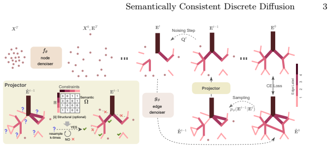
The central object is the semantic-consistency projector, a sampling-time module that checks candidate edges against an edge-label incompatibility matrix $\Omega \in \{0,1\}^{c\times c}$, where $\Omega[c_i,c_j]=1$ means edge types $c_i$ and $c_j$ cannot be adjacent around the same node. The projector works with an edge-deletion noising transition $Q_t = \alpha_t^e I + (1-\alpha_t^e)\mathbf{1} e_E^\top$, which treats "no edge" as an absorbing state so reverse sampling only adds edges. When the edge denoiser proposes an edge whose label is incompatible, the projector resamples that edge label up to $k$ times from the posterior $p_\phi(E^{t-1}_{ij}\mid E^t_{ij})$ and accepts the edge if any resampled label is compatible; otherwise it deletes the edge. This stochastic repair is what lets the method enforce semantics without the oversparsity caused by hard deletion.
What would settle it
Take a dataset with an independent expert-written set of forbidden vessel or airway label adjacencies that was not used to build the constraint matrix, generate a large batch of graphs, and check whether any generated graph contains a forbidden adjacency. One such graph would show the validity guarantee is only as good as the hand-written constraint list.
Extended reading notes
Core claim
The paper claims that anatomical validity in generated 3D biological graphs can be enforced as a byproduct of sampling rather than by post-processing. It achieves this by showing that semantic edge-label consistency is invariant under an edge-deletion noising process, so a stochastic projector can be applied at every reverse-diffusion step. The outcome on the circle-of-Willis and airway datasets is 100% semantically valid generated graphs with distribution statistics closer to real data than the uniform-noising baseline; on the circle of Willis, synthetic graphs used as training data push vessel-labeling balanced accuracy to 95.45 versus 93.77 for real data; and on airway trees the same model predicts missing links at 84.97 balanced accuracy while keeping all predictions valid. The paper also states its guarantee is limited to edge-deletion invariants, so properties such as a single connected component are not enforced.
Load-bearing premise
The method assumes the hand-built list of forbidden label pairs completely captures anatomical plausibility and that checking each node's local neighbors is enough, so a missing or wrongly included pair would make "valid" graphs anatomically wrong or artificially sparse.
Editorial extensions
If this is right
- All generated samples satisfy the semantic-consistency criterion defined by $\Omega$ (100% on both datasets), so downstream users can skip rejection sampling.
- Synthetic circle-of-Willis graphs train a vessel labeler that outperforms one trained on real data (95.45 vs 93.77 balanced accuracy, avg F1 0.967 vs 0.946).
- The same model can be used for link prediction on airway graphs with roughly 30% missing edges, reaching 84.97 balanced accuracy while keeping 100% semantic validity.
- Edge-deletion noising is better suited to sparse biological graphs than uniform-label noising, because early noising steps do not inject implausible long-range edges.
Reading between the lines
- Editorial extension: because the projector is applied at sampling time and does not depend on a specific architecture, the same stochastic-repair idea could be ported to other discrete diffusion models with pairwise compatibility constraints, such as molecular or material graphs.
- Editorial extension: the validity guarantee is only as strong as the hand-defined $\Omega$; learning this matrix from anatomical data or clinical ontologies would generalize the method to constraints the authors did not encode.
- Editorial extension: the paper's stated limitation that node coordinates are frozen during edge denoising suggests a testable extension where coordinates and edges are denoised jointly or alternately, which could improve anatomical fidelity further.
- Editorial extension: the reported low intervention rate (about 2% of generated edges) suggests the projector acts as a targeted safety valve rather than a wholesale override, which may explain why distribution statistics stay close to real data.
Editorial analysis
A structured set of objections, weighed in public.
Referee Report
Summary. The paper proposes a discrete diffusion model for generating 3D biological graphs, specifically circle of Willis vessel graphs and airway trees. The method has two stages: a point-cloud DDPM generates node coordinates, and an edge denoiser with an edge-deletion (absorbing-state) noising process generates edge labels. During sampling, a novel 'label-consistent projector' checks newly sampled edges against a manually defined edge-label adjacency incompatibility matrix Ω (Eq. 1) and, when a violation occurs, resamples the edge label up to k times before rejecting the edge. The authors report 100% semantic validity on both datasets, improved distribution statistics relative to baselines, and downstream gains in vessel labeling and link prediction.
Significance. If validated, the paper would make a useful contribution: it adapts edge-deletion discrete diffusion to sparse biological graphs, and it extends the hard-projection idea of Madeira et al. [9] to semantic edge-label constraints via stochastic resampling. The code is promised publicly, and the downstream applications (vessel labeling, link prediction) are practically relevant. However, the current evidence is weakened by the fact that the headline semantic-validity metric is the same predicate the projector enforces, and by a downstream comparison that confounds training-set size with data source. The central method is plausible and likely fixable, but several load-bearing evaluation points need to be reworked before the paper's claims are supported.
major comments (4)
- [Section 3, Eq. (1); Section 4, Tables 1 and 3] The semantic validity metric S.V. is defined by exactly the predicate P_Ω(E) in Eq. (1), and the projector is designed to enforce P_Ω at every denoising step. Consequently, the reported 100% S.V. scores in Tables 1 and 3 are guaranteed by construction and do not provide independent empirical evidence that the generated graphs are anatomically valid. The paper should either (a) report an independent anatomical validity measure, e.g., expert review or a held-out constraint set, or (b) explicitly frame the 100% S.V. as a correctness check of the projector rather than an empirical result.
- [Section 4, 'Downstream Tasks', Table 2] The claim that synthetic samples outperform real data for vessel labeling is confounded by training-set size: the synthetic condition trains on 1000 generated graphs while the real-data baseline trains on a 50-graph train split. A 20-fold difference in training volume could explain the 1.7-point balanced-accuracy gap even if the generated graphs are no more realistic than the real ones. The paper needs a matched comparison, e.g., training the real-data baseline on the same number of graphs (via augmentation or repeated sampling) or training all methods on equal-size synthetic corpora, and it should report error bars over multiple seeds.
- [Section 3, 'Semantically Consistent Projection'] The text states that the projector 'allows us to sample from the posterior distribution constrained by our semantic plausibility,' but this is not established. The k-resampling scheme is a heuristic: it samples a label from the model's posterior, checks P_Ω, and accepts the first valid label, which does not generally equal conditioning on the validity event. The theoretical result from [9] is about hard projection for invariant properties, not about stochastic resampling. The authors should either provide a correctness argument for the resampling distribution or soften the claim to say that the projector biases samples toward valid configurations in an empirically tested way.
- [Section 4, 'Semantic Validity (S.V.)' and Eq. (1)] The Ω matrix is manually constructed from the TopCoW annotation protocol, and the same protocol is used to define edge labels and the validity criterion. There is no evidence that Ω is complete (no omitted forbidden adjacencies) or not over-restrictive. If Ω is incomplete, the projector could certify graphs that are anatomically implausible; if it is over-restrictive, it could force unrealistic sparsity. The paper should validate Ω against independent anatomical knowledge or at least report sensitivity to its construction, e.g., by comparing S.V. under an expert-reviewed subset of Ω.
minor comments (4)
- [Abstract and Introduction] The abstract contains a typo, 'link predictior'; the Introduction also repeats this in the contributions list. Please correct.
- [Section 4, 'Datasets'] The sentence describing CROWN node counts says 'between 13 and 27 in CoW (average 8)', which is numerically inconsistent; likely the average should be near 18 or the range is a typo. Please clarify.
- [Section 4, 'Metrics'] There is an apparent typo 'A TM' for 'ATM' in the semantic validity paragraph. Please fix.
- [Section 3, Eq. (1)] Equation (1) is notationally dense and hard to parse; the tensor product over edges at a node would benefit from an explicit example or a rephrased definition, e.g., in terms of the number of pairs of incident edges whose labels are marked incompatible.
Circularity Check
The 100% semantic-validity result is entailed by the projector, and the validity standard is inherited from the authors' own TopCoW protocol; downstream labeling/link-prediction results are independent evidence.
-
self definitional
[Section 3 'Semantically Consistent Projection' and Section 4 'Semantic Validity (S.V.)']
"The projector begins with Et−1 ← Et and adds edges in Et−1 that satisfy the semantic-consistency criteria. Specifically, for each edge eij ∈ ˆEt−1 \ Et our projector adds Et−1 ← Et−1∪{eij} if PΩ(Et−1∪{eij}) = True. If not, we sample k new edge labels from eij ∼ pϕ(Et−1|Et). ... The projector accepts the edge if any of the k resampled edge labels satisfy the semantic consistency; otherwise, it rejects the edge."
The semantic-validity metric is exactly the predicate PΩ(E)=True that the projector uses as its acceptance test. Every edge that would make PΩ false is either relabeled until consistent or rejected before the final graph is emitted. Therefore the 'S.V.% 100' entries for Ours and Ours(k=0) in Table 1 are logically forced by the sampling procedure, not empirically discovered. The non-tautological content of Table 1 is restricted to the KL distribution statistics, which are measured against the real data independently of the projector's own constraint check.
-
self citation load bearing
[Section 4, 'Semantic Validity (S.V.)' and reference [24] (TopCoW)]
"Semantic Validity (S.V.): We use edge-label based criterion for semantic validity. CoW: The CoW graph has no structural constraints, and the TopCoW annotation protocol [24] determines its validity, please refer [24] for details. For CoW, we have 13x13 Ω matrix for 13 edge-labels."
The authority for anatomical validity is delegated entirely to the TopCoW annotation protocol [24], whose author list overlaps with the present paper (Li, Menze, and likely other co-authors). The Ω matrix that defines 'valid' in Eq. (1) is exactly the constraint set enforced by the projector, so the reported semantic validity measures agreement with the authors' own protocol rather than with an independent anatomical standard. This self-citation chain is load-bearing for the 'anatomically plausible graphs' claim, though softened by TopCoW being a public multi-author challenge dataset.
full rationale
The paper's core methodological claim, that stochastic projection enforces semantic consistency, is true by construction: the projector only emits graphs satisfying PΩ, and the S.V. metric is PΩ, making 100% S.V. a definitional outcome rather than a prediction. The Ω standard itself is inherited from the authors' own TopCoW protocol, a mild self-citation that further weakens the independence of the validity claim. However, the paper does contain substantial independent evaluation: KL distribution statistics are compared with real data, and the downstream vessel labeling and airway link-prediction tasks use external ground-truth labels and held-out TopCoW/ATM samples. Table 2's 'synthetic beats real' comparison is statistically confounded (1000 synthetic training graphs vs. 50 real graphs, no error bars), but a confound is not a circularity; the downstream result is not equivalent to the projector's input. On balance, the semantic-validity headline is circular to a moderate degree, while the utility claims have independent content, supporting a score of 4 rather than a higher score.
Assumptions & free parameters
free parameters (3)
- k (resampling attempts) =
4
- Omega constraint matrix =
13x13 for CoW, 4x4 for ATM
- Link prediction denoising steps =
100
assumptions (5)
- standard math Standard DDPM and discrete diffusion Markov-chain assumptions hold for coordinate and edge noising.
- standard math Edge-deletion noising preserves the P_Omega constraint because it only removes edges and never changes labels.
- domain assumption The manually defined Omega matrix correctly and completely characterizes anatomical plausibility for both datasets.
- domain assumption Training graphs satisfy the Omega constraint, so the denoiser learns to propose consistent edges.
- domain assumption The graph transformer edge denoiser, trained on fixed node coordinates, assigns meaningful posterior probabilities even when coordinates come from a separately trained point cloud generator.
Cite this review
Pith. "Pith review of Semantically Consistent Discrete Diffusion for 3D Biological Graph Modeling." pith.science (2026). https://pith.science/paper/66KAPZLO
@misc{pith2026250704856,
  author       = {Pith},
  title        = {Pith review of: Semantically Consistent Discrete Diffusion for 3D Biological Graph Modeling},
  year         = {2026},
  howpublished = {\url{https://pith.science/paper/66KAPZLO}},
  note         = {Machine review of arXiv:2507.04856}
}
read the original abstract
3D spatial graphs play a crucial role in biological and clinical research by modeling anatomical networks such as blood vessels,neurons, and airways. However, generating 3D biological graphs while maintaining anatomical validity remains challenging, a key limitation of existing diffusion-based methods. In this work, we propose a novel 3D biological graph generation method that adheres to structural and semantic plausibility conditions. We achieve this by using a novel projection operator during sampling that stochastically fixes inconsistencies. Further, we adopt a superior edge-deletion-based noising procedure suitable for sparse biological graphs. Our method demonstrates superior performance on two real-world datasets, human circle of Willis and lung airways, compared to previous approaches. Importantly, we demonstrate that the generated samples significantly enhance downstream graph labeling performance. Furthermore, we show that our generative model is a reasonable out-of-the-box link predictior.
Figures


Reference graph
Works this paper leans on
-
[9]
Advances in Neural Information Processing Systems 37, 137218–137262 (2025)
Madeira, M., Vignac, C., Thanou, D., Frossard, P.: Generative modelling of struc- turally constrained graphs. Advances in Neural Information Processing Systems 37, 137218–137262 (2025)
work page 2025
-
[1]
Advances in Neural Information Processing Systems 34, 17981–17993 (2021)
Austin, J., Johnson, D.D., Ho, J., Tarlow, D., Van Den Berg, R.: Structured de- noising diffusion models in discrete state-spaces. Advances in Neural Information Processing Systems 34, 17981–17993 (2021)
work page 2021
-
[2]
BMC bioinformatics22(1), 1–28 (2021)
Drees, D., Scherzinger, A., Hägerling, R., Kiefer, F., Jiang, X.: Scalable robust graph and feature extraction for arbitrary vessel networks in large volumetric datasets. BMC bioinformatics22(1), 1–28 (2021)
work page 2021
-
[3]
In: International Con- ference on Medical Image Computing and Computer-Assisted Intervention
Feldman, P., Fainstein, M., Siless, V., Delrieux, C., Iarussi, E.: Vesselvae: Recur- sive variational autoencoders for 3d blood vessel synthesis. In: International Con- ference on Medical Image Computing and Computer-Assisted Intervention. pp. 67–76. Springer (2023)
2023
-
[4]
Advances in neural information processing systems36, 12489–12517 (2023)
Gruver, N., Stanton, S., Frey, N., Rudner, T.G., Hotzel, I., Lafrance-Vanasse, J., Rajpal, A., Cho, K., Wilson, A.G.: Protein design with guided discrete diffusion. Advances in neural information processing systems36, 12489–12517 (2023)
work page 2023
-
[5]
Advances in neural information processing systems33, 6840–6851 (2020)
Ho, J., Jain, A., Abbeel, P.: Denoising diffusion probabilistic models. Advances in neural information processing systems33, 6840–6851 (2020)
2020
-
[6]
Journal of the Royal Society Interface 19(191), 20220087 (2022) 10 Prabhakar and Shit et al
Jessen, E., Steinbach, M.C., Debbaut, C., Schillinger, D.: Rigorous mathemati- cal optimization of synthetic hepatic vascular trees. Journal of the Royal Society Interface 19(191), 20220087 (2022) 10 Prabhakar and Shit et al
work page 2022
-
[7]
In: Medical Imaging with Deep Learning (2024)
Kuipers, T.P., Konduri, P.R., Marquering, H., Bekkers, E.J.: Generating cerebral vessel trees of acute ischemic stroke patients using conditional set-diffusion. In: Medical Imaging with Deep Learning (2024)
2024
Show all 26 references
-
[8]
arXiv preprint arXiv:2310.16834 (2023)
Lou, A., Meng, C., Ermon, S.: Discrete diffusion modeling by estimating the ratios of the data distribution. arXiv preprint arXiv:2310.16834 (2023)
2023 arXiv
-
[10]
Clinical Biomechanics66, 60–65 (2019)
Van de Moortele, T., Goerke, U., Wendt, C.H., Coletti, F.: Airway morphology and inspiratory flow features in the early stages of chronic obstructive pulmonary disease. Clinical Biomechanics66, 60–65 (2019)
2019
-
[11]
PloS one 15(4), e0230259 (2020)
Nousias,S.,Zacharaki,E.I.,Moustakas,K.:Avatree:Anopen-sourcecomputational modelling framework modelling anatomically valid airway tree conformations. PloS one 15(4), e0230259 (2020)
2020
-
[12]
In: Thirty-Fifth Conference on Neural Information Processing Systems Datasets and Benchmarks Track (Round 2) (2021)
Paetzold, J.C., McGinnis, J., Shit, S., Ezhov, I., Büschl, P., Prabhakar, C., Sekuboyina, A., Todorov, M., Kaissis, G., Ertürk, A., et al.: Whole brain ves- sel graphs: A dataset and benchmark for graph learning and neuroscience. In: Thirty-Fifth Conference on Neural Informati...
2021
-
[13]
Journal of microscopy199(2), 149–161 (2000)
Pothuaud, L., Porion, P., Lespessailles, E., Benhamou, C., Levitz, P.: A new method for three-dimensional skeleton graph analysis of porous media: applica- tion to trabecular bone microarchitecture. Journal of microscopy199(2), 149–161 (2000)
2000
-
[14]
In: Interna- tional Conference on Medical Image Computing and Computer-Assisted Interven- tion
Prabhakar, C., Shit, S., Musio, F., Yang, K., Amiranashvili, T., Paetzold, J.C., Li, H.B., Menze, B.: 3d vessel graph generation using denoising diffusion. In: Interna- tional Conference on Medical Image Computing and Computer-Assisted Interven- tion. pp. 3–13. Springer (2024)
2024
-
[15]
Pulmonary circulation6(1), 70–81 (2016)
Rahaghi, F.N., Ross, J.C., Agarwal, M., González, G., Come, C., Diaz, A., Vegas- Sánchez-Ferrero, G., Hunsaker, A., Estépar, R.S.J., Waxman, A., et al.: Pulmonary vascular morphology as an imaging biomarker in chronic thromboembolic pul- monary hypertension. Pulmonary circulat...
2016
-
[16]
In: Eurographics 2021 - Short Papers
Rauch, N., Harders, M.: Interactive Synthesis of 3D Geometries of Blood Ves- sels. In: Eurographics 2021 - Short Papers. The Eurographics Association (2021). https://doi.org/10.2312/egs.20211012
2021 doi
-
[17]
Journal of Cerebral Blood Flow & Metabolism29(8), 1429–1443 (2009)
Reichold,J.,Stampanoni,M.,Keller,A.L.,Buck,A.,Jenny,P.,Weber,B.:Vascular graph model to simulate the cerebral blood flow in realistic vascular networks. Journal of Cerebral Blood Flow & Metabolism29(8), 1429–1443 (2009)
2009
-
[18]
Mathematics 8(12), 2236 (2020)
Savinkov, R., Grebennikov, D., Puchkova, D., Chereshnev, V., Sazonov, I., Bocharov, G.: Graph theory for modeling and analysis of the human lymphatic system. Mathematics 8(12), 2236 (2020)
2020
-
[19]
In: Forty-first International Conference on Machine Learning (2024)
Sharma, K., Kumar, S., Trivedi, R.: Diffuse, sample, project: plug-and-play con- trollable graph generation. In: Forty-first International Conference on Machine Learning (2024)
2024
-
[20]
arXiv preprint arXiv:2209.14734 (2022)
Vignac, C., Krawczuk, I., Siraudin, A., Wang, B., Cevher, V., Frossard, P.: Digress: Discrete denoising diffusion for graph generation. arXiv preprint arXiv:2209.14734 (2022)
2022 arXiv
-
[21]
arXiv preprint arXiv:2302.09048 (2023)
Vignac, C., Osman, N., Toni, L., Frossard, P.: Midi: Mixed graph and 3d denoising diffusion for molecule generation. arXiv preprint arXiv:2302.09048 (2023)
2023 arXiv
-
[22]
In: Medical Imaging with Deep Learning (2024) Semantically Consistent Discrete Diffusion 11
Vos, I., Ruigrok, Y., Kuijf, H.: Results of the crown challenge on automated as- sessment of circle of willis morphology. In: Medical Imaging with Deep Learning (2024) Semantically Consistent Discrete Diffusion 11
2024
-
[23]
In: Proceedings of the Computer Vision and Pattern Recognition Conference
Wittmann, B., Wattenberg, Y., Amiranashvili, T., Shit, S., Menze, B.: vesselfm: A foundation model for universal 3d blood vessel segmentation. In: Proceedings of the Computer Vision and Pattern Recognition Conference. pp. 20874–20884 (2025)
2025
-
[24]
arXiv preprint arXiv:2312.17670 (2024)
Yang, K., Musio, F., Ma, Y., Juchler, N., Paetzold, J.C., Al-Maskari, R., Höher, L., Li, H.B., Hamamci, I.E., Sekuboyina, A., et al.: Benchmarking the cow with the topcow challenge: Topology-aware anatomical segmentation of the circle of willis for cta and mra. arXiv preprint ...
2024 arXiv
-
[25]
arXiv preprint arXiv:2401.09500 (2024)
Yang, N., Zeng, K., Lu, H., Wu, Y., Yuan, Z., Chen, D., Jiang, S., Wu, J., Wang, Y., Yan, J.: Morphgrower: A synchronized layer-by-layer growing approach for plausible neuronal morphology generation. arXiv preprint arXiv:2401.09500 (2024)
2024 arXiv
-
[26]
Medical image analysis 90, 102957 (2023)
Zhang, M., Wu, Y., Zhang, H., Qin, Y., Zheng, H., Tang, W., Arnold, C., Pei, C., Yu, P., Nan, Y., et al.: Multi-site, multi-domain airway tree modeling. Medical image analysis 90, 102957 (2023)
2023
Reviewed August 6, 2026 · model on record in the stance chip above.
Discussion (0). Sign in to comment.