REVIEW 4 major objections 6 minor 110 references
eegFloss: A Python package for refining sleep EEG recordings using machine learning models
T0 review · 4 major / 6 minor · reviewed 2026-08-06 · deepseek-v4-flash
Pith's one-line read A machine-learning model trained on 127 nights of sleep EEG can automatically flag artifact-contaminated 10-second segments, retaining about 94% of usable data and improving automatic sleep staging by up to 4.73% absolute F1 on tested…
desk verdict Useful open-source tool and new artifact-labeled sleep EEG dataset, but the headline F1 and 93.55% recall are measured on an artificially balanced test set and not on the real ~82% usable distribution. read the letter →
The pith
A machine-rendered reading of the paper's core claim, the machinery that carries it, and where it could break.
The reading
What carries the argument
The central object is the eegUsability model, a LightGBM gradient-boosting classifier that consumes 3,618 features per 10-second sample: a flattened windowed spectrogram of EEG plus accelerometer magnitude, and 780 statistical, temporal, and spectral features from the TSFEL library. It outputs one of five labels for each channel and epoch: usable, No Data, High Noise, Spiky Noise, or M-shaped Noise. A companion model, eegMobility, classifies accelerometer windows into Lying, Stationary, Mobile, or Idle to define Lights Out and Lights On moments. The mechanism that ties the package together is a three-step aggregation that binarizes channel-wise usability, combines channels by majority vote, and rescales to sleep-scoring epoch length so artifacts can be removed before or after autoscoring.
What would settle it
Re-label a random sample of Zmax nights with a second independent scorer and recompute eegUsability's confusion matrix, or obtain manual artifact labels for non-Zmax recordings and compare the model's F1; the central claim fails if agreement with a multi-scorer consensus drops substantially below the reported weighted F1 of roughly 0.85.
Extended reading notes
Core claim
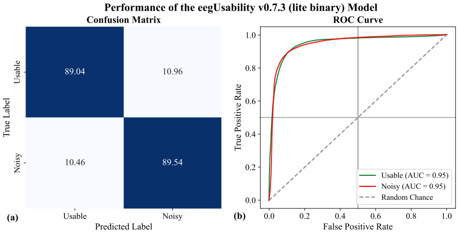
The authors claim that eegUsability, a LightGBM classifier over 3,618 spectrogram and statistical features per 10-second epoch, can identify artifact-contaminated sleep EEG with weighted F1 of approximately 0.85 and kappa of 0.78 while preserving 93.55% of usable epochs. The artifact taxonomy covers flat or disconnected signals (No Data), high-amplitude external noise, a high-frequency low-amplitude artifact called Spiky Noise, and a slow periodic M-shaped waveform; each was labeled by visual inspection of spectrograms, time-domain signals, and accelerometer data. Applied to autoscoring, rejecting flagged epochs improves agreement with manual PSG staging by 4.73 and 2.03 absolute F1 points on the Wearanize+ and QSci datasets. The authors further argue that the model extends beyond Zmax, although the non-Zmax evidence is presented through visual inspection of usability graphs rather than quantitative labels.
Load-bearing premise
The model's training labels were produced by a single human scorer, so the reported accuracy assumes those labels are a trustworthy standard; the cross-device claim further assumes the artifact types and signal ranges learned from Zmax carry over to other hardware, which the paper supports only by visual inspection.
Editorial extensions
If this is right
- Sleep studies using autoscorers can run eegUsability before or after staging and exclude artifact-contaminated epochs, improving agreement with manual staging by the measured amounts on datasets with similar artifact burden.
- With 93.55% recall for usable data, most clean EEG is retained, so preprocessing does not drastically shrink the analyzable dataset.
- Because the model consumes EEG plus accelerometer magnitude and standard statistical features, it can flag epochs for any sleep EEG device that keeps similar signal ranges, subject to the caveats the authors list.
- The eegMobility model makes automatic time-in-bed detection possible from accelerometer data, replacing the common assumption that the whole recording equals time in bed.
Reading between the lines
- A testable extension is to measure whether the reported autoscoring gains scale with artifact burden: the larger gain (4.73%) appeared on the dataset with 18.95% artifact-contaminated epochs, while the smaller gain (2.03%) appeared on the cleaner dataset with 4.73% contamination, so datasets with more artifacts should show larger improvements if the mechanism is correct.
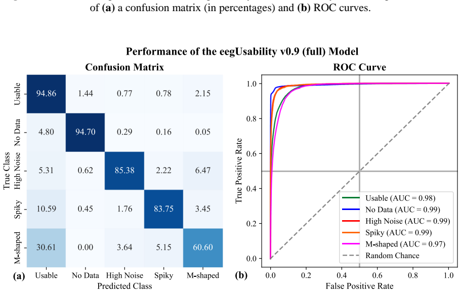
- The paper reports that 51.46% of M-shaped Noise instances are misclassified as good data, and that M-shaped Noise is rarely seen in non-Zmax recordings; this suggests the artifact may be device-specific, and a simpler model trained without that class could perform better on other hardware.
- The most direct next validation, which the authors identify as future work, is a multi-scorer labeling study on both Zmax and non-Zmax data; such a study would test whether single-scorer labels suffice or whether the model's uncertainty estimates are needed for deployment.
Editorial analysis
A structured set of objections, weighed in public.
Referee Report
Summary. The paper introduces eegFloss, an open-source Python package built around two LightGBM classifiers: eegUsability (artifact detection in 10-second sleep EEG epochs, trained on 127 manually labeled Zmax nights from 15 participants) and eegMobility (activity classification from Zmax accelerometer data for automatic time-in-bed detection). The authors report weighted F1 ≈ 0.85 and κ = 0.78 for eegUsability, with 93.55% recall for usable epochs, and near-perfect performance for eegMobility. The package aggregates usability labels with autoscores, filters Spiky Noise, generates hypnograms, and computes sleep statistics; the authors also report improved agreement between Dreamento autoscores and manual PSG scores after artifact rejection on two independent datasets. The paper includes model variants, processing times, and extensive supplementary visual outputs.
Significance. If the evaluation issues are resolved, the contribution is real and useful: the package is open-source (MIT license, DOI), the core model is trained on an unusually large manually labeled sleep-EEG corpus, and the subject-disjoint split is a sound design choice. The autoscoring improvement in §5.4 is a practical downstream demonstration, and the eegMobility results are strong. The paper is also transparent about several limitations (single-scorer labels, no quantitative non-Zmax validation, v0.9 training-set evaluation). The main weakness is that the headline eegUsability metrics are measured on a rebalanced test set and therefore do not directly reflect deployment on natural sleep recordings; this, plus the unvalidated cross-device claim, prevents acceptance in its current form.
major comments (4)
- [§3.1.2, Table 1] The headline metrics (weighted F1 = 84.87%, κ = 0.78, usable-data recall = 93.55%) are computed on a test set that was rebalanced to 50% usable data, whereas the natural corpus has more than 82% usable data (Figure 1). Because LightGBM probability outputs are fit to the training prior, applying the default decision threshold to full recordings will generally produce a different precision/recall tradeoff. Please report metrics on the original unbalanced held-out set (112,360 samples per Figure 1) and show how the usable-data retention rate and artifact F1 vary with the decision threshold on that natural distribution.
- [§4.7] The claim that eegUsability "extends beyond Zmax" is supported only by visual inspection of usability graphs (Figures 11–13); the paper explicitly states that no manual artifact labels are available for these recordings. This is not quantitative validation. If cross-device generalization is a central selling point, at least one of the following should be added: manual labels on a sample of non-Zmax nights with inter-rater reliability, a proxy validation such as comparing downstream PSG autoscoring with and without eegFloss rejection on non-Zmax data, or a clearly stated limitation that the cross-device claim is anecdotal.
- [§5.1, §3.1.1] The ground truth consists of single-scorer visual labels, as acknowledged in §5.1. Since all artifact-class definitions and merging rules (§3.1.1.1–§3.1.1.3) are derived from this subjective process, and since the central performance claims depend on label quality, the absence of any inter-rater reliability or consistency check is a load-bearing gap. Please provide at least a second-scorer agreement statistic on a subset of nights, or an objective signal-property validation of the artifact classes (e.g., spectral or amplitude criteria), before the F1/kappa numbers are taken at face value.
- [Table 5, §5.3] The v0.9 ("full") row reports weighted F1 = 90.11% and κ = 0.850, but the footnote states that these results are from a test set that is a subset of the training data. Including this row in the same comparison table as the other variants is misleading and should be removed or relegated to a clearly separated "training-set performance" table, since it cannot be compared with held-out results.
minor comments (6)
- [§3.1.5] The text says the classifier "identifies usable data with an impressive 93.55% accuracy"; this is row-wise recall for Class 0, not accuracy, and should be labeled as such.
- [§3.2.3] The text reports "a κ score of 99.38" while Table 2 lists κ = 0.994; please use one convention consistently.
- [Appendix 2, Table 2.1] The table caption reads "Performance of different versions of the eegUsability model" but the table contains eegMobility variants; the caption should name eegMobility.
- [§5.5] The two-minute threshold for TIB is described as chosen based on observations without a quantitative comparison; please add a short sensitivity analysis or state the manual-review protocol used to obtain the 119/122 accuracy figure.
- [§4.7] It would help to state explicitly how eegUsability handles missing ACC input (e.g., whether features are zero-filled or dropped) and whether the reported performance was obtained with or without ACC, since the non-Zmax PSG examples do not have ACC.
- [§3.1.1.4] The window-size rationale says M-shaped noise has a period of about 4 seconds, while §3.1.1.1 says 4–5 seconds; please align the two descriptions.
Circularity Check
No significant circularity: eegUsability's headline metrics are held-out subject evaluations, and the downstream autoscoring gains are independent external checks.
full rationale
The central artifact-detection claim rests on a subject-disjoint train/test split of 127 manually labeled nights; the confusion-matrix and Table 1 numbers are computed on unseen subjects, so they are not equivalent to the training fit. The manual artifact labels are the target by definition, but a supervised classifier predicting those labels is not circular. The §5.4 autoscoring improvement is measured on independent Wearanize+ and QSci datasets against Dreamento autoscores, and artifact rejection is the intervention rather than a fitted input. Self-citations ([53], [30], [105], [106]) supply datasets or device validation; none is used as a uniqueness theorem or to forbid alternatives, and the 'first of their kind' claim is unsupported rather than circular. The main in-scope weakness is Table 5's v0.9 row, which is evaluated on a subset of the training data; the footnote discloses this, and it is not the basis for the paper's headline claims. Single-scorer labeling (§5.1) and unquantified cross-device transfer (§4.7) are correctness and validation risks, not circular reductions. I therefore find no step in which a claimed prediction reduces to its own inputs by construction.
Assumptions & free parameters
free parameters (4)
- LightGBM hyperparameters =
not specified in detail
- TIB two-minute threshold =
120 s
- Test class-ratio undersampling =
50% usable, 28.23% No Data, 6.61% High Noise, 5.81% Spiky, 9.34% M-Shaped
- Epoch window length =
10 s
assumptions (5)
- domain assumption Manual visual labels by a single scorer are correct ground truth.
- domain assumption The five artifact classes are exhaustive and mutually exclusive after merging.
- domain assumption Zmax data properties (amplitude ±100 µV, ACC in g) transfer to other devices with normalization.
- ad hoc to paper A 10-second epoch captures the defining features of all artifact classes.
- ad hoc to paper The 2-minute consecutive Lying threshold defines TIB.
Cite this review
Pith. "Pith review of eegFloss: A Python package for refining sleep EEG recordings using machine learning models." pith.science (2026). https://pith.science/paper/VA7E7WDN
@misc{pith2026250706433,
  author       = {Pith},
  title        = {Pith review of: eegFloss: A Python package for refining sleep EEG recordings using machine learning models},
  year         = {2026},
  howpublished = {\url{https://pith.science/paper/VA7E7WDN}},
  note         = {Machine review of arXiv:2507.06433}
}
read the original abstract
Electroencephalography (EEG) allows monitoring of brain activity, providing insights into the functional dynamics of various brain regions and their roles in cognitive processes. EEG is a cornerstone in sleep research, serving as the primary modality of polysomnography, the gold standard in the field. However, EEG signals are prone to artifacts caused by both internal (device-specific) factors and external (environmental) interferences. As sleep studies are becoming larger, most rely on automatic sleep staging, a process highly susceptible to artifacts, leading to erroneous sleep scores. This paper addresses this challenge by introducing eegFloss, an open-source Python package to utilize eegUsability, a novel machine learning (ML) model designed to detect segments with artifacts in sleep EEG recordings. eegUsability has been trained and evaluated on manually artifact-labeled EEG data collected from 15 participants over 127 nights using the Zmax headband. It demonstrates solid overall classification performance (F1-score is approximately 0.85, Cohens kappa is 0.78), achieving a high recall rate of approximately 94% in identifying channel-wise usable EEG data, and extends beyond Zmax. Additionally, eegFloss offers features such as automatic time-in-bed detection using another ML model named eegMobility, filtering out certain artifacts, and generating hypnograms and sleep statistics. By addressing a fundamental challenge faced by most sleep studies, eegFloss can enhance the precision and rigor of their analysis as well as the accuracy and reliability of their outcomes.
Figures


Figures from the paper (21 more)






Reference graph
Works this paper leans on
-
[1]
Berger, Über das Elektrenkephalogramm des Menschen, Arch
H. Berger, Über das Elektrenkephalogramm des Menschen, Arch. Für Psychiatr. Nervenkrankh. 87 (1929) 527–570. https://doi.org/10.1007/BF01797193
-
[2]
R. Sutter, P.W. Kaplan, D.L. Schomer, Historical Aspects of Electroencephalography, Oxford University Press, 2017. https://doi.org/10.1093/med/9780190228484.003.0001
arXiv 2017
-
[3]
Niedermeyer, F.H
E. Niedermeyer, F.H. Lopes da Silva, eds., Electroencephalography: basic principles, clinical applications, and related fields, 5th ed, Lippincott Williams & Wilkins, Philadelphia, 2005
2005
-
[4]
Luck, An introduction to the event-related potential technique, 2nd ed, MIT Press, Cambridge, Mass., 2014
S.J. Luck, An introduction to the event-related potential technique, 2nd ed, MIT Press, Cambridge, Mass., 2014
2014
-
[5]
Dickter, P.D
C.L. Dickter, P.D. Kieffaber, EEG methods for social and per sonality psychology, Online-Ausg, SAGE Publications, Thousand Oaks, California, 2014
2014
-
[6]
D.J. McFarland, J.R. Wolpaw, EEG -based brain–computer interfaces, Curr. Opin. Biomed. Eng. 4 (2017) 194–200. https://doi.org/10.1016/j.cobme.2017.11.004
-
[7]
Müller, M
K.-R. Müller, M. Krauledat, G. Dornhege, G. Curio, B. Blankertz, Machine learning techniques for brain-computer interfaces, Biomed Tech 49 (2004) 11–22
2004
-
[8]
M. Aljalal, S. Ibrahim, R. Djemal, W. Ko, Comprehensive review on brain -controlled mobile robots and robotic arms based on electroencephalography signals, Intell. Serv. Robot. 1 3 (2020) 539–563. https://doi.org/10.1007/s11370-020-00328-5
Show all 110 references
-
[9]
Soufineyestani, D
M. Soufineyestani, D. Dowling, A. Khan, Electroencephalography (EEG) Technology Applications and Available Devices, Appl. Sci. 1 0 (2020) 7453. https://doi.org/10.3390/app10217453
2020 doi
-
[10]
Thompson, T
T. Thompson, T. Steffert, T. Ros, J. Leach, J. Gruzelier, EEG applications for sport and performance, Methods 45 (2008) 279–288. https://doi.org/10.1016/j.ymeth.2008.07.006
2008 doi
-
[11]
Belkofer, A.V
C.M. Belkofer, A.V. Van Hecke, L.M. Konopka, Effects of Drawing on Alpha Activity: A Quantitative EEG Study With Implications for Art Therapy, Art Ther. 31 (2014) 61 –68. https://doi.org/10.1080/07421656.2014.903821
2014
-
[12]
Zheng, L
H. Zheng, L. Niu, W. Qiu, D. Liang, X. Long, G. Li, Z. Liu, L. Meng, The Emergence of Functional Ultrasound for Noninvasive B rain–Computer Interface, Research 6 (2023) 0200. https://doi.org/10.34133/research.0200
2023 doi
-
[13]
Malmivuo, Comparison of the Properties of EEG and MEG in Detecting the Electric Activity of the Brain, Brain Topogr
J. Malmivuo, Comparison of the Properties of EEG and MEG in Detecting the Electric Activity of the Brain, Brain Topogr. 25 (2012) 1–19. https://doi.org/10.1007/s10548-011-0202-1
2012 doi
-
[14]
Crosson, A
B. Crosson, A. Ford, K.M. McGregor, M. Meinzer, S. Cheshkov, X. Li, D. Walker-Batson, R.W. Briggs, Functional imaging and related techniques: An introduction for rehabilitation researchers, J. Rehabil. Res. Dev. 47 (2010) vii. https://doi.org/10.1682/JRRD.2010.02.0017
2010 doi
-
[15]
Logothetis, What we can do and what we cannot do with fMRI, Nature 453 (2008) 869–878
N.K. Logothetis, What we can do and what we cannot do with fMRI, Nature 453 (2008) 869–878. https://doi.org/10.1038/nature06976
2008 doi
-
[16]
Marino, D
M. Marino, D. Mantini, Human brain imaging with high‐density electroencephalography: Techniques and applications, J. Physiol. (2024) JP286639. https://doi.org/10.1113/JP286639
2024 doi
-
[17]
Lustenberger, R
C. Lustenberger, R. Huber, High Density Electroencephalography in Sleep Research: Potential, Problems, Future Perspective, Front. Neurol. 3 (2012). https://doi.org/10.3389/fneur.2012.00077
2012 arXiv
-
[18]
Prakash, D
V. Prakash, D. Kumar, Artifact Detection and Removal in EEG: A Review of Methods and Contemporary Usage, in: K.C. Santosh, S.K. Sood, H.M. Pandey, C. Virmani (Eds.), Adv. Artif.- Bus. Anal. Quantum Mach. Learn., Springer Nature Singapore, Singapore, 2024: pp. 263 –274. https:/...
2024 doi
-
[19]
Jiang, G
X. Jiang, G. -B. Bian, Z. Tian, Removal of Artifacts from EEG Signals: A Review, Sensors 19 (2019) 987. https://doi.org/10.3390/s19050987
2019 doi
-
[20]
Dizeux, M
A. Dizeux, M. Gesnik, H. A hnine, K. Blaize, F. Arcizet, S. Picaud, J. -A. Sahel, T. Deffieux, P. Pouget, M. Tanter, Functional ultrasound imaging of the brain reveals propagation of task-related 44 brain activity in behaving primates, Nat. Commun. 10 (2019) 1400. https://doi....
2019 doi
-
[21]
L.-A. Sieu, A. Bergel, E. Tiran, T. Deffieux, M. Pernot, J. -L. Gennisson, M. Tanter, I. Cohen, EEG and functional ultrasound imaging in mobile rats, Nat. Methods 12 (2 015) 831 –834. https://doi.org/10.1038/nmeth.3506
-
[22]
Srinivasan, W.R
R. Srinivasan, W.R. Winter, P.L. Nunez, Source analysis of EEG oscillations using high-resolution EEG and MEG, in: Prog. Brain Res., Elsevi er, 2006: pp. 29 –42. https://doi.org/10.1016/S0079- 6123(06)59003-X
2006 doi
-
[23]
Struck, M.B
A.F. Struck, M.B. Westover, L.T. Hall, G.M. Deck, A.J. Cole, E.S. Rosenthal, Metabolic Correlates of the Ictal-Interictal Continuum: FDG-PET During Continuous EEG, Neurocrit. Care 24 (2016) 324–331. https://doi.org/10.1007/s12028-016-0245-y
2016 doi
-
[24]
Abreu, A
R. Abreu, A. Leal, P. Figueiredo, EEG -Informed fMRI: A Review of Dat a Analysis Methods, Front. Hum. Neurosci. 12 (2018) 29. https://doi.org/10.3389/fnhum.2018.00029
2018
-
[25]
Iber, The AASM manual for the scoring of sleep and associated events: Rules, Terminol
C. Iber, The AASM manual for the scoring of sleep and associated events: Rules, Terminol. Tech. Specif. (2007)
2007
-
[26]
Berry, The AASM manual for the scoring of sleep and associated events, Rules Terminol
R. Berry, The AASM manual for the scoring of sleep and associated events, Rules Terminol. Tech. Specif. Version 2 (2012)
2012
-
[27]
Lendner, R.F
J.D. Lendner, R.F. Helfrich, B.A. Mander, L. Romundstad, J.J. Lin, M.P. Walker, P.G. Larsson, R.T. Knight, An electrophysiological marker of arousal level in humans, eLife 9 (2020) e55092. https://doi.org/10.7554/eLife.55092
2020 doi
-
[28]
Sleep Med
Manual Versus Automated Sleep Scoring for Diagnosis of Obstructive Sleep Apnoea, Indian J. Sleep Med. 11 (2016) 5–7. https://doi.org/10.5958/0974-0155.2016.00002.4
2016
-
[29]
Lee, J.Y
Y.J. Lee, J.Y. Lee, J.H. Cho, J.H. Choi, Interrater reliability of sleep stage scoring: a meta-analysis, J. Clin. Sleep Med. 18 (2022) 193–202. https://doi.org/10.5664/jcsm.9538
2022 doi
-
[30]
Sikder, L
N. Sikder, L. Verkaar, A. Paltarzhytskaya, S. Acan, E. Krugliakova, Y. Rosenblum, M. Krauledat, M. Dresler, P. Zerr, Wearanize+: A Multimodal Dataset for Evaluating Wearable Technologies in Sleep Research, (2025). https://doi.org/10.31219/osf.io/dth8y_v1
2025 doi
-
[31]
Bzdok, J.P.A
D. Bzdok, J.P.A. Ioannidis, Exploration, Inference, and Prediction in Neuroscience and Biomedicine, Trends Neurosci. 42 (2019) 251–262. https://doi.org/10.1016/j.tins.2019.02.001
2019 doi
-
[32]
Sporns, The human connectome: a complex network, Ann
O. Sporns, The human connectome: a complex network, Ann. N. Y. Acad. Sci. 1224 (2011) 109–
2011
-
[33]
Wolpaw, N
J.R. Wolpaw, N. Birbaumer, D.J. McFarland, G. Pfurtscheller, T.M. Vaughan, Brain –computer interfaces for communication and control, Clin. Neurophysiol. 113 (2002) 767 –791. https://doi.org/10.1016/S1388-2457(02)00057-3
2002 doi
-
[34]
Shalaby, A
A. Shalaby, A. Soliman, S. Elaskary, A. Refaey, M. Abdelazim, F. Khalifa, Editorial: Artificial intelligence based computer -aided diagnosis applications for brain disorders from medical imaging data, Front. Neurosci. 17 (2023) 998818. https://doi.org/10.3389/fnins.2023.998818
2023
-
[35]
Thieme, D
A. Thieme, D. Belgrave, G. Doherty, Machine Learning in Mental Health: A Systematic Review of the HCI Literature to Support the Development of Effective and Implementable ML Systems, ACM Trans. Comput.-Hum. Interact. 27 (2020) 1–53. https://doi.org/10.1145/3398069
2020 doi
-
[36]
H. Yue, Z. Chen, W. Guo, L. Sun, Y. Dai, Y. Wang, W. Ma, X. Fan, W. Wen, W. Lei, Research and application of deep learning -based sleep staging: Data, modeling, validation, and clinical practice, Sleep Med. Rev. 74 (2024) 101897. https://doi.org/10.1016/j.smrv.2024.101897
2024
-
[37]
Perslev, S
M. Perslev, S. Darkner, L. Kempfner, M. Nikolic, P.J. Jennum, C. Igel, U -Sleep: resilient high- frequency sleep staging, Npj Digit. Med. 4 (2021) 72. https://doi.org/10.1038/s41746-021-00440- 5
2021 doi
-
[38]
Vallat, M.P
R. Vallat, M.P. Walker, An open-source, high-performance tool for automated sleep staging, eLife 10 (2021) e70092. https://doi.org/10.7554/eLife.70092
2021 doi
-
[39]
Kevat, R
A. Kevat, R. Steinkey, S. Suresh, W.R. Ruehland, J. Chawla, P.I. Terrill, A. Collaro, K. Iyer, Evaluation of automated pediatric sleep stage classification using U-Sleep: a convolutional neural 45 network, J. Clin. Sleep Med. 21 (2025) 277–285. https://doi.org/10.5664/jcsm.11362
2025 doi
-
[40]
Benedetti, E
D. Benedetti, E. Frati, O. Kiss, D. Yuksel, U. Faraguna, B.P. Hasler, P.L. Franzen, D.B. Clark, F.C. Baker, M. De Zambotti, Performance ev aluation of the open -source Yet Another Spindle Algorithm sleep staging algorithm against gold standard manual evaluation of polysomnogra...
2023 doi
-
[41]
V. Muto, C. Berthomier, Looking for a balance between visual and automatic sleep scoring, Npj Digit. Med. 6 (2023) 165. https://doi.org/10.1038/s41746-023-00915-7
2023 doi
-
[42]
De Gans, P
C.J. De Gans, P. Burger, E.S. Van Den Ende, J. Hermanides, P.W.B. Nanayakkara, R.J.B.J. Gemke, F. Rutters, D.J. Stenvers, Sleep assessment using EEG -based wearables – A systematic review, Sleep Med. Rev. 76 (2024) 101951. https://doi.org/10.1016/j.smrv.2024.101951
2024
-
[43]
Jafarzadeh Esfahani, N
M. Jafarzadeh Esfahani, N. Sikder, R. Ter Horst, A.H. Daraie, K. Appel, F.D. Weber, K.E. Bevelander, M. Dresler, Citizen neuroscience: Wearable technolo gy and open software to study the human brain in its natural habitat, Eur. J. Neurosci. (2024) ejn.16227. https://doi.org/10...
2024 doi
-
[44]
Biosignals (n.d.)
The Dreem Headband, Beac. Biosignals (n.d.). htt ps://beacon.bio/dreem-headband/ (accessed November 28, 2024)
2024
-
[45]
https://choosemuse.com/products/muse-s-gen-2 (accessed November 28, 2024)
Muse S | Muse TM EEG-Powered Meditation & Sleep Headband, (2024). https://choosemuse.com/products/muse-s-gen-2 (accessed November 28, 2024)
2024
-
[46]
http://hypnodynecorp.com/ (accessed November 28, 2024)
Hypnodyne ZMax is an advanced and simple to use EEG home sleep monitor used by researchers but available to everyone, (n.d.). http://hypnodynecorp.com/ (accessed November 28, 2024)
2024
-
[47]
h ttps://www.sleeploop.ch/ (accessed January 25, 2025)
SleepLoop – Healthy sleeping made in Switzerland, (n.d.). h ttps://www.sleeploop.ch/ (accessed January 25, 2025)
2025
-
[48]
https://somneesleep.com/products/smart -sleep-headband (accessed November 28, 2024)
Smart Sleep Headband, Somnee (n.d.). https://somneesleep.com/products/smart -sleep-headband (accessed November 28, 2024)
2024
-
[49]
https://www.advancedbrainmonitoring.com/products/sleep -profiler (accessed December 7, 2024)
Sleep Profiler, (n.d.). https://www.advancedbrainmonitoring.com/products/sleep -profiler (accessed December 7, 2024)
2024
-
[50]
https://www.bitbrain.com/neurotechnology -products/textile-eeg/ikon (accessed December 7, 2024)
Ikon, Bitbrain (n.d .). https://www.bitbrain.com/neurotechnology -products/textile-eeg/ikon (accessed December 7, 2024)
2024
-
[51]
https://shop.openbci.com/products/openbci-eeg-headband-kit (accessed December 7, 2024)
OpenBCI EEG Headband Kit – OpenBCI Online Store, (n.d.). https://shop.openbci.com/products/openbci-eeg-headband-kit (accessed December 7, 2024)
2024
-
[52]
Cannard, H
C. Cannard, H. Wahbeh, A. Delorme, Validating the wearable MUSE headset for EEG spectral analysis and Frontal Alpha Asymmetry, in: 20 21 IEEE Int. Conf. Bioinforma. Biomed. BIBM, IEEE, Houston, TX, USA, 2021: pp. 3603 –3610. https://doi.org/10.1109/BIBM52615.2021.9669778
2021
-
[53]
Esfahani, F.D
M.J. Esfahani, F.D. Weber, M. Boon, S. Anthes, T. A lmazova, M.V. Hal, Y. Keuren, C. Heuvelmans, E. Simo, L. Bovy, N. Adelhöfer, M.M.T. Avest, M. Perslev, R.T. Horst, C. Harous, T. Sundelin, J. Axelsson, M. Dresler, Validation of the sleep EEG headband ZMax, Neuroscience,
-
[54]
Ferster, C
M.L. Ferster, C. Lustenberger, W. Karlen, Configurable Mobile System for Autonomous High - Quality Sleep Monitoring and Closed-Loop Acoustic Stimulation, IEEE Sens. Lett. 3 (2019) 1–4. https://doi.org/10.1109/LSENS.2019.2914425
2019
-
[55]
Ferster, G
M.L. Ferster, G. Da Poian, K. Menachery, S.J. Schreiner, C. Lustenberger, A. Maric, R. Huber, C.R. Baumann, W. Karlen, Benchmarking Real -Time Algorithms for In -Phase Auditory Stimulation of Low Amplitude Slow Waves With Wearable EEG Devices During Sleep, IEEE Trans. Biomed. ...
2022
-
[56]
Esparza-Iaizzo, M
M. Esparza-Iaizzo, M. Sierra-Torralba, J.G. Klinzing, J. Minguez, L. Montesano, E. López-Larraz, Automatic sleep scoring for real-time monitoring and stimulation in individuals with and without sleep apnea, (2024). https://doi.org/10.1101/2024.06.12.597764
2024 doi
-
[57]
Hinrichs, M
H. Hinrichs, M. Scholz, A.K. Baum, J.W.Y. Kam, R.T. Knight, H.-J. Heinze, Comparison between 46 a wireless dry electrode EEG system with a conventional wired wet electrode EEG system for clinical applications, Sci. Rep. 10 (2020) 5218. https://doi.org/10.1038/s41598-020-62154-0
2020 doi
-
[58]
Vitazkova, H
D. Vitazkova, H. Kosnacova, D. Turonova, E. Foltan, M. Jagelka, M. Berki, M. Micj an, O. Kokavec, F. Gerhat, E. Vavrinsky, Transforming Sleep Monitoring: Review of Wearable and Remote Devices Advancing Toward Home Polysomnography and Their Role in Predicting Neurological Disor...
2024
-
[59]
P. Liu, W. Qian, H. Zhang, Y. Zhu, Q. Hong, Q. Li, Y. Yao, Automatic sleep stage classification using deep learning: signals, data representation, and neural networks, Artif. Intell. Rev. 57 (2024)
2024
-
[60]
Malafeev, D
A. Malafeev, D. Laptev, S. Bauer, X. Omlin, A. Wierzbicka, A. Wichniak, W. Jernajczyk, R. Riener, J. Buhmann, P. Achermann, Auto matic Human Sleep Stage Scoring Using Deep Neural Networks, Front. Neurosci. 12 (2018) 781. https://doi.org/10.3389/fnins.2018.00781
2018
-
[61]
Korkalainen, J
H. Korkalainen, J. Aakko, B. Duce, S. Kainulainen, A. L eino, S. Nikkonen, I.O. Afara, S. Myllymaa, J. Töyräs, T. Leppänen, Deep learning enables sleep staging from photoplethysmogram for patients with suspected sleep apnea, Sleep 43 (2020) zsaa098. https://doi.org/10.1093/sle...
2020 doi
-
[62]
H. Zhu, Y. Wu, N. Shen, J. Fan, L. Tao, C. Fu, H. Yu, F. Wan, S.H. Pun, C. Chen, W. Chen, The Masking Impact of Intra -Artifacts in EEG on Deep Learning -Based Sleep Staging Systems: A Comparative Study, IEEE Trans . Neural Syst. Rehabil. Eng. 30 (2022) 1452 –1463. https://doi...
2022
-
[63]
Rayan, A.B
A. Rayan, A.B. Szabo, L. Genzel, The pros and cons of using automated sleep scoring in sleep research, SLEEP 47 (2024) zsad275. https://doi.org/10.1093/sleep/zsad275
2024 doi
-
[64]
https://lightgbm.readthedocs.io/en/stable/ (accessed February 19, 2025)
Welcome to LightGBM’s documentation! — LightGBM 4.6.0 documentation, (n.d.). https://lightgbm.readthedocs.io/en/stable/ (accessed February 19, 2025)
2025
-
[65]
Mannan, M.A
M.M.N. Mannan, M.A. Kamran, M.Y. Jeong, Identification and Removal of Physiological Artifacts From Electroencephalogram Signals: A Review, IEEE Access 6 (20 18) 30630–30652. https://doi.org/10.1109/ACCESS.2018.2842082
2018
-
[66]
’T Wallant, V
D.C. ’T Wallant, V. Muto, G. Gaggioni, M. Jaspar, S.L. Chellappa, C. Meyer, G. Vandewalle, P. Maquet, C. Phillips, Automatic ar tifacts and arousals detection in whole -night sleep EEG recordings, J. Neurosci. Methods 258 (2016) 124 –133. https://doi.org/10.1016/j.jneumeth.2015.11.005
2016 doi
-
[67]
Gharbali, J.M
A.A. Gharbali, J.M. Fonseca, S. Najdi, T.Y. Rezaii, Automatic EOG and EMG Artifact Removal Method for Sleep Stage Classification, in: L.M. Camarinha-Matos, A.J. Falcão, N. Vafaei, S. Najdi (Eds.), Technol. Innov. Cyber -Phys. Syst., Springer International Publishing, Cham, 201...
2016 doi
-
[68]
Malafeev, X
A. Malafeev, X. Omlin, A. Wierzbicka, A. Wichniak, W. Jernajczyk, R. Riener, P. Achermann, Automatic artefact detection in single -channel s leep EEGrecordings, J. Sleep Res. 28 (2019) e12679. https://doi.org/10.1111/jsr.12679
2019 doi
-
[69]
Leach, G
S. Leach, G. Sousouri, R. Huber, ‘High-Density-SleepCleaner’: An open-source, semi-automatic artifact removal routine tailored to high -density sleep EEG, J. Neurosci. Methods 391 (2023) 109849. https://doi.org/10.1016/j.jneumeth.2023.109849
2023
-
[70]
Cox, F.D
R. Cox, F.D. Weber, E.J.W. Van Someren, Customiza ble automated cleaning of multichannel sleep EEG in SleepTrip, Front. Neuroinformatics 18 (2024) 1415512. https://doi.org/10.3389/fninf.2024.1415512
2024
-
[71]
Anandanadarajah, A
N. Anandanadarajah, A. Talukder, D. Yeu ng, Y. Li, D.M. Umbach, Z. Fan, L. Li, Detection of Movement and Lead -Popping Artifacts in Polysomnography EEG Data, Signals 5 (2024) 690 –
2024
-
[72]
Delorme, T
A. Delorme, T. Sejnowski, S. Makeig, Enhanced detection of artifacts in EEG data using higher - order statistics and independent component analysis, NeuroImage 34 (2007) 1443 –1449. https://doi.org/10.1016/j.neuroimage.2006.11.004
2007 doi
-
[73]
P.P. Ujma, M. Dresler, R. Bódizs, Comparing Manual and Automatic Artifact Detection in Sleep 47 EEG Recordings, Psychophysiology 62 (2025) e70016. https://doi.org/10.1111/psyp.70016
2025 doi
-
[74]
P. Lima, J. Leitao, T. Paiva, Artifact detection in sleep EEG recording, in: Proc. Electrotech. Conf. Integrating Res. Ind. Educ. Energy Commun. Eng., IEEE, Lisbon, Portugal, 1989: pp. 273 –277. https://doi.org/10.1109/MELCON.1989.50035
1989
- [75]
-
[76]
Saifutdinova, D.U
E. Saifutdinova, D.U. Dudysova, L. Lhotska, V. Gerla, M. Macas, Artifact Detection in Multichannel Sleep EEG using Random Forest Classifier, in: 2018 IEEE Int. Conf. Bioinforma. Biomed. BIBM, IEEE, Madrid, Spain, 2018: pp. 2803 –2805. https://doi.org/10.1109/BIBM.2018.8621374
2018
-
[77]
https://lightgbm.readthedocs.io/en/stable/ (accessed February 9, 2025)
Welcome to LightGBM’s documentation! — LightGBM 4.5.0 documentation, (n.d.). https://lightgbm.readthedocs.io/en/stable/ (accessed February 9, 2025)
2025
-
[78]
https://hypnodynecorp.com/lineup.png (accessed November 28, 2024)
lineup.png (1988×1528), (n.d.). https://hypnodynecorp.com/lineup.png (accessed November 28, 2024)
1988
-
[79]
Lampert, S.E.M
T.A. Lampert, S.E.M. O’Keefe, A survey of spectrogram track detection algorithms, Appl. Acoust. 71 (2010) 87–100. https://doi.org/10.1016/j.apacoust.2009.08.007
2010 doi
-
[80]
Grigg-Damberger, Polysomnographic Artifacts, in: W.O
M. Grigg-Damberger, Polysomnographic Artifacts, in: W.O. Tatum (Ed.), Atlas Artifacts Clin. Neurophysiol., Springer Publishing Company, New York, NY, 2018. https://doi.org/10.1891/9780826169358.0011
2018
-
[81]
Attarian, N.S
H.P. Attarian, N.S. Undevia, Atlas of Electroencephalography in Sleep Medicine, Springer US, Boston, MA, 2012. https://doi.org/10.1007/978-1-4614-2293-8
2012 doi
-
[82]
Amin, F.A
U. Amin, F.A. Nascimento, I. Karakis, D. Schomer, S.R. Benbadis, Normal variants and artifacts: Importance in EEG interpretation, Epileptic. Disord. 25 (2023) 591 –648. https://doi.org/10.1002/epd2.20040
2023 doi
-
[83]
Varotto, G
G. Varotto, G. Susi, L. Tassi, F. Gozzo, S. Franceschetti, F. Panzica, Comparison of Resampling Techniques for Imbalanced Datasets in Machine Learning: Application to Epileptogenic Zone Localization From Interictal Intracranial EEG Recordings in Patients With Focal Epilepsy, F...
2021
-
[84]
Barandas, D
M. Barandas, D. Folgado, L. Fernandes, S. Santos, M. Abreu, P. Bota, H. Liu, T. Schultz, H. Gamboa, TSFEL: Time Series Feature Extraction Library, SoftwareX 11 (2020) 100456. https://doi.org/10.1016/j.softx.2020.100456
2020
-
[85]
https://tsfel.readthedocs.io/en/latest/index.html (accessed February 17, 2025)
Welcome to TSFEL documentation! — TSFEL 0.1.9 documentation, (n.d.). https://tsfel.readthedocs.io/en/latest/index.html (accessed February 17, 2025)
2025
-
[86]
https://tsfel.readthedocs.io/en/latest/descriptions/feature_list.html (accessed February 18, 2025)
List of available features — TSFEL 0.1.9 documentation, (n.d.). https://tsfel.readthedocs.io/en/latest/descriptions/feature_list.html (accessed February 18, 2025)
2025
-
[87]
G. Ke, Q. Meng, T. Finley, T. Wang, W. Chen, W. Ma, Q. Ye, T. -Y. Liu, LightGBM: A Highly Efficient Gradient Boosting Decision Tree, in: Adv. Neural Inf. Process. Syst., Curran Associates, Inc., 2017. https://proceedings.neurips.cc/paper/2017/hash/6449f44a102fde848669bdd9eb6b7...
2017
- [88]
-
[89]
Japkowicz, Z
N. Japkowicz, Z. Boukouvalas, Machine Learning Evaluation: Towards Reliable and Responsible AI, Cambridge University Press, Cambridge, 2024. https://doi.org/10.1017/9781009003872
2024 doi
-
[90]
Sleep Dictionary, Sleep Found. (2020). https://www.sleepfoundation.org/how-sleep-works/sleep- dictionary (accessed February 20, 2025)
2020
-
[91]
Ariza -Colpas, E
P.P. Ariza -Colpas, E. Vicario, A.I. Oviedo -Carrascal, S. Butt Aziz, M.A. Piñeres -Melo, A. Quintero-Linero, F. Patara, Human Activity Recognition Data Analysis: History, Evolutions, and New Trends, Sensors 22 (2022) 3401. https://doi.org/10.3390/s22093401. 48
2022 doi
-
[92]
Welch, The use of fast Fourier transform for the estimation of power spectra: A method based on time averaging over short, modified periodograms, IEEE Trans
P. Welch, The use of fast Fourier transform for the estimation of power spectra: A method based on time averaging over short, modified periodograms, IEEE Trans. Audio Electroacoustics 15 (1967) 70–73. https://doi.org/10.1109/TAU.1967.1161901
1967
-
[93]
G. Wang, Q. Li, L. Wang, W. Wang, M. Wu, T. Liu, Impact of Sliding Window Length in Indoor Human Motion Modes and Pose Pattern Recognition Based on Smartphone Sensors, Sensors 18 (2018) 1965. https://doi.org/10.3390/s18061965
2018 doi
-
[94]
Sikder, A.A
N. Sikder, A.A. Nahid, KU-HAR: An open dataset for heterogeneous human activity recognition, Pattern Recognit. Lett. 146 (2021) 46–54. https://doi.org/10.1016/j.patrec.2021.02.024
2021 doi
-
[95]
Sikder, M.A.R
N. Sikder, M.A.R. Ahad, A. -A. Nahid, Human Action Recognition Based on a Sequential Deep Learning Model, in: 2021 Jt. 10th Int. Conf. Inform. Electron. Vis. ICIEV 2021 5th Int. Conf. Imaging Vis. Pa ttern Recognit. IcIVPR, IEEE, Kitakyushu, Japan, 2021: pp. 1 –7. https://doi....
2021
-
[96]
Lyons, Understanding digital signal processing, 3rd ed, Prentice Hall, Upper Saddle River, NJ, 2011
R.G. Lyons, Understanding digital signal processing, 3rd ed, Prentice Hall, Upper Saddle River, NJ, 2011
2011
-
[97]
Lanquart, Contribution to the Definition of the Power Bands Limits of Sleep EEG by Linear Prediction, Comput
J.-P. Lanquart, Contribution to the Definition of the Power Bands Limits of Sleep EEG by Linear Prediction, Comput. Biomed. Res. 31 (1998) 100–111. https://doi.org/10.1006/cbmr.1998.1474
1998
-
[98]
Sateia, D.J
M.J. Sateia, D.J. Buysse, A.D. Krystal, D.N. Neubauer, J.L. Heald, Clinical Practice Guideline for the Pharmacologic Treatment of Chronic Insomnia in Adul ts: An American Academy of Sleep Medicine Clinical Practice Guideline, J. Clin. Sleep Med. 13 (2017) 307 –349. https://doi...
2017 doi
-
[99]
https://github.com/raphaelvallat/yasa/blob/master/src/yasa/sleepstats.py (accessed March 2, 2025)
Vallat, Raphael, yasa/src/yasa/sleepstats.py at master · raphaelvallat/yasa, GitHub (n.d.). https://github.com/raphaelvallat/yasa/blob/master/src/yasa/sleepstats.py (accessed March 2, 2025)
2025
-
[100]
https://natus.com/neuro/brain-quick-eeg-system/ (accessed February 3, 2025)
BRAIN QUICK® EEG System - Video EEG Monit oring, Natus (n.d.). https://natus.com/neuro/brain-quick-eeg-system/ (accessed February 3, 2025)
2025
-
[101]
https://somnomedics.de/enus/somnomedics-diagnostic-devices/sleep-diagnostics/in-lab- polysomnography/somnoscreen-plus/ (accessed November 28, 2024)
SOMNOscreen® plus - In-Lab Polysomnography, Somnomedics (n.d.). https://somnomedics.de/enus/somnomedics-diagnostic-devices/sleep-diagnostics/in-lab- polysomnography/somnoscreen-plus/ (accessed November 28, 2024)
2024
-
[102]
López-Larraz, M
E. López-Larraz, M. Sierra-Torralba, S. Clemente, G. Fierro, D. Oriol, J. Minguez, L. Montesano, J.G. Klinzing, Bitbrain Open Access Sleep Dataset, (2024). https://doi.org/10.18112/OPENNEURO.DS005555.V1.0.0
2024 doi
-
[103]
https://www.bitbrain.co m/neurotechnology-products/textile-eeg/ikon (accessed February 3, 2025)
Ikon, Bitbrain (n.d.). https://www.bitbrain.co m/neurotechnology-products/textile-eeg/ikon (accessed February 3, 2025)
2025
-
[104]
López-Larraz, C
E. López-Larraz, C. Escolano, A. Robledo-Menéndez, L. Morlas, A. Alda, J. Minguez, A garment that measures brain ac tivity: proof of concept of an EEG sensor layer fully implemented with smart textiles, Front. Hum. Neurosci. 17 (2023) 1135153. https://doi.org/10.3389/fnhum.202...
2023
-
[105]
Sikder, R
N. Sikder, R. Te r Horst, P. Zerr, M. Krauledat, M. Dresler, The quantified scientist: A six -year- long n= 1 sleep study, in: J. SLEEP Res., WILEY 111 RIVER ST, HOBOKEN 07030 -5774, NJ USA, 2024
2024
-
[106]
Sikder, M
N. Sikder, M. Jafarzadeh Esfahani, M. van Bakel, S. Idesis, L. Bovy, F.D. Weber, R. ter Horst, M. Krauledat, Matthias Dresler, The quantified scientist: a longitudinal study to explore the interdependencies between sleep, stress, the gut and other bodily functions, in: Suppl. ...
2022 doi
-
[125]
https://doi.org/10.1111/j.1749-6632.2010.05888.x
2010
-
[301]
https://doi.org/10.1007/s10462-024-10926-9
-
[704]
https://doi.org/10.3390/signals5040038
-
[2023]
https://doi.org/10.1101/2023.08.18.553744
2023 doi
Reviewed August 6, 2026 · model on record in the stance chip above.
Discussion (0). Continue with ORCID to comment.