REVIEW 5 major objections 6 minor 48 references
An empirical study for the early detection of Mpox from skin lesion images using pretrained CNN models leveraging XAI technique
T0 review · 5 major / 6 minor · reviewed 2026-08-06 · deepseek-v4-flash
Pith's one-line read InceptionV3 hits 95% on Mpox images; MobileNetV2 93% on six classes
desk verdict Routine transfer-learning benchmark whose headline accuracies are unreliable due to ambiguous augmentation/split order and internally inconsistent AUC metrics. read the letter →
The pith
A machine-rendered reading of the paper's core claim, the machinery that carries it, and where it could break.
The reading
What carries the argument
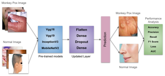
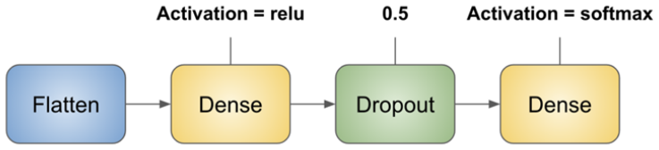
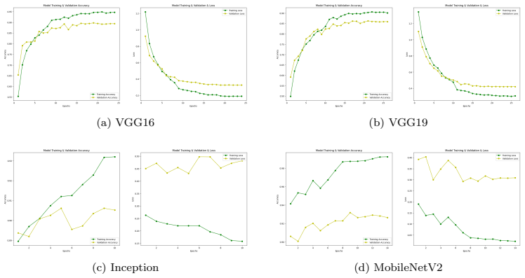
The mechanism is transfer learning from ImageNet: every pretrained convolutional base is frozen, and only a custom classification head is trained. The head consists of a flatten layer, a dense layer with 256 ReLU units, a dropout layer at rate 0.5, and a softmax output; early stopping and model checkpointing cap training at 30 epochs. Grad-CAM then computes gradient-weighted activations from the final convolutional layer to produce a heatmap of the image regions that most influenced the prediction. This combination is what the paper claims makes the models lightweight, fast, and interpretable.
What would settle it
Rerun the same four-model pipeline on the two Kaggle datasets with augmentation applied strictly after the train/validation/test split, and report accuracy on the untouched original test images; if the figures fall materially below 95% on binary and 93% on six-class data, the reported numbers were inflated by leakage of augmented copies into training.
Extended reading notes
Core claim
On the paper's own terms, the core discovery is that a frozen pretrained feature extractor plus a small custom head—a flatten layer, a dense layer of 256 ReLU units, a dropout layer at 0.5, and a softmax output—is enough to reach near-clinical accuracy on both datasets. InceptionV3 achieves 0.95 accuracy, precision, recall, and F1 on the binary Monkeypox Skin Lesion Dataset, and MobileNetV2 achieves 0.93 accuracy and precision with 0.90 recall on the six-class MSLD v2.0. The paper further reports that freezing the ImageNet base shrinks trainable parameters substantially, with VGG16 dropping from about 21 million total parameters to about 6.4 million trainable, and that Grad-CAM highlights regions corresponding to lesion areas. The stated conclusion is that these lightweight, interpretable models generalize across two independent datasets and are competitive with prior state-of-the-art results.
Load-bearing premise
The reported accuracies are only meaningful as generalization estimates if augmented copies of the same original photograph never appear in both the training and test sets, and the paper does not show that the data split was performed before augmentation.
Editorial extensions
If this is right
- If correct, InceptionV3 provides a binary Mpox-versus-others screen at 95% accuracy and MobileNetV2 a six-class differential at 93% accuracy on these Kaggle datasets.
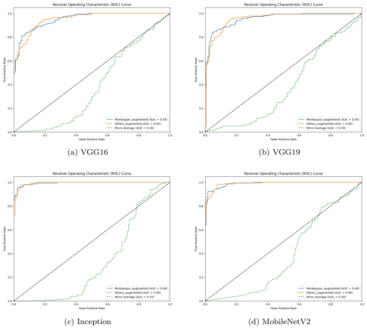
- Per-class AUC values up to 0.99 on binary and 1.00 on some multi-class categories indicate these models separate Mpox from common look-alikes on the images tested.
- Grad-CAM heatmaps give a clinician a visual region to check, which is the core of the paper's interpretability claim.
- Wall times of about 16 minutes for InceptionV3 on binary and 57 minutes for MobileNetV2 on multi-class suggest the fine-tuned heads can be retrained on moderate hardware.
- The reduced trainable parameter counts, such as VGG16 shrinking from about 21M to 6.4M parameters, support deployment on resource-limited devices.
Reading between the lines
- The paper's own future-work statement admits the link between Grad-CAM heatmaps and model performance was not explicitly established, so the heatmaps should be treated as illustrative rather than validated clinical evidence.
- If the augmentation-before-split pipeline inflated the reported accuracies, the true ranking of the four models on unseen patient images is unknown and could differ from InceptionV3/MobileNetV2 leading.
- A direct deployment test would classify only the original, non-augmented images, one per patient, to measure the accuracy a clinician would actually experience.
- The method's applicability to other rash-like diseases could be tested by fine-tuning the same frozen bases on an independent dermatology dataset with a held-out patient split.
Editorial analysis
A structured set of objections, weighed in public.
Referee Report
Summary. The paper presents an empirical benchmark of four pretrained CNN architectures (VGG16, VGG19, InceptionV3, MobileNetV2) fine-tuned with a small custom head for early detection of mpox from skin lesion images. Two public Kaggle datasets are used: a two-class MSLD dataset and a six-class MSLD v2.0 dataset. The authors report that InceptionV3 achieves 95% accuracy on the binary dataset and MobileNetV2 achieves 93% accuracy on the multi-class dataset, and they use Grad-CAM to visualize which image regions drive predictions. The paper also reports precision, recall, F1-score, loss, wall time, and CPU time, and compares the results with selected prior work.
Significance. If the reported numbers were reliable, this would be a useful but incremental empirical confirmation that standard transfer learning with frozen pretrained backbones can classify skin-lesion images on these specific public datasets. The comparison across four architectures, the reporting of wall/CPU time, and the use of two datasets are positive features, as is the candid discussion of some limitations in Sections 6 and 7. However, the contribution is primarily an evaluation rather than a methodological advance, and the paper provides no code or evaluation scripts to support reproducibility. The significance is therefore conditional on the trustworthiness of the reported metrics, and the concerns below put that trustworthiness in question.
major comments (5)
- [§3.1 (Figure 3), §3.2, §3.3.2] The reported test accuracies are intended to measure generalization to unseen images, but the pipeline described in Figure 3 and Section 3.3.2 applies data augmentation before the train/validation/test split, and Section 3.2 states that only the pre-augmented Kaggle versions are used. Consequently, augmented copies of the same original lesion image can appear in both the training and test partitions, and the manuscript does not state that the split was performed on original-image identity (nor is code provided to rule this out). This directly threatens the central claims of 0.95 accuracy for InceptionV3 in Table 8 and 0.93 accuracy for MobileNetV2 in Table 9. The authors must re-run the experiments with augmentation contained within the training split, or provide evidence that no source-image overlap exists across splits.
- [§4.1, §4.2 (ROC analysis)] The micro-average AUC values are mathematically inconsistent with the other reported metrics. In a binary problem the micro-average ROC is identical to the overall ROC, so values of 0.33–0.39 for models with per-class AUCs of 0.95–0.99 cannot arise from a correct ROC computation. In the multi-class setting, micro-average AUCs of 0.37–0.61 alongside accuracies of 0.85–0.93 and per-class AUCs of 0.93–1.00 are likewise implausible. This indicates a systematic error in the ROC/AUC computation and raises doubt about all metrics produced by the same evaluation pipeline.
- [§3.3.7 (Eq. 1)] The paper's metric definitions are incorrect: Eq. (1) defines accuracy as (TP+FP)/Total instead of (TP+TN)/Total, and the surrounding text defines FP as "Actual Positive but predicted negative" and FN as "Actual negative but predicted positive", interchanging the two. Because accuracy, precision, recall, and F1-score are the load-bearing outcomes of the study, the authors need to correct these definitions and confirm that the reported numbers in Tables 8 and 9 were computed with a standard implementation.
- [§7 (Future work) and §5 (Discussion)] The authors themselves state in Section 7 that "the relationship between Grad-CAM outputs and the CNN model's performance was not explicitly established, leaving the relevance of CNN predictions and Grad-CAM visualizations unclear." This admission undercuts the interpretability component of the contribution: Section 5 describes the Grad-CAM heatmaps as validating "the clinical relevance of predictions," which is stronger than the evidence supports. The paper should either provide a quantitative or structured evaluation linking Grad-CAM localizations to model decisions, or substantially weaken the claim. If this is not done, the XAI claim should be treated as illustrative rather than validated.
- [§3.3.2, Tables 4–5] All results are reported from a single 75/15/10 split, with no repeated runs, cross-validation, confidence intervals, or significance tests. Given the small number of original images (roughly 400 and 1000 before augmentation, as noted in Section 3.2 and Table 3), the 0.93–0.95 accuracies could vary substantially across splits. The authors should report variability, for example through repeated stratified splits or k-fold cross-validation, before claiming that one architecture demonstrated the best performance.
minor comments (6)
- [§3.3.6, Table 6, §8] The learning rate is stated as 0.001 in the text of Section 3.3.6 but as 0.0001 in Table 6, and the Conclusion repeats 0.0001; these values should be reconciled.
- [Table 3 vs Tables 1–2] Table 3 reports roughly 1,200 augmented images for MSLD and more than 3,000 for MSLD v2.0, but Tables 1 and 2 and the text in Section 3.2 use 3,192 and 7,532 images for the augmented versions; the counts should be made consistent.
- [Tables 8 and 11] In Section 4.1, the CPU time for InceptionV3 is reported as "3m 60s" in Table 8, while Table 11 lists 3m 16s for the same model; the entry should be corrected, and "3m 60s" should be written as 4m.
- [§3.1.2, §5] Several cross-references are inaccurate: Section 5 refers to confusion matrices in "figure 7 and figure 10," and the research-question roadmap in Section 3.1.2 points to sections that do not match the actual numbering; these need to be updated.
- [Throughout] The manuscript contains many typographical and grammatical errors (for example, "this models were basically trained," "videnced," "Orthodox virus genus" for Orthopoxvirus, and "Figure 5 and 6 displays") that should be corrected by a thorough language edit.
- [§9] No code or training/evaluation script is provided, and the only availability statement is a Kaggle link; providing the split and evaluation code would materially improve reproducibility and would help resolve the leakage concern raised in the first major comment.
Circularity Check
No significant circularity: the empirical benchmark is measured on external public datasets, with no fitted parameter, derivation chain, or load-bearing self-citation reused as evidence.
full rationale
This paper is an empirical benchmark, not a derivation. It fine-tunes four pretrained CNN models (VGG16, VGG19, InceptionV3, MobileNetV2) on two public Kaggle datasets and reports measured accuracy, precision, recall, F1, loss, and Grad-CAM visualizations. No equation or construction step equates an output to an input: the reported 95% and 93% accuracies are empirical measurements made on held-out portions of those external datasets, and no parameter is fitted to test outcomes and then renamed as a prediction. The dataset sources cited as [49] and [50] are works by other research groups, not the present authors, so the paper does not lean on a load-bearing self-citation chain. Likewise, no ‘uniqueness theorem’ from the authors' prior work is imported, and the transfer-learning procedure and Grad-CAM technique are standard external methods. The paper does contain serious validity threats: Figure 3 and Section 3.3.2 describe augmentation being applied before the train/validation/test split, which can allow augmented copies of the same source image to appear in both training and test partitions, plausibly inflating the reported accuracies; the micro-average AUC values of 0.33–0.39 for binary models with per-class AUCs of 0.95–0.99 are also internally inconsistent. However, data leakage and metric inconsistencies are not circularity: they concern whether the test set measures generalization, not whether the paper's conclusions reduce by construction to its own inputs. The paper itself admits in Section 7 that the relationship between Grad-CAM outputs and model performance was not explicitly established, which is a limitation statement but again not a circular step. There is no fitted parameter reused as evidence, no self-citation carrying the central claim, and no definitional equivalence between input and output. Accordingly, the circularity burden is minimal and the honest finding is 'no significant circularity' with score 0.
Assumptions & free parameters
free parameters (7)
- Dense layer size (256) =
256
- Dropout rate (0.5) =
0.5
- Learning rate =
0.0001 (Table 6) or 0.001 (Section 3.3.6 text)
- Batch size (16) =
16
- Early stopping patience (5) =
5
- Train/validation/test split ratio (75/15/10) =
75/15/10
- MSLD v2.0 fold selection =
fifth fold of the augmented dataset
assumptions (4)
- domain assumption ImageNet-pretrained features transfer to skin lesion classification
- domain assumption The two Kaggle datasets are correctly labeled and representative
- domain assumption Data augmentation preserves labels and does not leak information across the train/test split
- domain assumption Grad-CAM heatmaps correspond to clinically meaningful features
Cite this review
Pith. "Pith review of An empirical study for the early detection of Mpox from skin lesion images using pretrained CNN models leveraging XAI technique." pith.science (2026). https://pith.science/paper/6YLW2QEZ
@misc{pith2026250715915,
  author       = {Pith},
  title        = {Pith review of: An empirical study for the early detection of Mpox from skin lesion images using pretrained CNN models leveraging XAI technique},
  year         = {2026},
  howpublished = {\url{https://pith.science/paper/6YLW2QEZ}},
  note         = {Machine review of arXiv:2507.15915}
}
read the original abstract
Context: Mpox is a zoonotic disease caused by the Mpox virus, which shares similarities with other skin conditions, making accurate early diagnosis challenging. Artificial intelligence (AI), especially Deep Learning (DL), has a strong tool for medical image analysis; however, pre-trained models like CNNs and XAI techniques for mpox detection is underexplored. Objective: This study aims to evaluate the effectiveness of pre-trained CNN models (VGG16, VGG19, InceptionV3, MobileNetV2) for the early detection of monkeypox using binary and multi-class datasets. It also seeks to enhance model interpretability using Grad-CAM an XAI technique. Method: Two datasets, MSLD and MSLD v2.0, were used for training and validation. Transfer learning techniques were applied to fine-tune pre-trained CNN models by freezing initial layers and adding custom layers for adapting the final features for mpox detection task and avoid overfitting. Models performance were evaluated using metrics such as accuracy, precision, recall, F1-score and ROC. Grad-CAM was utilized for visualizing critical features. Results: InceptionV3 demonstrated the best performance on the binary dataset with an accuracy of 95%, while MobileNetV2 outperformed on the multi-class dataset with an accuracy of 93%. Grad-CAM successfully highlighted key image regions. Despite high accuracy, some models showed overfitting tendencies, as videnced by discrepancies between training and validation losses. Conclusion: This study underscores the potential of pre-trained CNN models in monkeypox detection and the value of XAI techniques. Future work should address dataset limitations, incorporate multimodal data, and explore additional interpretability techniques to improve diagnostic reliability and model transparency
Figures


Figures from the paper (13 more)








Reference graph
Works this paper leans on
-
[1]
Le ´on-Figueroa, D.A., Barboza, J.J., Garcia-Vasquez, E.A., Bonilla- Aldana, D.K., Diaz-Torres, M., Salda˜na-Cumpa, H.M., Diaz-Murillo, M.T., Cruz, O.C.S., Rodriguez-Morales, A.J., 2022. Epidemiologi- cal situation of mon- keypox transmission by possible sexual contact: a systematic review. Trop- ical Medicine and Infectious Disease 7, 267.https://doi.org...
-
[2]
de Noordhout, C.M., Devleesschauwer, B., Haagsma, J.A., Havelaar, A.H., Bertrand, S., Vandenberg, O., Quoilin, S., Brandt, P.T., Spey- broeck, N., 2017. Burden of salmonellosis, campylobacteriosis and liste- riosis: a time series analysis, belgium, 2012 to 2020 . Eurosurveillance 22, 30615. https://doi.org/10.2807/1560-7917
-
[3]
Genomic variability of monkeypox virus among hu- mans, democratic republic of the congo
Kugelman, J.R., Johnston, S.C., Mulembakani, P.M., Kisalu, N., Lee, M.S., Koroleva, G., McCarthy, S.E., Gestole, M.C., Wolfe, N.D., Fair, J.N., et al., 2014. Genomic variability of monkeypox virus among hu- mans, democratic republic of the congo. Emerging infectious diseases 20, 232
work page 2014
-
[4]
Mon- keypox: a comprehensive review of transmission, pathogenesis, and man- ifestation
Kaler, J., Hussain, A., Flores, G., Kheiri, S., Desrosiers, D., 2022. Mon- keypox: a comprehensive review of transmission, pathogenesis, and man- ifestation. Cureus 14
work page 2022
-
[5]
Monkeypox virus emerges from the shadow of its more infamous cousin: fam- ily biology matters
Xiang, Y., White, A., 2022. Monkeypox virus emerges from the shadow of its more infamous cousin: fam- ily biology matters . Emerging microbes & infections 11, 1768–1777.https://doi.org/10.1080/22221751.2022.2095309 39
arXiv 2022
-
[6]
Re-emerging human monkeypox: a major public-health debacle
Saxena, S.K., Ansari, S., Maurya, V.K., Kumar, S., Jain, A., Paweska, J.T., Tri- pathi, A.K., Abdel-Moneim, A.S., 2023. Re-emerging human monkeypox: a major public-health debacle. Journal of medical virology 95, e27902. https://doi.org/10.1002/jmv.27902
-
[7]
Farahat, R.A., Sah, R., El-Sakka, A.A., Benmelouka, A.Y., Kundu, M., Labieb, F., Shaheen, R.S., Abdelaal, A., Abdelazeem, B., Bonilla- Aldana, D.K., et al., 2022. Human monkeypox disease (mpx) . Le in- fezioni in Medicina 30, 372.https://doi.org/10.53854/liim-3003-6
-
[8]
A global up- date of mpox (monkeypox) in children
Sam-Agudu, N.A., Martyn-Dickens, C., Ewa, A.U., 2023. A global up- date of mpox (monkeypox) in children. Current Opinion in Pediatrics 35, 193–200
work page 2023
Show all 48 references
-
[9]
Outbreak of human monkeypox in nigeria in 2017–18: a clinical and epidemiological report
Yinka-Ogunleye, A., Aruna, O., Dalhat, M., Ogoina, D., McCollum, A., Disu, Y., Mamadu, I., Akinpelu, A., Ahmad, A., Burga, J., et al., 2019. Outbreak of human monkeypox in nigeria in 2017–18: a clinical and epidemiological report. The Lancet Infectious Diseases 19, 872–879.htt...
2019 doi
-
[10]
Recent outbreak of monkeypox: implications for pub- lic health recommendations and crisis management in india
Kumar, A., Borkar, S.K., Choudhari, S.G., Mendhe, H.G., Bankar, N.J., Mendhe, H., 2023. Recent outbreak of monkeypox: implications for pub- lic health recommendations and crisis management in india. Cureus 15
2023
-
[11]
Monkey- pox virus infection in humans across 16 coun- tries—april–june 2022
Thornhill, J.P., Barkati, S., Walmsley, S., Rockstroh, J., Antinori, A., Harrison, L.B., Palich, R., Nori, A., Reeves, I., Habibi, M.S., et al., 2022. Monkey- pox virus infection in humans across 16 coun- tries—april–june 2022. New England Journal of Medicine 387, 679–691. htt...
2022 doi
-
[12]
Monkeypox virus: a re- emergent threat to humans
Gong, Q., Wang, C., Chuai, X., Chiu, S., 2022. Monkeypox virus: a re- emergent threat to humans . Virologica Sinica 37, 477–482.https://doi.org/10.1016/j.virs.2022.07.006
2022 doi
-
[13]
Use of artificial intelligence in infectious diseases, in: Artificial intelligence in precision health
Agrebi, S., Larbi, A., 2020. Use of artificial intelligence in infectious diseases, in: Artificial intelligence in precision health. Elsevier, pp. 415–438.https://doi.org/10.1016/B978-0-12-817133-2.00018-5
2020 doi
-
[14]
Machine-learning-based disease diagnosis: A comprehensive review , in: Healthcare, MDPI
Ahsan, M.M., Luna, S.A., Siddique, Z., 2022. Machine-learning-based disease diagnosis: A comprehensive review , in: Healthcare, MDPI. p. 541.https://doi.org/10.3390/healthcare10030541 40
2022 doi
-
[15]
Ex- plainable artificial intelligence (xai) in deep learning-based medical image analysis
Van der Velden, B.H., Kuijf, H.J., Gilhuijs, K.G., Viergever, M.A., 2022. Ex- plainable artificial intelligence (xai) in deep learning-based medical image analysis . Medical Image Analysis 79, 102470.https://doi.org/10.1016/j.media.2022.102470
2022
-
[16]
A cnn-based methodology for breast can- cer diagnosis using thermal images
Zuluaga-Gomez, J., Al Masry, Z., Benaggoune, K., Meraghni, S., Zerhouni, N., 2021. A cnn-based methodology for breast can- cer diagnosis using thermal images. Computer Methods in Biome- chanics and Biomedical Engineering: Imaging & Visualization 9, 131–145.https://doi.org/10.1...
2021
-
[17]
Medical image analysis using deep learning algorithms
Li, M., Jiang, Y., Zhang, Y., Zhu, H., 2023. Medical image analysis using deep learning algorithms. Frontiers in public health 11, 1273253. https://doi.org/10.3389/fpubh.2023.1273253
2023
-
[18]
Transfer learning for medical im- age classification: a litera- ture review
Kim, H.E., Cosa-Linan, A., Santhanam, N., Jannesari, M., Maros, M.E., Gans- landt, T., 2022. Transfer learning for medical im- age classification: a litera- ture review. BMC medical imaging 22, 69.https://doi.org/10.1186/s12880-022-00793-7
2022 doi
-
[19]
Transformers in medical image analysis
He, K., Gan, C., Li, Z., Rekik, I., Yin, Z., Ji, W., Gao, Y., Wang, Q., Zhang, J., Shen, D., 2023. Transformers in medical image analysis. In- telligent Medicine 3, 59–78.https://doi.org/10.1016/j.imed.2022.07.002
2023 doi
-
[21]
Poxnet22: A fine-tuned model for the classification of monkeypox disease using transfer learning
Yasmin, F., Hassan, M.M., Hasan, M., Zaman, S., Kaushal, C., El- Shafai, W., Soliman, N.F., 2023. Poxnet22: A fine-tuned model for the classification of monkeypox disease using transfer learning. Ieee Access 11, 24053–24076.https://doi.org/10.1109/ACCESS.2023.3253868
2023
-
[22]
Monkey- pox diagnosis with inter- pretable deep learning
Ahsan, M.M., Ali, M.S., Hassan, M.M., Abdullah, T.A., Gupta, K.D., Bagci, U., Kaushal, C., Soliman, N.F., 2023. Monkey- pox diagnosis with inter- pretable deep learning. IEEE Access 11, 81965–81980.https://doi.org/10.1016/j.eswa.2022.119483
2023
-
[23]
Attention to monkeypox: An 41 inter- pretable monkeypox detection technique using attention mecha- nism
Raha, A.D., Gain, M., Debnath, R., Adhikary, A., Qiao, Y., Hassan, M.M., Bairagi, A.K., Islam, S.M.S., 2024. Attention to monkeypox: An 41 inter- pretable monkeypox detection technique using attention mecha- nism. IEEE Access .https://doi.org/10.1109/ACCESS.2024.3385099
2024
-
[24]
Monkeypox virus detection using pre- trained deep learning-based approaches
Sitaula, C., Shahi, T.B., 2022. Monkeypox virus detection using pre- trained deep learning-based approaches. Journal of Medical Systems 46, 78.https://doi.org/10.1007/s10916-022-01868-2
2022 doi
-
[25]
Monkeypox de- tection using deep neural networks
Sorayaie Azar, A., Naemi, A., Babaei Rikan, S., Bagherzadeh Mohasefi, J., Pirnejad, H., Wiil, U.K., 2023. Monkeypox de- tection using deep neural networks. BMC Infectious Diseases 23, 438.https://doi.org/10.1186/s12879-023-08408-4
2023 doi
-
[26]
Hyper-parameter tuned deep learning approach for ef- fective human monkeypox disease detection
Dahiya, N., Sharma, Y.K., Rani, U., Hussain, S., Nabilal, K.V., Mohan, A., Nuristani, N., 2023. Hyper-parameter tuned deep learning approach for ef- fective human monkeypox disease detection . Scientific reports 13, 15930.https://doi.org/10.1038/s41598-023-43236-1
2023 doi
-
[28]
Attention learning models using local zernike moments-based normalized images and convolutional neural networks for skin lesion classification
Singh, C., Ranade, S.K., Singh, S.P., 2024. Attention learning models using local zernike moments-based normalized images and convolutional neural networks for skin lesion classification. Biomedical Signal Process- ing and Control 96, 106512.https://doi.org/10.1016/j.bspc.2024.106512
2024
-
[29]
Mox-net: Multi-stage deep hybrid feature fusion and selection frame- work for mon- keypox classification
Maqsood, S., Dama ˇseviˇcius, R., Shahid, S., Forkert, N.D., 2024. Mox-net: Multi-stage deep hybrid feature fusion and selection frame- work for mon- keypox classification. Expert Systems with Applications 255, 124584. https://doi.org/10.1016/J.ESW A.2024.124584
2024
-
[30]
S., Hossain, M
Bala, D., Hossain, M. S., Hossain, M. A., Abdullah, M. I., Rah- man, M. M., Manavalan, B., Gu, N., Islam, M. S., & Huang, Z. (2023). MonkeyNet: A robust deep convolutional neural network for monkeypox disease detection and classification. Neural Networks, 161, 757–775.https://...
2023 doi
-
[31]
Meta- heuristics optimization-based ensemble of deep neural net- works for mpox disease de- tection
Asif, S., Zhao, M., Tang, F., Zhu, Y., Zhao, B., 2023. Meta- heuristics optimization-based ensemble of deep neural net- works for mpox disease de- tection. Neural Networks 167, 342–359.https://doi.org/10.1016/j.neunet.2023.08.035 42
2023 doi
-
[32]
Y.,& Sahin, V
Oztel, I., Oztel, G. Y.,& Sahin, V. H. (2023). Deep Learning-Based Skin Diseases Classification using Smartphones. Advanced Intelligent Sys- tems, 5(12). https://doi.org/10.1002/aisy.202300211
2023 doi
-
[33]
B.G., Rombach, H.D., 1994
Caldiera, V.R. B.G., Rombach, H.D., 1994. The goal question metric approach. Encyclopedia of software engineering , 528–532
1994
-
[34]
The effectiveness of data augmenta- tion in image classification using deep learning
Wang, J., Perez, L., et al., 2017. The effectiveness of data augmenta- tion in image classification using deep learning. Convolutional Neural Networks Vis. Recognit 11, 1–8
2017
-
[35]
Improving deep learning with generic data aug- mentation , in: 2018 IEEE sympo- sium series on computational intelligence (SSCI), IEEE
Taylor, L., Nitschke, G., 2018. Improving deep learning with generic data aug- mentation , in: 2018 IEEE sympo- sium series on computational intelligence (SSCI), IEEE. pp. 1542–1545.https://doi.org/10.1109/SSCI.2018.8628742
2018
-
[37]
Very deep convolutional networks for large-scale image recognition
Simonyan, K., Zisserman, A., 2014. Very deep convolutional networks for large-scale image recognition. arXiv preprint arXiv:1409.1556
2014 arXiv
-
[38]
4510– 4520.https://doi.org/10.1109/CVPR.2018.00474
Sandler, M., Howard, A., Zhu, M., Zhmoginov, A., Chen, L.C., 2018.Mo- bilenetv2: Inverted residuals and linear bottlenecks , in: Proceedings of the IEEE conference on computer vision and pattern recognition, pp. 4510– 4520.https://doi.org/10.1109/CVPR.2018.00474
2018
-
[39]
Re- thinking the inception architecture for computer vision , in: Proceedings of the IEEE conference on computer vision and pattern recognition, pp
Szegedy, C., Vanhoucke, V., Ioffe, S., Shlens, J., Wojna, Z., 2016. Re- thinking the inception architecture for computer vision , in: Proceedings of the IEEE conference on computer vision and pattern recognition, pp. 2818–2826.https://doi.org/10.1109/CVPR.2016.308
2016 doi
-
[40]
Ahsan, M.M., Li, Y., Zhang, J., Ahad, M.T., Yazdan, M.M.S.,
-
[41]
Optimization methods for large- scale machine learning
Bottou, L., Curtis, F.E., Nocedal, J., 2018. Optimization methods for large- scale machine learning . SIAM review 60, 223–311
2018
-
[42]
Adam: A method for stochastic optimiza- tion
Kingma, D.P., Ba, J., 2015. Adam: A method for stochastic optimiza- tion. in- ternational conference on learning representations (2015) . San Diego, Cali- fornia .https://arxiv.org/abs/1412.6980v9
2015 arXiv
-
[43]
R., Cogswell, M., Das, A., Vedantam, R., Parikh, D., & Ba- tra, D
Selvaraju, R. R., Cogswell, M., Das, A., Vedantam, R., Parikh, D., & Ba- tra, D. (2016).Grad-CAM: Visual Explanations from Deep Networks via Gradient-based Localization.https://doi.org/10.1007/s11263-019-01228- 7
2016 doi
-
[44]
Multimedia Tools and Applications 83, 71909– 71923
Yolcu Oztel, G., 2024.Vision transformer and cnn-based skin lesion anal- ysis: classification of monkeypox. Multimedia Tools and Applications 83, 71909– 71923. https://doi.org/10.1007/s11042-024-19757-w
2024 doi
-
[45]
The effectiveness of data augmentation in im- age classification using deep learning
Perez, L., Wang, J., 2017. The effectiveness of data augmentation in im- age classification using deep learning. arXiv preprint arXiv:1712.04621
2017 arXiv
-
[46]
External validity
Findley, M.G., Kikuta, K., Denly, M., 2021. External validity. Annual review of political science 24, 365–393.https://doi.org/10.1146/annurev- polisci-041719-102556
2021 doi
-
[47]
The precision-recall plot is more informative than the roc plot when evaluating bi- nary classifiers on imbalanced datasets
Saito, T., Rehmsmeier, M., 2015. The precision-recall plot is more informative than the roc plot when evaluating bi- nary classifiers on imbalanced datasets. PloS one 10, e0118432. https://doi.org/10.1371/JOURNAL.PONE.0118432
2015 doi
-
[48]
The class imbalance problem in deep learning
Ghosh, K., Bellinger, C., Corizzo, R., Branco, P., Krawczyk, B., Jap- kowicz, N., 2024. The class imbalance problem in deep learning. Machine Learning 113, 4845–4901. https://doi.org/10.1007/S10994-022-06268- 8/FIGURES/27
2024 doi
-
[49]
N., Ahmed, Md
Ali, S. N., Ahmed, Md. T., Jahan, T., Paul, J., Sani, S. M. S., Noor, N., Asma, A. N., & Hasan, T. (2023). A Web-based Mpox Skin Lesion Detection System Using State-of-the-art Deep Learning Models Consid- ering Racial Diversity . https://arxiv.org/abs/2306.14169v1 44
2023 arXiv
-
[50]
N., Ahmed, Md
Ali, S. N., Ahmed, Md. T., Paul, J., Jahan, T., Sani, S. M. S., Noor, N., & Hasan, T. (2022).Monkeypox Skin Lesion Detection Using Deep Learn- ing Models: A Feasibility Study . https://arxiv.org/abs/2207.03342v1 45
2022 arXiv
-
[2020]
Journal of Sensor and Actuator Networks 9, 54.https://doi.org/10.3390/jsan9040054 43
Face recognition in an unconstrained and real-time envi- ronment using novel bmc- lbph methods incorporates with dji vision sensor . Journal of Sensor and Actuator Networks 9, 54.https://doi.org/10.3390/jsan9040054 43
Reviewed August 6, 2026 · model on record in the stance chip above.
Discussion (0). Continue with ORCID to comment.