REVIEW 3 major objections 7 minor 37 references
SP-Mamba: Spatial-Perception State Space Model for Unsupervised Medical Anomaly Detection
T0 review · 3 major / 7 minor · reviewed 2026-08-15 · deepseek-v4-flash
Pith's one-line read SP-Mamba claims state-of-the-art unsupervised medical anomaly detection on three benchmarks, using roughly half the compute of transformer-based rivals by making anatomical spatial structure explicit.
desk verdict Solid incremental method with a real test-set-selection problem: the ZhangLab SOTA is partly an artifact of tuning scoring weights on the test set, but the core architecture is worth reviewing. read the letter →
The pith
A machine-rendered reading of the paper's core claim, the machinery that carries it, and where it could break.
The reading
What carries the argument
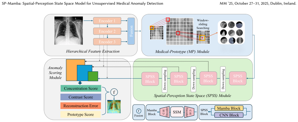
The load-bearing mechanism is the Circular-Hilbert scanning Mamba block inside a pyramidal auto-encoder. For each feature map, the inner $\frac{h'}{2}\times\frac{w'}{2}$ patches are serialized along an $n$-order Hilbert curve, which keeps spatially adjacent patches adjacent in the one-dimensional sequence, while the outer ring is scanned circularly; scanning clockwise or counterclockwise, from corners, and inward or outward yields eight directions in the experiments and up to sixteen in principle. This concentrates the state-space model's long-range modeling on the lesion-relevant image center and reduces interference from structurally random borders. The second mechanism is the Medical-Prototype module, which learns $K$ prototypes and matches each prototype patch through a sliding $p\times p$ window, taking the minimum cosine distance, so that normal anatomical variation is absorbed rather than flagged. The third is the scoring rule $S_{\mathrm{total}}=S_{\mathrm{org}}+\alpha S_{\mathrm{p-dist}}+\beta S_{\mathrm{concen}}+\gamma S_{\mathrm{contra}}$, where $\beta$ is negative and $\gamma$ positive in the selected configuration, encoding the observed fact that abnormal anomaly maps are more concentrated and higher-contrast than normal ones. These mechanisms carry the argument because each one turns an anatomical or statistical regularity into an explicit inductive bias.
What would settle it
On a held-out dataset of diffuse chest disease (for example, widespread ground-glass opacities rather than a focal pneumonia), compute the total score with and without the $S_{\mathrm{concen}}$ and $S_{\mathrm{contra}}$ terms while keeping the ZhangLab-tuned weights fixed; if the two terms do not raise image-level AUC, or if their class separation collapses when the lesion is spread out, the scoring premise fails. A second check: re-select $\beta$ and $\gamma$ on the CheXpert training set and compare with the fixed ZhangLab values; if fixed values are clearly worse, the reported state of the art rests on test-set tuning.
Extended reading notes
Core claim
The central claim is that the spatial regularity of medical images can be converted into a measurable anomaly-detection advantage. Trained only on normal images, the model is supposed to reconstruct normal features accurately and fail on anomalies; the paper's contribution is to steer that failure toward lesions. The window-sliding prototype learning computes, for each patch of a learned prototype, the minimum cosine distance against all patches in a $p\times p$ window around the corresponding position, so shifted anatomy is matched instead of scored as anomalous. The Circular-Hilbert scanning method applies a Hilbert curve to the central half of each feature map and circular scans to the surrounding region, producing eight scan directions used by the Mamba decoder. At test time the anomaly score combines reconstruction distance, prototype distance, the concentration score $S_{\mathrm{concen}}$, and the difference-of-Gaussians contrast score $S_{\mathrm{contra}}$. On the paper's benchmarks, the full system is reported to reach 92.0% AUC on the ZhangLab chest X-ray dataset, 80.4% AUC on CheXpert, and the best mean image-level and pixel-level results on Uni-Medical.
Load-bearing premise
The load-bearing premise is that the concentration and contrast statistics of the anomaly map separate normal from abnormal images in every modality the model is applied to, and that the scoring weights $\beta=-0.025$ and $\gamma=400$, chosen by searching on the ZhangLab test set, keep working on CheXpert and Uni-Medical without retuning; the paper supports the statistical separation with two visual examples and reports no validation-set-based selection of the weights.
Editorial extensions
If this is right
- If the reported numbers hold, Mamba-based detectors become a practical choice for chest X-ray screening: higher AUC and accuracy than transformer-based baselines with roughly half the parameters and FLOPs.
- The same three-component recipe (prototype matching, locality-preserving scan, and anomaly-map statistics) transfers across modalities: the paper reports gains on brain MRI, liver CT, and retinal OCT under the Uni-Medical protocol.
- The anomaly scoring module is additive: the ablation shows that adding the contrast score and then the concentration score improves AUC and F1 on both chest X-ray datasets, implying the two statistics capture complementary signal.
- Because the model runs at 25.8M parameters and 8.3 GFLOPs, deployment on modest hardware or higher-resolution inputs becomes plausible without the quadratic attention cost of transformers.
Reading between the lines
- A testable extension the paper leaves implicit: the concentration and contrast scores are post-hoc statistics computed from whatever anomaly map the model produces, so they could be attached to other reconstruction-based detectors and evaluated for the same gain.
- The window-sliding prototype distance is close to a soft registration step; one could push this further by reading out the displacement that achieves the minimum distance as an explicit anomaly signal, which might catch lesions that manifest as gross anatomical displacement rather than intensity change.
- The Circular-Hilbert scan's division of labor (locality-preserving center, circular periphery) suggests a natural 3D extension for volumetric CT or MRI: Hilbert-scan in-plane and sweep slices circularly, preserving linear complexity.
Signed reviews
Editorial analysis
A structured set of objections, weighed in public.
Referee Report
Summary. The paper introduces SP-Mamba, an unsupervised medical anomaly detection model based on a Mamba state-space decoder. It contributes (i) a Circular-Hilbert scanning scheme that prioritizes central image regions for the Mamba blocks, (ii) a window-sliding medical prototype module that matches normal features against learned prototypes within a local window, and (iii) an anomaly scoring function that combines reconstruction error, prototype distance, and two post-hoc statistics of the anomaly map: a concentration score and a DoG-based contrast score. The method is evaluated on ZhangLab Chest X-ray, CheXpert, and Uni-Medical (brain/liver/retinal), reporting state-of-the-art AUC/Acc/F1 or AUC/AP/F1 with about 25.8M parameters and 8.3 GFLOPs. The central empirical claim is that SP-Mamba outperforms all compared methods, including SQUID, SimSID, and MambaAD, on all three benchmarks.
Significance. If the reported results are reproducible, SP-Mamba is a valuable contribution: it achieves competitive or better accuracy than transformer-based methods at roughly half the parameters and FLOPs, and it offers a set of design ideas (scanning order, prototypes, scoring statistics) that are clearly described and ablated. The paper ships code and the ablations in Table 4 isolate the contribution of each component. However, the main empirical claim is currently weakened by the fact that key hyperparameters, in particular the weights beta and gamma of the concentration and contrast scores, are selected on the ZhangLab test set with no validation split; this makes the reported SOTA on ZhangLab an optimistic, selection-biased estimate, and the transfer of those weights to CheXpert and Uni-Medical is not demonstrated. The significance of the work therefore depends on fixing this evaluation protocol.
major comments (3)
- [Section 4.4, Figure 6] Hyperparameters K, alpha, p, sigma, k_sigma, beta, and gamma are selected using AUC on the ZhangLab test set, and no validation split or cross-validation is described. Consequently, the ZhangLab numbers in Table 1 are not an independent estimate of generalization; the reported 92.0 AUC is the result of test-set selection. Please add a validation protocol (e.g., a held-out split of the training set, or repeated cross-validation on the training set) and report test-set performance only after fixing all hyperparameters. Also state explicitly whether the same hyperparameters are used for CheXpert and Uni-Medical or whether they were re-tuned per dataset.
- [Table 4, Eqs. (4), (5), (7)] The ablation shows that adding S_concen and S_contra is the largest single improvement on CheXpert (AUC 73.1 to 80.4), yet the premise that anomalous maps are concentrated and high-contrast while normal maps are dispersed and low-contrast is supported only by two visual examples in Figure 4. Because beta and gamma are tuned on the ZhangLab test set, the CheXpert and Uni-Medical gains could reflect overfitting to the tuning statistic rather than a general property of medical anomaly maps. Please provide quantitative evidence: per-dataset distributions of S_concen and S_contra for normal versus abnormal images (e.g., box plots or histograms), and a sensitivity analysis of the Table 2 and Table 3 results to beta and gamma over a grid, to show the reported ranking is not an artifact of the chosen weights.
- [Table 2, Section 4.2] The statement that SP-Mamba "outperforms all the comparative methods" on CheXpert is not supported by the F1 column: SP-Mamba reports F1 = 75.8±1.5 while SQUID reports 75.9±5.7, so SP-Mamba's F1 is not higher. Please restrict the SOTA claim to AUC and Acc, or provide a statistical comparison (e.g., paired tests across runs) that justifies a claim of overall superiority.
minor comments (7)
- [Eq. (3)] The recurrence for the Hilbert matrix H_{n+1} is typeset in a garbled, hard-to-read way; please rewrite it cleanly and verify the formula against a standard Hilbert curve construction.
- [Section 3.4, Eq. (5)] The text says G is "a two-dimensional Gaussian function with mean sigma," but the formula has zero mean and standard deviation sigma; please correct the wording to "standard deviation sigma."
- [Section 5] The conclusion claims SP-Mamba "attempts to firstly apply Mamba in medical AD," but MambaAD [11] already applies Mamba to medical anomaly detection on the Uni-Medical benchmark; please reword this claim to avoid inaccuracy.
- [Table 3] Uni-Medical results are reported as means without standard deviations or error bars; please add standard deviations across runs or specify that results come from a single run.
- [Section 4.4 / Eq. (2)] The choice of the loss weight epsilon (set to 25) is stated but not analyzed; please include epsilon in the hyperparameter study or provide a brief justification for the selected value.
- [Figure 6] The captions for Figure 6 should state the dataset and metric (AUC) and should specify which hyperparameters are held fixed while the plotted one is varied.
- [Section 4.4 heading] The heading "Affect of Medical-Prototype Module" should read "Effect of Medical-Prototype Module."
Circularity Check
ZhangLab SOTA is partly a test-set-selected result: the anomaly-score weights beta and gamma in Eq. 7 are tuned on the ZhangLab test set and then reported on that same test set; the rest of the pipeline is self-contained.
-
fitted input called prediction
[Section 4.4, Eq. (7), Table 1 (ZhangLab Chest X-ray)]
"On the ZhangLab Chest X-ray dataset, we select the weight β of S_concen as -0.025 and the weight γ of S_contra as 400 in the final model."
The final anomaly score is S_total = S_org + α·S_p−dist + β·S_concen + γ·S_contra (Eq. 7). Section 4.4 states that β and γ, together with K, α, p, σ and kσ, are selected by evaluating on the ZhangLab test set, and Table 1 then reports SP-Mamba's 92.0 AUC on that same ZhangLab test set as the headline SOTA. No held-out validation split is described. The concentration/contrast terms enter linearly in Eq. 7, so their coefficients can be tuned to maximize the very metric being reported; the ablation jump from 90.5 to 92.0 on ZhangLab when adding S_concen and S_contra is therefore partly a selected rather than predicted gain. The CheXpert and Uni-Medical results use the ZhangLab-selected weights as transfer and are not forced by the same construction, so the circularity is partial.
full rationale
SP-Mamba's core derivation is not circular: the pretrained ResNet34 encoder, MSE reconstruction loss, window-sliding prototype distance, and Circular-Hilbert Mamba decoder are defined by the paper's own equations and evaluated against external baselines such as SQUID, SimSID, and MambaAD. There is no load-bearing self-citation chain and no imported uniqueness theorem; the Circular-Hilbert ablation alone improves ZhangLab AUC from 86.2 to 90.0, and the CheXpert and Uni-Medical tables use hyperparameters selected on a different dataset, so those results retain independent content. The one genuine circularity-like step is the ZhangLab test-set tuning of the anomaly-scoring weights β and γ (and other scoring hyperparameters), followed by reporting the ZhangLab test-set result as an out-of-sample state of the art. Because the ZhangLab SOTA claim is in part a fitted result while the architecture and cross-dataset results are not definitionally forced, a moderate score of 4 is appropriate rather than a higher score.
Assumptions & free parameters
free parameters (8)
- beta (weight of concentration score) =
-0.025 on ZhangLab
- gamma (weight of contrast score) =
400 on ZhangLab
- alpha (weight of prototype distance) =
1 on ZhangLab
- K (number of prototypes) =
10 on ZhangLab; different per dataset
- p (sliding window side length) =
3 on ZhangLab
- sigma (Gaussian std for contrast score) =
0.6 on ZhangLab
- k_sigma (DoG scale multiplier) =
1.2 on ZhangLab
- epsilon (loss weight for prototype distance) =
25
assumptions (5)
- domain assumption Normal images can be reconstructed accurately while abnormal images produce large reconstruction errors.
- domain assumption Radiography images have consistent anatomical patterns and spatial position is informative for normality.
- domain assumption Anomaly maps of abnormal images are more concentrated and higher contrast than those of normal images.
- ad hoc to paper Lesions are concentrated in the central region of the image, so the C-H scan should prioritize the central h'/2 x w'/2 patches.
- standard math Hilbert curves preserve 2D locality when mapped to 1D sequences.
Cite this review
Pith. "Pith review of SP-Mamba: Spatial-Perception State Space Model for Unsupervised Medical Anomaly Detection." pith.science (2026). https://pith.science/paper/BKVSN4WL
@misc{pith2026250719076,
  author       = {Pith},
  title        = {Pith review of: SP-Mamba: Spatial-Perception State Space Model for Unsupervised Medical Anomaly Detection},
  year         = {2026},
  howpublished = {\url{https://pith.science/paper/BKVSN4WL}},
  note         = {Machine review of arXiv:2507.19076}
}
read the original abstract
Radiography imaging protocols target on specific anatomical regions, resulting in highly consistent images with recurrent structural patterns across patients. Recent advances in medical anomaly detection have demonstrated the effectiveness of CNN- and transformer-based approaches. However, CNNs exhibit limitations in capturing long-range dependencies, while transformers suffer from quadratic computational complexity. In contrast, Mamba-based models, leveraging superior long-range modeling, structural feature extraction, and linear computational efficiency, have emerged as a promising alternative. To capitalize on the inherent structural regularity of medical images, this study introduces SP-Mamba, a spatial-perception Mamba framework for unsupervised medical anomaly detection. The window-sliding prototype learning and Circular-Hilbert scanning-based Mamba are introduced to better exploit consistent anatomical patterns and leverage spatial information for medical anomaly detection. Furthermore, we excavate the concentration and contrast characteristics of anomaly maps for improving anomaly detection. Extensive experiments on three diverse medical anomaly detection benchmarks confirm the proposed method's state-of-the-art performance, validating its efficacy and robustness. The code is available at https://github.com/Ray-RuiPan/SP-Mamba.
Figures


Figures from the paper (8 more)


Reference graph
Works this paper leans on
-
[1]
Jinan Bao, Hanshi Sun, Hanqiu Deng, Yinsheng He, Zhaoxiang Zhang, and Xingyu Li. 2024. BMAD: Benchmarks for Medical Anomaly Detection. http: //arxiv.org/abs/2306.11876 arXiv:2306.11876 [eess]
arXiv 2024
-
[2]
Yu Cai, Hao Chen, Xin Yang, Yu Zhou, and Kwang-Ting Cheng. 2022. Dual- distribution discrepancy for anomaly detection in chest x-rays. InInternational Conference on Medical Image Computing and Computer-Assisted Intervention. Springer, 584–593
work page 2022
-
[3]
Yuanhong Chen, Yu Tian, Guansong Pang, and Gustavo Carneiro. 2022. Deep One-Class Classification via Interpolated Gaussian Descriptor. arXiv:2101.10043 [cs.CV] https://arxiv.org/abs/2101.10043
work page Pith review arXiv 2022
-
[4]
Thomas Defard, Aleksandr Setkov, Angelique Loesch, and Romaric Audigier
-
[5]
Hanqiu Deng and Xingyu Li. 2022. Anomaly Detection via Reverse Distilla- tion From One-Class Embedding. InProceedings of the IEEE/CVF Conference on Computer Vision and Pattern Recognition (CVPR). 9737–9746
work page 2022
-
[6]
Tharindu Fernando, Harshala Gammulle, Simon Denman, Sridha Sridharan, and Clinton Fookes. 2021. Deep learning for medical anomaly detection–a survey. ACM Computing Surveys (CSUR)54, 7 (2021), 1–37
2021
-
[8]
Albert Gu and Tri Dao. 2024. Mamba: Linear-Time Sequence Modeling with Selective State Spaces. arXiv:2312.00752 [cs.LG] https://arxiv.org/abs/2312.00752
arXiv 2024
-
[9]
Hang Guo, Yong Guo, Yaohua Zha, Yulun Zhang, Wenbo Li, Tao Dai, Shu-Tao Xia, and Yawei Li. 2025. MambaIRv2: Attentive State Space Restoration. doi:10. 48550/arXiv.2411.15269 arXiv:2411.15269 [eess]
Show all 37 references
-
[10]
Fatemeh Haghighi, Mohammad Reza Hosseinzadeh Taher, Zongwei Zhou, Michael B Gotway, and Jianming Liang. 2021. Transferable visual words: Ex- ploiting the semantics of anatomical patterns for self-supervised learning.IEEE transactions on medical imaging40, 10 (2021), 2857–2868
2021
-
[11]
Haoyang He, Yuhu Bai, Jiangning Zhang, Qingdong He, Hongxu Chen, Zhenye Gan, Chengjie Wang, Xiangtai Li, Guanzhong Tian, and Lei Xie. 2024. MambaAD: Exploring State Space Models for Multi-class Unsupervised Anomaly Detection. http://arxiv.org/abs/2404.06564 arXiv:2404.06564 [cs]
2024 arXiv
-
[12]
Haoyang He, Jiangning Zhang, Hongxu Chen, Xuhai Chen, Zhishan Li, Xu Chen, Yabiao Wang, Chengjie Wang, and Lei Xie. 2023. DiAD: A Diffusion- based Framework for Multi-class Anomaly Detection. arXiv:2312.06607 [cs.CV] https://arxiv.org/abs/2312.06607
2023 arXiv
-
[13]
Chaoqin Huang, Aofan Jiang, Jinghao Feng, Ya Zhang, Xinchao Wang, and Yan- feng Wang. 2024. Adapting Visual-Language Models for Generalizable Anomaly Detection in Medical Images. InProceedings of the IEEE/CVF Conference on Com- puter Vision and Pattern Recognition (CVPR). 11375–11385
2024
-
[14]
Mong, Safwan S
Jeremy Irvin, Pranav Rajpurkar, Michael Ko, Yifan Yu, Silviana Ciurea-Ilcus, Chris Chute, Henrik Marklund, Behzad Haghgoo, Robyn Ball, Katie Shpanskaya, Jayne Seekins, David A. Mong, Safwan S. Halabi, Jesse K. Sandberg, Ricky Jones, David B. Larson, Curtis P. Langlotz, Bhavik ...
2019 arXiv
-
[15]
Kermany, Michael Goldbaum, Wenjia Cai, Carolina C
Daniel S. Kermany, Michael Goldbaum, Wenjia Cai, Carolina C. S. Valentim, Huiying Liang, Sally L. Baxter, Alex McKeown, Ge Yang, Xiaokang Wu, Fangbing Yan, Justin Dong, Made K. Prasadha, Jacqueline Pei, Magdalene Y. L. Ting, Jie Zhu, Christina Li, Sierra Hewett, Jason Dong, Ia...
2018
-
[16]
Chun-Liang Li, Kihyuk Sohn, Jinsung Yoon, and Tomas Pfister. 2021. CutPaste: Self-Supervised Learning for Anomaly Detection and Localization. InProceedings of the IEEE/CVF Conference on Computer Vision and Pattern Recognition (CVPR). 9664–9674
2021
-
[17]
Yue Liu, Yunjie Tian, Yuzhong Zhao, Hongtian Yu, Lingxi Xie, Yaowei Wang, Qixiang Ye, Jianbin Jiao, and Yunfan Liu. 2024. Vmamba: Visual state space model. Advances in neural information processing systems37 (2024), 103031–103063
2024
-
[18]
Yue Liu, Yunjie Tian, Yuzhong Zhao, Hongtian Yu, Lingxi Xie, Yaowei Wang, Qixiang Ye, and Yunfan Liu. 2024. VMamba: Visual State Space Model. http: //arxiv.org/abs/2401.10166 arXiv:2401.10166 [cs]
2024 arXiv
-
[19]
Zhikang Liu, Yiming Zhou, Yuansheng Xu, and Zilei Wang. 2023. SimpleNet: A Simple Network for Image Anomaly Detection and Localization. InProceedings of the IEEE/CVF Conference on Computer Vision and Pattern Recognition (CVPR). 20402–20411
2023
-
[20]
Shuai Lu, Weihang Zhang, He Zhao, Hanruo Liu, Ningli Wang, and Huiqi Li
-
[21]
Sergio Naval Marimont and Giacomo Tarroni. 2021. Implicit field learning for unsupervised anomaly detection in medical images. arXiv:2106.05214 [eess.IV] https://arxiv.org/abs/2106.05214
2021 arXiv
-
[22]
Tal Reiss, Niv Cohen, Liron Bergman, and Yedid Hoshen. 2021. PANDA: Adapting Pretrained Features for Anomaly Detection and Segmentation. InProceedings of the IEEE/CVF Conference on Computer Vision and Pattern Recognition (CVPR). 2806–2814
2021
-
[23]
Jiacheng Ruan, Jincheng Li, and Suncheng Xiang. 2024. Vm-unet: Vision mamba unet for medical image segmentation.arXiv preprint arXiv:2402.02491(2024)
2024 arXiv
-
[24]
Rohban, and Hamid R
Mohammadreza Salehi, Niousha Sadjadi, Soroosh Baselizadeh, Mohammad H. Rohban, and Hamid R. Rabiee. 2021. Multiresolution Knowledge Distillation for Anomaly Detection. InProceedings of the IEEE/CVF Conference on Computer Vision and Pattern Recognition (CVPR). 14902–14912
2021
-
[25]
Waldstein, Georg Langs, and Ursula Schmidt-Erfurth
Thomas Schlegl, Philipp Seeböck, Sebastian M. Waldstein, Georg Langs, and Ursula Schmidt-Erfurth. 2019. f-AnoGAN: Fast unsupervised anomaly detection with generative adversarial networks.Medical Image Analysis54 (2019), 30–44. doi:10.1016/j.media.2019.01.010
2019 doi
-
[26]
Waldstein, Ursula Schmidt- Erfurth, and Georg Langs
Thomas Schlegl, Philipp Seeböck, Sebastian M. Waldstein, Ursula Schmidt- Erfurth, and Georg Langs. 2017. Unsupervised Anomaly Detection with Genera- tive Adversarial Networks to Guide Marker Discovery. arXiv:1703.05921 [cs.CV] https://arxiv.org/abs/1703.05921
2017 arXiv
-
[27]
Yuan Shi, Bin Xia, Xiaoyu Jin, Xing Wang, Tianyu Zhao, Xin Xia, Xuefeng Xiao, and Wenming Yang. 2024. VmambaIR: Visual State Space Model for Image Restoration. arXiv:2403.11423 [cs.CV] https://arxiv.org/abs/2403.11423
2024 arXiv
-
[28]
Yuan Shi, Bin Xia, Xiaoyu Jin, Xing Wang, Tianyu Zhao, Xin Xia, Xuefeng Xiao, and Wenming Yang. 2025. Vmambair: Visual state space model for image restoration.IEEE Transactions on Circuits and Systems for Video Technology(2025)
2025
-
[29]
Ziyang Wang, Jian-Qing Zheng, Yichi Zhang, Ge Cui, and Lei Li. 2024. Mamba- unet: Unet-like pure visual mamba for medical image segmentation.arXiv preprint arXiv:2402.05079(2024)
2024 arXiv
- [30]
-
[31]
Tiange Xiang, Yixiao Zhang, Yongyi Lu, Alan Yuille, Chaoyi Zhang, Weidong Cai, and Zongwei Zhou. 2024. Exploiting Structural Consistency of Chest Anatomy for Unsupervised Anomaly Detection in Radiography Images.IEEE Transactions on Pattern Analysis and Machine Intelligence46, ...
2024
-
[32]
Yuille, Chaoyi Zhang, Weidong Cai, and Zongwei Zhou
Tiange Xiang, Yixiao Zhang, Yongyi Lu, Alan L. Yuille, Chaoyi Zhang, Weidong Cai, and Zongwei Zhou. 2023. SQUID: Deep Feature In-Painting for Unsupervised Anomaly Detection. http://arxiv.org/abs/2111.13495 arXiv:2111.13495 [cs]
2023 arXiv
-
[33]
Jianpeng Zhang, Yutong Xie, Guansong Pang, Zhibin Liao, Johan Verjans, Wenx- ing Li, Zongji Sun, Jian He, Yi Li, Chunhua Shen, et al. 2020. Viral pneumonia screening on chest X-rays using confidence-aware anomaly detection.IEEE transactions on medical imaging40, 3 (2020), 879–890
2020
-
[34]
Xuan Zhang, Shiyu Li, Xi Li, Ping Huang, Jiulong Shan, and Ting Chen. 2023. DeSTSeg: Segmentation Guided Denoising Student-Teacher for Anomaly Detec- tion. InProceedings of the IEEE/CVF Conference on Computer Vision and Pattern Recognition (CVPR). 3914–3923
2023
-
[36]
He Zhao, Yuexiang Li, Nanjun He, Kai Ma, Leyuan Fang, Huiqi Li, and Yefeng Zheng. 2021. Anomaly Detection for Medical Images Using Self-Supervised and Translation-Consistent Features.IEEE Transactions on Medical Imaging40, 12 (2021), 3641–3651. doi:10.1109/TMI.2021.3093883
2021
-
[37]
Lianghui Zhu, Bencheng Liao, Qian Zhang, Xinlong Wang, Wenyu Liu, and Xinggang Wang. 2024. Vision Mamba: Efficient Visual Representation Learn- ing with Bidirectional State Space Model. http://arxiv.org/abs/2401.09417 arXiv:2401.09417 [cs]. A More Qualitative Results MM ’25, O...
2024 arXiv
-
[2020]
arXiv:2011.08785 [cs.CV] https://arxiv.org/abs/2011.08785
PaDiM: a Patch Distribution Modeling Framework for Anomaly Detection and Localization. arXiv:2011.08785 [cs.CV] https://arxiv.org/abs/2011.08785
2011 arXiv
-
[2024]
IEEE Transactions on Image Processing33 (2024), 2770–2782
Anomaly Detection for Medical Images Using Heterogeneous Auto-Encoder. IEEE Transactions on Image Processing33 (2024), 2770–2782. doi:10.1109/TIP.2024. 3381435 Conference Name: IEEE Transactions on Image Processing
2024 doi
Reviewed August 15, 2026 · model on record in the stance chip above.
Discussion (0). Continue with ORCID to comment.