REVIEW 4 major objections 6 minor 39 references
M-Net: MRI Brain Tumor Sequential Segmentation Network via Mesh-Cast
T0 review · 4 major / 6 minor · reviewed 2026-08-15 · deepseek-v4-flash
Pith's one-line read M-Net treats MRI slice stacks as temporal sequences and, via a dimension-swapping Mesh-Cast mechanism plus a shuffle-then-order training schedule, reports the best tumor segmentation scores on BraTS2019 and BraTS2023.
desk verdict The Mesh-Cast/TPS ideas are real and worth a look, but the SOTA claim is unsupported as presented because the evaluation rests on one private split with no error bars, tiny margins, and TPS not applied to baselines. read the letter →
The pith
A machine-rendered reading of the paper's core claim, the machinery that carries it, and where it could break.
The reading
What carries the argument
Mesh-Cast is a transpose-and-sweep wrapper around any Sequential Module. Input features have shape T frames by C channels by D flattened pixels; the module first runs the sequence model with T as the sequence length and C treated like a batch dimension, then swaps the T and C roles with a transpose, runs the same sequence model along the channel axis, and transposes back. This repeated mesh of temporal and channel sweeps lets correlations across slices and across MRI modalities both be captured. The Two-Phase Sequential training strategy first trains on global frame-level shuffles and then fine-tunes on ordered sequences, which the paper argues lets the model learn common lesion patterns before sequence-specific continuity.
What would settle it
Run the released M-Net and all twelve baselines from Table 4 under the paper's exact protocol (8:2 split, 160x160 cropping, Z-score normalization, 300 epochs with early stopping at 30) and check whether M-Net still wins every metric on both datasets; if any baseline reaches or exceeds its Dice or Hausdorff95 values, the universal-superiority claim fails.
Extended reading notes
Core claim
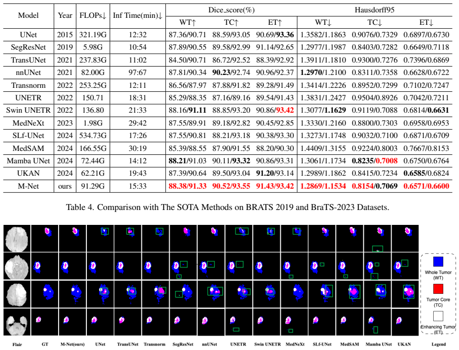
The central claim is that treating a stack of T MRI slices as a sequence, and pushing a sequential model first along the slice axis and then along the channel axis, is sufficient to reach the top of brain tumor segmentation benchmarks. On BraTS2019, M-Net reports Dice scores of 88.38 for whole tumor, 90.52 for tumor core, and 91.43 for enhancing tumor, with corresponding Hausdorff95 distances of 1.2869, 0.8154, and 0.6571; on BraTS2023, it reports 91.33, 93.55, and 93.42, leading every baseline on both datasets. The paper attributes the gain to the Mesh-Cast dual-axis sweep rather than to any particular sequence model, since the same wrapper improves LSTM, ConvLSTM, xLSTM, Transformer, and Mamba variants over their slice-only backbones by up to 1.2 Dice points.
Load-bearing premise
The evaluation protocol is fair and reproducible: all methods were retrained on the same 8:2 split, the same cropping and normalization, the same loss, and the same early-stopping rule, with no hidden hyperparameter tuning that favours M-Net.
Editorial extensions
If this is right
- Plugging Mesh-Cast into an existing 2D encoder-decoder should produce immediate gains on slice-stack medical data, because every tested sequence model improved over the slice-only backbone.
- Sequence models become interchangeable components: the best variant uses Mamba SSM, but LSTM, ConvLSTM, xLSTM, and Transformer variants all improve, so future backbones can swap in newer sequence models without redesign.
- Volumetric segmentation becomes feasible on limited hardware: M-Net's 91.29 GFLOPs and roughly 15-minute inference time compare with nnU-Net's 82 GFLOPs and roughly 97-minute inference time under the paper's setup.
- The TPS schedule is a separable contribution: phase-one shuffling alone already beats the ordered backbone on most metrics, and the full shuffle-then-order schedule is better than either phase alone.
Reading between the lines
- Mesh-Cast is potentially a general plug-in for any consistent-pattern sequence of images, such as CT slice stacks, video frames, or multi-view scans; the paper states this generality but does not test it.
- The channel-axis sweep may be doing more of the work than the paper's ablation fully isolates, because co-registered MRI modalities make channel-order correlations a proxy for inter-modal alignment; a test on single-modality sequences would separate the two.
- The TPS shuffle phase could act as a data augmentation that increases effective dataset diversity, which would predict larger gains on small datasets and diminishing returns as data grows; this is testable by scaling the training set.
- A direct ablation that replaces Mesh-Cast with simple concatenation of adjacent slices would clarify whether the dual-axis sweep itself, rather than the added sequence model, drives the reported gains.
Signed reviews
Editorial analysis
A structured set of objections, weighed in public.
Referee Report
Summary. The paper proposes M-Net, a 2D slice-sequence segmentation framework that treats the MRI slice stack as a temporal sequence. A Mesh-Cast mechanism repeatedly transposes the temporal and channel dimensions so that arbitrary sequential models (LSTM, ConvLSTM, xLSTM, Transformer, Mamba SSM) can process both inter-slice and inter-modality relationships, and a Two-Phase Sequential (TPS) training strategy first trains on globally shuffled sequences then fine-tunes on ordered sequences. The method is evaluated on BraTS2019 and BraTS2023 using an 8:2 split of the official training sets, with Dice and Hausdorff95 metrics, and the authors claim state-of-the-art performance on all regions. The paper also includes ablations across sequential backbones and training phases.
Significance. The conceptual contribution—reinterpreting adjacent MRI slices as a temporal spectrum and using a dimension-swapping mechanism to feed both slice and channel information into any sequence model—is interesting and could offer a computationally cheaper alternative to full 3D convolutions while retaining volumetric context. The TPS training strategy is simple, clearly described, and potentially generalizable to other sequence-based segmentation tasks. The ablation study across five sequential modules is a useful practical comparison. However, the central claim of state-of-the-art performance is not verifiable as presented because the evaluation relies on a private split, all baselines are retrained with unreported hyperparameters, no code or seeds are released, no repeated-run statistics are given, and the reported margins are very small. If the authors make the protocol reproducible and add statistical grounding, the method could be a valuable contribution; in its current form the empirical evidence is insufficient to support the strong SOTA claim.
major comments (4)
- [Section 4.4, Tables 2–4] The state-of-the-art claim rests on a single private 8:2 split of the BraTS training sets, with all comparison methods retrained by the authors under unreported hyperparameters, no fixed random seeds, no repeated runs, and no statistical tests. The margins over the strongest baselines are very small (e.g., BraTS2019 WT Dice 88.38 vs. 88.21 for Mamba UNet; BraTS2023 ET Dice 93.42 vs. 93.42 for Swin UNETR, a tie), so the reported superiority could plausibly be within run-to-run and split-to-split variability. Please release code and exact training configurations for every baseline, report mean±std over multiple seeds, and provide statistical significance tests, or explicitly reposition the results as preliminary.
- [Section 4.4, Table 3] The comparison conflates the architecture contribution with the training-strategy contribution because TPS is applied only to M-Net and not to any baseline. Table 3 shows that Backbone(Shuffled) alone already achieves 88.21/90.11/90.86 Dice on BraTS2019, which is comparable to or better than most of the baselines in Table 4; the full M-Net with TPS reaches 88.38/90.52/91.43. To support the claim that the architecture is responsible for the gains, the authors should apply TPS (at least the shuffle phase) to the baseline models, or otherwise separate the architecture and training-strategy effects in the comparison.
- [Section 4.1, Eq. (1), Table 1] The sequence length T, which is central to the method, is never specified for either dataset, and Table 1 is internally inconsistent. For BraTS2019, 2483+275+702=3460 sequences but the slice counts sum to 51925, which at 155 slices per case corresponds to exactly 335 cases, not 3460 sequences. For BraTS2023 the sequence counts (11250/3750/3763) do not reconcile with the slice counts divided by 155 either. This makes the input construction unverifiable and suggests a mismatch between the reported 'sequences' and actual MRI series. Please state T explicitly for each experiment and correct the data count table.
- [Section 4.4, Table 4, Abstract] The paper's claim of outperforming 'all key metrics' is contradicted by its own results: on BraTS2023 ET Dice, M-Net (93.42) ties with Swin UNETR (93.42), so the correct summary is 'almost all' metrics. Additionally, the BraTS2019 pattern in which ET Dice (91.43) exceeds WT Dice (88.38) is highly atypical—the enhancing tumor region is normally the most difficult—and the paper offers no explanation for this ordering. Please correct the overclaim and provide an analysis of the unusual region-wise performance pattern, as it may indicate an artifact of the evaluation protocol.
minor comments (6)
- [Section 4.2, Eq. (23)] Eq. (23) defines the standard Hausdorff distance, not the 95th percentile Hausdorff distance (HD95) that is used in the experiments; please provide the correct definition of HD95.
- [Table 4] The nnUNet row reports inference time as '97:67', which is not a valid mm:ss format (likely 97 minutes 67 seconds, i.e., 98:07); the UNETR FLOPs value '150.71' is missing the unit (presumably G); and the 'SegResNet' label is inconsistently spaced.
- [Author affiliation block] The author email 'sh1yuzh@163.con' contains a typo ('.con' instead of '.com').
- [Table 2] The column header 'Dicescore(%)' should be 'Dice score (%)', and the FLOPs column should state the unit for all rows consistently (e.g., 'G').
- [Section 3.3] The notation for sequence length is inconsistent: Eq. (16) uses T, but the text says 'each sequence Xi contain t elements'; please unify the symbols.
- [Figure 8] The caption for Figure 8 reads 'Multi-sequential Module (TPS)' but the text discusses different M-Net configurations; please align the caption with the actual configurations shown.
Circularity Check
No significant circularity: M-Net's contributions are architectural and empirical; the only self-citation is a non-load-bearing baseline reference.
full rationale
The paper does not derive its results from first principles; it proposes an architecture (Mesh-Cast), a training strategy (TPS), and evaluates them empirically on BraTS 2019 and 2023. The central comparison is against external, published baselines on public benchmark data, so the headline SOTA claim is a benchmark result rather than a fitted identity. The Mesh-Cast mechanism is defined as a dimensional transpose-and-process operation over temporal and channel axes; it is not defined in terms of the reported Dice or Hausdorff95 numbers, and no equation equates a predicted metric to a fitted parameter. The TPS training strategy is a training schedule, not a fitted parameter renamed as a prediction; Table 3 openly shows how much of the gain comes from the shuffle phase. The only self-citation is SLf-UNet [9], whose authors overlap with the current paper, but it appears solely as one of many comparison baselines in Table 4 and is not used to justify any methodological premise or to rule out alternatives. Evaluation concerns raised elsewhere, such as the private 8:2 split, the absence of error bars, TPS not being applied to baselines, and reporting inconsistencies (e.g., nnUNet inference time '97:67'), are reproducibility and fairness risks, not circularity by construction under the definitions used here. No load-bearing step reduces to its own inputs, and no uniqueness theorem or cited prior result from the same authors is used to force the design choice.
Assumptions & free parameters
free parameters (3)
- sequence length T
- loss weighting lambda
- layer attention coefficients beta_i =
less than 1
assumptions (3)
- domain assumption Adjacent MRI slices exhibit sufficient 'temporal-like' spatial correlation to be modeled as sequences.
- domain assumption The BraTS ground truth annotations and MRI preprocessing (cropping to 160x160, Z-score normalization) are accurate and do not disadvantage any method.
- ad hoc to paper Retraining all baseline models with the same loss function and the same early-stopping criterion is sufficient for a fair comparison.
Cite this review
Pith. "Pith review of M-Net: MRI Brain Tumor Sequential Segmentation Network via Mesh-Cast." pith.science (2026). https://pith.science/paper/FURMH4AZ
@misc{pith2026250720582,
  author       = {Pith},
  title        = {Pith review of: M-Net: MRI Brain Tumor Sequential Segmentation Network via Mesh-Cast},
  year         = {2026},
  howpublished = {\url{https://pith.science/paper/FURMH4AZ}},
  note         = {Machine review of arXiv:2507.20582}
}
read the original abstract
MRI tumor segmentation remains a critical challenge in medical imaging, where volumetric analysis faces unique computational demands due to the complexity of 3D data. The spatially sequential arrangement of adjacent MRI slices provides valuable information that enhances segmentation continuity and accuracy, yet this characteristic remains underutilized in many existing models. The spatial correlations between adjacent MRI slices can be regarded as "temporal-like" data, similar to frame sequences in video segmentation tasks. To bridge this gap, we propose M-Net, a flexible framework specifically designed for sequential image segmentation. M-Net introduces the novel Mesh-Cast mechanism, which seamlessly integrates arbitrary sequential models into the processing of both channel and temporal information, thereby systematically capturing the inherent "temporal-like" spatial correlations between MRI slices. Additionally, we define an MRI sequential input pattern and design a Two-Phase Sequential (TPS) training strategy, which first focuses on learning common patterns across sequences before refining slice-specific feature extraction. This approach leverages temporal modeling techniques to preserve volumetric contextual information while avoiding the high computational cost of full 3D convolutions, thereby enhancing the generalizability and robustness of M-Net in sequential segmentation tasks. Experiments on the BraTS2019 and BraTS2023 datasets demonstrate that M-Net outperforms existing methods across all key metrics, establishing itself as a robust solution for temporally-aware MRI tumor segmentation.
Figures


Figures from the paper (6 more)


Reference graph
Works this paper leans on
-
[1]
N. M. AboElenein, S. Piao, A. Noor, and P. N. Ahmed. Mirau-net: An improved neural network based on u-net for gliomas segmentation. Signal Processing: Image Communi- cation, 101:116553, 2022. 1
work page 2022
-
[2]
R. Azad, M. T. Al-Antary, M. Heidari, and D. Merhof. Transnorm: Transformer provides a strong spatial normal- ization mechanism for a deep segmentation model. IEEE Access, 10:108205–108215, 2022. 2, 7
work page 2022
- [3]
- [4]
-
[5]
M. Beck, K. P ¨oppel, M. Spanring, A. Auer, O. Prudnikova, M. Kopp, and S. Hochreiter. xlstm: Extended long short- term memory. arXiv preprint, 2024. 2, 3
work page 2024
-
[6]
Sequential 3D U-Nets for Biologically-Informed Brain Tumor Segmentation
A. Beers, K. Chang, J. Brown, E. Sartor, C. P. Mammen, E. Gerstner, and J. Kalpathy-Cramer. Sequential 3d u-nets for biologically-informed brain tumor segmentation. arXiv preprint arXiv:1709.02967, 2017. 2
work page Pith review arXiv 2017
-
[7]
J. Chen, Y . Lu, Q. Yu, X. Luo, E. Adeli, Y . Wang, and Y . Zhou. Transunet: Transformers make strong en- coders for medical image segmentation. arXiv preprint arXiv:2102.04306, 2021. 2, 7
arXiv 2021
-
[8]
E. Dandıl and S. Karaca. Detection of pseudo brain tumors via stacked lstm neural networks using mr spectroscopy sig- nals. Biocybernetics and Biomedical Engineering , 41(1): 173–195, 2021. 2
work page 2021
Show all 39 references
-
[9]
H. Ding, J. Lu, J. Cai, Y . Zhang, and Y . Shang. Slf-unet: Im- proved unet for brain mri segmentation by combining spatial and low-frequency domain features. In Computer Graphics International Conference, pages 415–426. Cham: Springer Nature Switzerland, 2023. 7
2023
-
[10]
X. Feng, N. J. Tustison, S. H. Patel, and C. H. Meyer. Brain tumor segmentation using an ensemble of 3d u-nets and over- all survival prediction using radiomic features. Frontiers in computational neuroscience, 14:25, 2020. 2
2020
-
[11]
Fukushima
K. Fukushima. Neocognitron: A self-organizing neural net- work model for a mechanism of pattern recognition unaf- fected by shift in position. Biological cybernetics , 36(4): 193–202, 1980. 1
1980
-
[12]
A. Graves. Long short-term memory. 2012. 2
2012
-
[13]
Gu and T
A. Gu and T. Dao. Mamba: Linear-time sequence modeling with selective state spaces. arXiv preprint arXiv:2312.00752, 2023. 2, 3
2023 arXiv
-
[14]
R. Gu, G. Wang, T. Song, R. Huang, M. Aertsen, J. Deprest, and S. Zhang. Ca-net: Comprehensive attention convolu- tional neural networks for explainable medical image seg- mentation. IEEE transactions on medical imaging , 40(2): 699–711, 2020. 1
2020
-
[15]
K. Han, Y . Wang, H. Chen, X. Chen, J. Guo, Z. Liu, and D. Tao. A survey on vision transformer. IEEE transactions on pattern analysis and machine intelligence, 45(1):87–110,
-
[16]
Hatamizadeh, V
A. Hatamizadeh, V . Nath, Y . Tang, D. Yang, H. R. Roth, and D. Xu. Swin unetr: Swin transformers for semantic segmen- tation of brain tumors in mri images. In International MIC- CAI brainlesion workshop, pages 272–284. Cham: Springer International Publishing, 2021. 2, 7
2021
-
[17]
Hatamizadeh, Y
A. Hatamizadeh, Y . Tang, V . Nath, D. Yang, A. Myronenko, B. Landman, and D. Xu. Unetr: Transformers for 3d medical image segmentation. In Proceedings of the IEEE/CVF winter conference on applications of computer vision , pages 574– 584, 2022. 2, 7
2022
-
[18]
Jaeger, Simon A
Fabian Isensee, Paul F. Jaeger, Simon A. A. Kohl, and et al. nnu-net: a self-configuring method for deep learning- based biomedical image segmentation. Nature Methods, 18 (2):203–211, 2021. 7
2021
-
[19]
Video polyp segmentation: A deep learning perspective
Guoping Ji, Guoli Xiao, Yung-Chieh Chou, and et al. Video polyp segmentation: A deep learning perspective. Machine Intelligence Research, 19(6):531–549, 2022. 2
2022
-
[20]
C. Li, X. Liu, W. Li, C. Wang, H. Liu, and Y . Yuan. U-kan makes strong backbone for medical image segmentation and generation. arXiv preprint, 2024. 1, 7
2024
-
[21]
X. Li, X. Sun, Y . Meng, J. Liang, F. Wu, and J. Li. Dice loss for data-imbalanced nlp tasks. arXiv preprint, 2019. 6
2019
-
[22]
Z. P. Liang and P. C. Lauterbur. Principles of magnetic res- onance imaging. SPIE Optical Engineering Press, Belling- ham, 2000. 1
2000
-
[23]
Vmamba: Visual state space model
Yujun Liu, Yuxin Tian, Yujie Zhao, et al. Vmamba: Visual state space model. Advances in Neural Information Process- ing Systems, 37:103031–103063, 2024. 2
2024
-
[24]
Z. Liu, Y . Wang, S. Vaidya, F. Ruehle, J. Halverson, M. Soljaˇci´c, and M. Tegmark. Kan: Kolmogorov-arnold net- works. arXiv preprint, 2024. 2
2024
-
[25]
Mehta and T
R. Mehta and T. Arbel. 3d u-net for brain tumour segmenta- tion. In International MICCAI Brainlesion Workshop, pages 254–266. Cham: Springer International Publishing, 2018. 2
2018
-
[26]
B. H. Menze, A. Jakab, S. Bauer, J. Kalpathy-Cramer, K. Farahani, J. Kirby, and K. Van Leemput. The multimodal brain tumor image segmentation benchmark (brats). IEEE transactions on medical imaging , 34(10):1993–2024, 2014. 6
1993
-
[27]
Myronenko
A. Myronenko. 3d mri brain tumor segmentation using au- toencoder regularization. In Brainlesion: Glioma, Multiple Sclerosis, Stroke and Traumatic Brain Injuries: 4th Interna- tional Workshop, BrainLes 2018, Held in Conjunction with MICCAI 2018, Granada, Spain, September 16, 2...
2018
-
[28]
Pfeuffer, K
A. Pfeuffer, K. Schulz, and K. Dietmayer. Semantic segmen- tation of video sequences with convolutional lstms. In 2019 IEEE intelligent vehicles symposium (IV), pages 1441–1447. IEEE, 2019. 3
2019
-
[29]
Ronneberger, P
O. Ronneberger, P. Fischer, and T. Brox. U-net: Convo- lutional networks for biomedical image segmentation. In Medical image computing and computer-assisted interven- tion–MICCAI 2015: 18th international conference, Munich, Germany, October 5-9, 2015, proceedings, part III 18,...
2015
-
[30]
S. Roy, G. Koehler, C. Ulrich, M. Baumgartner, J. Petersen, F. Isensee, and K. H. Maier-Hein. Mednext: transformer- driven scaling of convnets for medical image segmentation. In International Conference on Medical Image Computing and Computer-Assisted Intervention, pages 405–4...
-
[31]
Shahzadi, T
I. Shahzadi, T. B. Tang, F. Meriadeau, and A. Quyyum. Cnn- lstm: Cascaded framework for brain tumour classification. In 2018 IEEE-EMBS Conference on Biomedical Engineering and Sciences (IECBES), pages 633–637. IEEE, 2018. 2
2018
-
[32]
A. C. Tan, D. M. Ashley, G. Y . L ´opez, M. Malinzak, H. S. Friedman, and M. Khasraw. Management of glioblastoma: State of the art and future directions. CA: a cancer journal for clinicians, 70(4):299–312, 2020. 1
2020
-
[33]
A. Vaswani. Attention is all you need. In Advances in Neural Information Processing Systems, 2017. 2
2017
-
[34]
Z. Wang, J. Q. Zheng, Y . Zhang, G. Cui, and L. Li. Mamba- unet: Unet-like pure visual mamba for medical image seg- mentation. arXiv preprint, 2024. 2, 7
2024
-
[35]
J. Wu, W. Ji, Y . Liu, H. Fu, M. Xu, Y . Xu, and Y . Jin. Medi- cal sam adapter: Adapting segment anything model for med- ical image segmentation. arXiv preprint arXiv:2304.12620,
-
[36]
F. Xu, H. Ma, J. Sun, R. Wu, X. Liu, and Y . Kong. Lstm multi-modal unet for brain tumor segmentation. In 2019 IEEE 4th international conference on image, vision and com- puting (ICIVC), pages 236–240. IEEE, 2019. 2
2019
-
[37]
Zhang, Y
J. Zhang, Y . Xie, Y . Wang, and Y . Xia. Inter-slice context residual learning for 3d medical image segmentation. IEEE Transactions on Medical Imaging, 40(2):661–672, 2020. 2
2020
-
[38]
Crandall, and et al
Tianfei Zhou, Fatih Porikli, David J. Crandall, and et al. A survey on deep learning technique for video segmentation. IEEE Transactions on Pattern Analysis and Machine Intelli- gence, 45(6):7099–7122, 2022. 2
2022
-
[2023]
Springer Nature Switzerland. 2, 7
Reviewed August 15, 2026 · model on record in the stance chip above.
Discussion (0). Continue with ORCID to comment.