REVIEW 1 major objections 4 minor 1 cited by
Coherence of dipole-forbidden Rydberg excitons in Cu$_2$O measured by polarization- and time-resolved multi-photon spectroscopy
T0 review · 1 major / 4 minor · reviewed 2026-08-06 · deepseek-v4-flash
Pith's one-line read 2PE-DFG, a two-photon pump plus difference-frequency probe, directly measures coherence of electric-dipole-forbidden excitons: about 3 ns for the Cu2O 1S orthoexciton and a few picoseconds for n=3,4 Rydberg S and D states.
desk verdict Nice new technique for measuring coherence of dipole-forbidden excitons, but the extracted T2* values likely need a factor-of-2 correction from the intensity-decay analysis. read the letter →
The pith
A machine-rendered reading of the paper's core claim, the machinery that carries it, and where it could break.
The reading
What carries the argument
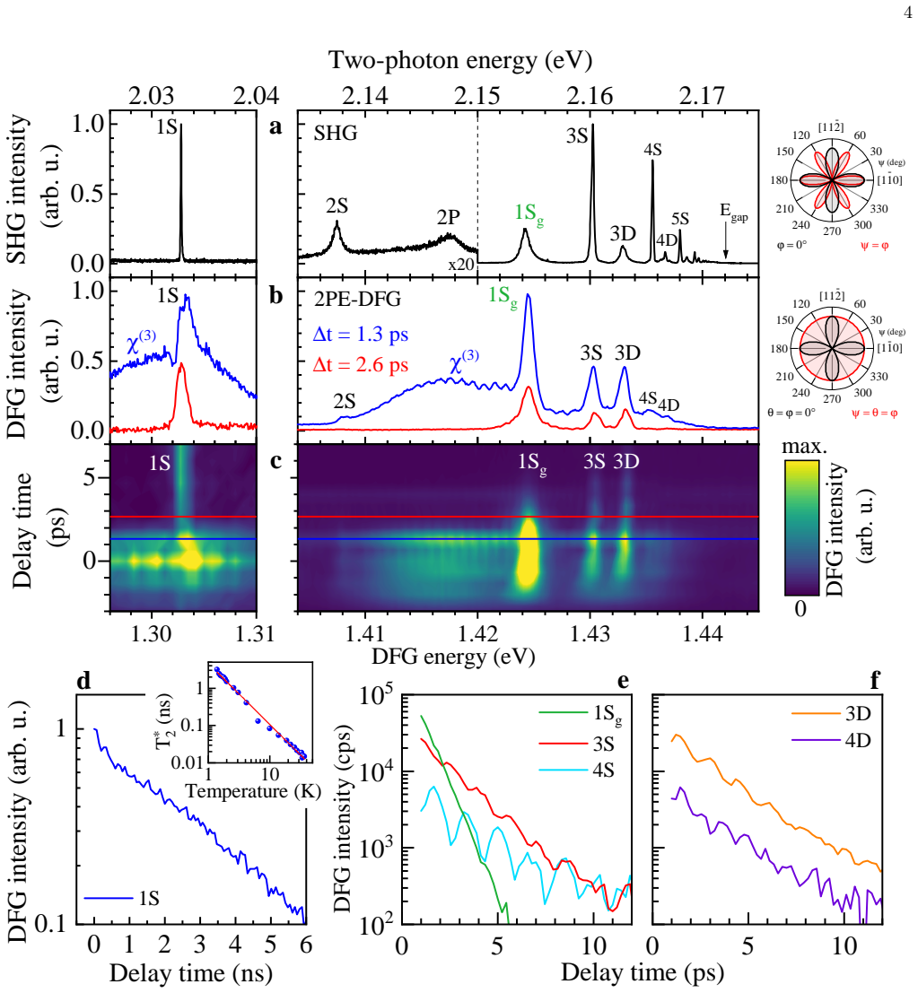
The load-bearing mechanism is the 2PE-DFG sequence: a spectrally broad femtosecond pulse at $\hbar\omega_1$ drives a two-photon transition into an even-parity exciton with $\Gamma_5^+$ symmetry, creating a coherent macroscopic polarization; a delayed picosecond pulse at $\hbar\omega_2$ converts that polarization into difference-frequency light at $\hbar\omega_3 = 2\hbar\omega_1 - \hbar\omega_2$, so the DFG intensity traces the surviving coherent polarization of the same state. Because both excitation and readout are two-photon processes, the method addresses the same exciton component in both channels, avoiding the cross-relaxation dynamics of earlier one-photon/two-photon schemes. Polarization tomography over the three linear angles $\psi$, $\theta$, and $\varphi$ is modeled from group-theoretical coupling coefficients for $\Gamma_5^+$ states and from the magnetic-field Hamiltonian of the $1S$ exciton system, and the signal decay is converted to $T_2^*$ using Eq. (1).
What would settle it
Measure, on the same crystal and at the same temperature, both the DFG decay and the population lifetime $T_1$ of the $1S$ orthoexciton (for example by time-resolved two-photon emission), and check whether $T_2^* \le 2T_1$ holds as Eq. (1) requires; then fire a strong dephasing pulse between pump and probe: if the DFG signal survives at delays where all coherent polarization should have been destroyed, the long-delay signal is not purely coherent.
Extended reading notes
Core claim
The central claim is that 2PE-DFG measures the coherent dynamics of electric-dipole-forbidden excitons directly in the time domain, without relying on cross-relaxation between exciton components or on photoluminescence. The signal at $\hbar\omega_3 = 2\hbar\omega_1 - \hbar\omega_2$ is generated only while the exciton polarization produced by the two-photon pump remains coherent, so its exponential decay gives $T_2^*$ through the standard relation involving the population time $T_1$, pure dephasing $T_2'$, and inhomogeneous dephasing $T_2^{\mathrm{inh}}$. In Cu$_2$O at 1.4 K the $1S$ orthoexciton shows $T_2^* \approx 3$ ns, the $S$ and $D$ Rydberg excitons with $n=3,4$ show $T_2^*$ between about 1.8 and 2.9 ps, and the green-series $1S_g$ state dephases in about 0.77 ps. In a magnetic field the $1S$ orthoexciton splits into a triplet, and quantum beats appear whose frequencies match the Zeeman splittings; choosing the linear polarization angles $(\psi,\theta,\varphi)$ selects one, two, or three of the $M$ states, giving three distinct beating regimes and a spectral resolution of magnetic splittings below 1 GHz, roughly an order of magnitude better than the spectrometer-limited SHG resolution.
Load-bearing premise
The load-bearing assumption is that the signal the detector sees comes entirely from the coherent collective excitation the pump creates, so watching that signal fade is the same as watching the excitation lose coherence, with no extra light from ordinary excited-state populations or from the broad nonlinear background at long delays; the paper states this in Section II but gives no control experiment isolating the coherent contribution at long delay.
Editorial extensions
If this is right
- Electric-dipole-forbidden exciton states, which linear optics cannot address, become measurable for their coherence rather than only their population; the paper demonstrates this on Cu$_2$O and states the technique is extendable to other semiconductors.
- The about 3 ns coherence time of the $1S$ orthoexciton, comparable to the narrow-linewidth limit set by the lifetime, means this state can hold a coherent polarization for nanoseconds at 1.4 K, a useful scale for coherent storage or manipulation.
- For $n=3$ and $4$ Rydberg excitons, the observed quantum beats show that coherence survives for at least the short population lifetime, so the few-picosecond dephasing is set by relaxation to lower states rather than by inhomogeneous broadening.
- Magnetic-field-induced beats read out Zeeman splittings in the time domain with sub-GHz precision, about an order of magnitude better than the 60 $\mu$eV spectrometer resolution, so small energy splittings can be mapped without a narrow-band laser.
- By varying incidence angles, the same two-photon pump plus DFG readout can be extended to momentum-resolved ($K$-space) spectroscopy of excitons, as the paper states in its conclusions.
Reading between the lines
- Beyond the paper: subtracting an independently measured population lifetime $T_1$ from the same crystal could isolate the pure dephasing rate $T_2'$ via Eq. (1), turning 2PE-DFG into a three-channel measurement of homogeneous, lifetime, and inhomogeneous dephasing contributions.
- Beyond the paper: the power-dependent shortening of $T_2^*$ at high pump intensities, including a fast 130-ps component, suggests 2PE-DFG could serve as a quantitative probe of exciton-exciton or exciton-carrier scattering, a topic the paper raises but does not develop.
- Beyond the paper: the sub-GHz beat resolution in weak magnetic fields implies that the same polarization-selective quantum-beat protocol could map local strain fields or small internal fields in inhomogeneous crystals by scanning the two beams spatially.
- Beyond the paper: if the transfer to long-lived spin-triplet excitons in other materials succeeds, the technique may provide a direct way to benchmark candidate quantum memories among dark excitons; the paper names candidate materials but does not test them.
Editorial analysis
A structured set of objections, weighed in public.
Referee Report
Summary. The manuscript introduces a two-photon-excitation difference-frequency-generation (2PE-DFG) technique for time-resolved measurement of the coherence of electric-dipole-forbidden excitons, and demonstrates it on Cu2O. A femtosecond pump pulse creates a coherent exciton polarization via two-photon absorption; a delayed picosecond probe pulse generates a DFG signal whose decay is interpreted as the exciton ensemble dephasing time T2*. The authors report T2* ≈ 3 ns for the 1S orthoexciton, a few picoseconds for n=3 and n=4 S/D Rydberg states, and observe magnetic-field-induced quantum beats among the 1S spin sublevels with frequencies matching SHG-measured splittings. Polarization tomography of the pump, probe, and signal allows selective addressing of M=0 and M=±1 states, and the experimental maps agree with a group-theory model. The manuscript claims the technique as a general tool for ED-forbidden excitons.
Significance. The proposed 2PE-DFG technique addresses a genuine gap: ED-forbidden excitons are difficult to excite and probe coherently, and the demonstrated polarization control is a valuable addition to nonlinear spectroscopy. The quantum-beat frequencies are read directly from time traces and match independent SHG splittings, and the polarization tomography maps are compared with a group-theory model using coupling parameters from earlier work rather than fitted to the data. These checks make the qualitative picture—few-picosecond dephasing for Rydberg states and nanosecond-scale coherence for the 1S state—convincing. However, the absolute calibration of T2* from the DFG intensity decay is ambiguous by a factor of 2, which affects the central quantitative claim and the comparison with spectral linewidths. The paper is likely correctable and would then be a solid contribution.
major comments (1)
- [Section II, Eq. (1), Fig. 2d, Table I] The central quantitative claim that the fitted decay of the 2PE-DFG signal directly equals T2* is ambiguous by a factor of 2. The measured quantity is the DFG intensity, which for direct (square-law) CCD detection is proportional to |P(t)|^2, where P(t) is the coherent exciton polarization amplitude. Equation (1) defines T2* via the decay of the polarization amplitude. If P(t) ∝ exp(-t/T2*), the detected intensity decays as exp(-2t/T2*), i.e., with a time constant of T2*/2. The paper does not state that the extracted exponential time constant was multiplied by 2, nor does it describe any heterodyne detection that would make the signal linear in P(t). The caption of Fig. 2d says the exponential decay 'corresponds to the dephasing time of 3 ns', and Table I converts fitted values to linewidths using Γ_DFG = 2ħ/T2*. If the factor of 2 is missing, every listed T2* is a factor of 2 too small. The 3D entry is a sharp test: with the reported T2* = 2.33 ps, Γ_DFG = 565 µeV matches Γ_SHG = 560 µeV; with the standard quadratic intensity mapping the same trace would imply T2* = 4.66 ps and Γ_DFG = 283 µeV, a factor-of-2 discrepancy. Please clarify the extraction: either explicitly apply the factor-of-2 correction between the measured intensity decay and the polarization-amplitude decay, or provide evidence for a detection scheme that is linear in the coherent polarization. This is essential for the validity of all absolute dephasing times and their comparison with spectral linewidths.
minor comments (4)
- [Section II and Methods] The spectral resolution of the DFG experiment is stated as 1.1 meV in Section II, while the Methods section gives the ps-pulse FWHM as 0.7 meV and the spectrometer resolution as 800 µeV; please reconcile these numbers.
- [Abstract and Table I] The abstract states that the n=2, 3, and 4 Rydberg states have short dephasing times, but Table I reports DFG dephasing times only for 3S, 3D, 4S, and 4D; the 2S state is missing. Please clarify whether a 2S DFG measurement was attempted and why it is omitted.
- [Section IV, Fig. 5b] The frequency resolution of the FFT is determined by the total scan range of 6 ns; please quote the nominal frequency resolution or the number of points used, to support the claim of resolving peaks below 1 GHz.
- [Table I] For the 1S state, the DFG-derived linewidth (0.42 µeV) is about three times narrower than the single-photon transmission value (1.35 µeV). The text calls these 'comparable'; a brief comment on this factor-of-three difference would help the reader evaluate the consistency.
Circularity Check
No circularity found: central results are direct time-domain measurements cross-checked against independent SHG spectra.
full rationale
The derivation chain is self-contained and no circular reduction is found. The central quantitative outputs—the ~3 ns dephasing time of the 1S orthoexciton, the few-picosecond T2* values of the Rydberg S and D states, and the quantum-beat frequencies—are extracted by exponential fits and FFT analysis of the measured 2PE-DFG time traces (Figs. 2d-2f, 4, 5), not by any equation that re-inserts a fitted input. The beat frequencies are read directly from FFT spectra and compared in Table I and Figs. 4e/5b with energy splittings taken from the SHG spectrum, an independent observable, and the two agree, which is exactly the kind of external check that precludes circularity. The T2* values are cross-checked against SHG linewidths via the relation Gamma_DFG = 2 hbar / T2* (Table I), and the paper explicitly reports that for most Rydberg states the DFG-derived coherence times are two to three times shorter than the SHG-derived expectation, attributing the gap to free-carrier effects; reporting this mismatch demonstrates that the comparison is not forced to agree. The polarization tomography model (SI Sec. S3) takes the coupling parameters a = 91 ueV/T and b = 48.1 ueV/T from the same group's Ref. [S17] as fixed inputs rather than fitting them to the present data, and the modeled maps are checked against the independently measured polarization maps of Fig. 3d, while the FFT beat assignment does not depend on those parameters. Eq. (1) defines T2* in the standard way, and the measured decay is interpreted as T2*; a possible calibration issue (whether the detected DFG intensity scales as |P|^2 or linearly in P) is a correctness risk rather than circularity, because the 3D entry's DFG-derived width of 565 ueV is validated against the independent SHG width of 560 ueV rather than being defined by it. The same-group citations [10,13,22,S17] supply the symmetry-analysis framework, but that framework rests on externally checkable group-theory tables and is tested against the paper's own data, so it does not raise the circularity score. Verdict: no significant circularity (score 0).
Assumptions & free parameters
free parameters (4)
- 1S orthoexciton dephasing time T2* =
3.07 +/- 0.07 ns (3 ns in main text)
- Rydberg exciton dephasing times T2* =
1.84 +/- 0.04 ps (3S), 2.33 +/- 0.09 ps (3D), 2.90 +/- 0.33 ps (4S), 2.58 +/- 0.16 ps (4D), 0.77 +/- 0.01 ps (1Sg)
- Temperature exponent b =
-1.6
- Magnetic coupling parameters a and b in the Hamiltonian MB =
a = 91 ueV/T, b = 48.1 ueV/T
assumptions (5)
- domain assumption The DFG signal originates from the coherent exciton polarization, and its decay is governed by Eq. (1) for the ensemble dephasing time T2*.
- domain assumption Two-photon excitation and two-photon DFG probe the same exciton component, so no cross-relaxation between exciton spin components is involved.
- standard math Group-theoretical selection rules for the O_h point group and the Gamma5+ component of S and D excitons (Eqs. S2, S4, S5 in the SI).
- domain assumption The four-state Hamiltonian MB in Eq. (S11), with parameters a and b from Ref. [S17], describes the 1S exciton in a magnetic field in Voigt geometry.
- ad hoc to paper Three-photon absorption generates free carriers that shorten the coherence time of Rydberg excitons.
Cite this review
Pith. "Pith review of Coherence of dipole-forbidden Rydberg excitons in Cu$_2$O measured by polarization- and time-resolved multi-photon spectroscopy." pith.science (2026). https://pith.science/paper/M7L6PUXV
@misc{pith2026250722717,
  author       = {Pith},
  title        = {Pith review of: Coherence of dipole-forbidden Rydberg excitons in Cu$_2$O measured by polarization- and time-resolved multi-photon spectroscopy},
  year         = {2026},
  howpublished = {\url{https://pith.science/paper/M7L6PUXV}},
  note         = {Machine review of arXiv:2507.22717}
}
abstract
Quantum applications of solid state systems base upon generation and control of coherent electronic excitations. Prominent examples are exciton states in semiconductors excitable by photons. The high oscillator strength of electric-dipole (ED) allowed exciton states favors their efficient coherent generation, but limits also their lifetime. ED-forbidden exciton states with long recombination times might maintain long-lived coherence, especially in highly-quality crystals with suppressed exciton scattering. Here, we propose a multi-photon technique combining two-photon excitation with difference frequency generation (2PE-DFG) for time-resolved measurements of exciton coherence. The technique utilizes polarization tomography for state-selective control in both the pump and probe processes. Its potential is demonstrated by measuring the coherent dynamics of the ED-forbidden $S$ and $D$ excitons in Cu$_2$O crystals. The excited states of the Rydberg excitons with principal quantum number $n=2$, $3$, and $4$ have short dephasing times of a few picoseconds, limited by their relaxation to lower lying states. The dephasing time reaches 3 ns for the $1S$ state. In an external magnetic field up to 10 T, the $1S$ exciton splits into a triplet so that quantum beats are observed after coherent excitation, for which three distinct regimes are found depending on the chosen polarization tomography scheme. These results establish the 2PE-DFG technique as a powerful tool to assess the coherent dynamics of ED-forbidden excitons.
Figures


Figures from the paper (2 more)


Forward citations
Cited by 1 Pith paper
-
Polarization- and time-resolved nonlinear multi-photon spectroscopy for confocal microscopy of semiconductor nanostructures
A confocal nonlinear microscope with full polarization control, cryostat, and magnet is demonstrated on Cu2O, ZnSe, MoS2, and Cs2AgBiBr6.
Reference graph
Works this paper leans on
- [1]
-
[2]
1 T ∗ 2 = 1 2T1 + 1 T ′ 2 + 1 T inh 2
This dephasing time is contributed by the pure dephasing time T ′ 2, which characterizes the loss of coherence of individual excitons due to scattering on phonons, impurities, or charge carriers; by the exciton population time (lifetime)T1, limited by radiative recombination or, for excited exciton states, by energy relaxation to lower-lying states; and b...
-
[3]
and QB periods (τQB) are evaluated from the time-resolved data presented in Figs. 2d-2f. Linewidths ΓSHG as FWHM and spectral separations ∆ESHG of the exciton states are obtained from the SHG spectrum in Fig. 2a. Exciton lines in Fig. 2a are fitted with a Voigt function which is a convolution of a Lorentzian (exciton line shape) and Gaussian (accounting f...
-
[4]
1S exciton linewidth is obtained from single-photon transmission spectra measured in Ref
The measured spectral linewidth of corresponding excitons via SHG are converted into coherence times for comparison according toT ∗ 2,SHG = 2ℏ/ΓSHG. 1S exciton linewidth is obtained from single-photon transmission spectra measured in Ref. [26]. dephasing time quantum beats exciton state T ∗ 2,DFG (ps) ΓDFG (µeV) ΓSHG (µeV) T ∗ 2,SHG (ps) τQB (ps) ∆EQB (me...
-
[5]
T. Kazimierczuk, D. Fr¨ ohlich, S. Scheel, H. Stolz, and M. Bayer, Giant Rydberg excitons in the copper oxide Cu2O, Nature 514, 343–347 (2014)
work page 2014
-
[6]
M. A. M. Versteegh, S. Steinhauer, J. Bajo, T. Lettner, A. Soro, A. Romanova, S. Gyger, L. Schweickert, A. Mysyrowicz, and V. Zwiller, Giant Rydberg excitons in Cu2O probed by photoluminescence excitation spectroscopy, Phys. Rev. B104, 245206 (2021)
work page 2021
-
[7]
J. P. Rogers et al., High-resolution nanosecond spectroscopy of even-parity Rydberg excitons in Cu2O, Phys. Rev. B105, 115206 (2022)
work page 2022
-
[8]
E. F. Gross and N.A. Karryev, Optical spectrum of exciton, Dokl. Akad. Nauk SSSR84, 471 (1952)
work page 1952
Show all 33 references
-
[9]
E. F. Gross, Optical spectrum of excitons in the crystal lattice, Il Nuovo Cimento3, 672–701 (1956)
1956
-
[10]
Morita, K
Y. Morita, K. Yoshioka, and M. Kuwata-Gonokami, Observation of Bose-Einstein condensates of excitons in a bulk semi- conductor, Nat. Commun.13, 5388 (2022)
2022
-
[11]
Heck¨ otter, A
J. Heck¨ otter, A. Farenbruch, D. Fr¨ ohlich, M. Aßmann, D. R. Yakovlev, M. Bayer, M. A. Semina, M. M. Glazov, P. Rommel, J. Ertl, J. Main, and H. Stolz, The energy level spectrum of the yellow excitons in cuprous oxide, Physics Reports1100, 1–69 (2025). 10
2025
-
[12]
Fr¨ ohlich, R
D. Fr¨ ohlich, R. Kenklies, C. Uihlein, and C. Schwab, Assignment of the even-parity excitons in Cu2O, Phys. Rev. Lett. 43, 1260–1263 (1979)
1979
-
[13]
J. Mund, D. Fr¨ ohlich, D. R. Yakovlev, and M. Bayer, High-resolution second harmonic generation spectroscopy with femtosecond laser pulses on excitons in Cu2O, Phys. Rev. B98, 085203 (2018)
2018
-
[14]
J. Mund, C. Uihlein, D. Fr¨ ohlich, D. R. Yakovlev, and M. Bayer, Second harmonic generation on the yellow 1S exciton in Cu2O in symmetry-forbidden geometries, Phys. Rev. B99, 195204 (2019)
2019
-
[15]
Farenbruch, D
A. Farenbruch, D. Fr¨ ohlich, D. R. Yakovlev, and M. Bayer, Rydberg series of dark excitons in Cu2O, Phys. Rev. Lett. 125, 207402 (2020)
2020
-
[16]
Farenbruch, D
A. Farenbruch, D. Fr¨ ohlich, D. R. Yakovlev, and M. Bayer, Two-photon absorption and second harmonic generation of 1S para- and orthoexcitons in Cu2O coupled by a magnetic field, Phys. Rev. B102, 115203 (2020)
2020
-
[17]
J. S. Weiner, N. Caswell, P.Y.Yu, and A. Mysyrowicz, Ortho- to para-exciton conversion in Cu2O: A subnanosecond time-resolved photoluminescence study, Solid State Commun.46, 105–108 (1983)
1983
-
[18]
J. S. Weiner and P.Y.Yu, Time-resolved hot luminescence and resonant Raman scattering Cu2O revisited, Solid State Commun. 50, 493–496 (1984)
1984
-
[19]
Stolz, Quantum beats and exciton coherence in time-resolved resonant light scattering, Phys
H. Stolz, Quantum beats and exciton coherence in time-resolved resonant light scattering, Phys. Stat. Sol.b 173, 99 (1992)
1992
-
[20]
Fr¨ ohlich, K
D. Fr¨ ohlich, K. Reimann, and R. Wille, Time-resolved two-photon emission in Cu2O, Europhys. Lett.3, 853 (1987)
1987
-
[21]
J. I. Jang, K. E. O’Hara, and J. P. Wolfe, Spin-exchange kinetics of excitons in Cu2O: Transverse acoustic phonon mechanism, Phys. Rev. B70, 195205 (2004)
2004
-
[22]
Karpinska, M
K. Karpinska, M. Mostovoy, M. A. van der Vegte, A. Revcolevschi, and P. H. M. van Loosdrecht, Decay and coherence of two-photon excited yellow orthoexcitons in Cu2O, Phys. Rev. B72, 155201 (2005)
2005
-
[23]
K.YoshiokaandM.Kuwata-Gonokami, DarkexcitonsinCu 2Ocrystalsfortwo-photoncoherencestorageinsemiconductors, Phys. Rev. B73, 081202 (2006)
2006
-
[24]
Chakrabarti, K
P. Chakrabarti, K. Morin, D. Lagarde, X. Marie, and T. Boulier, Direct measurement of the lifetime and coherence time of Cu2O Rydberg excitons, Phys. Rev. Lett.134, 126902 (2025)
2025
-
[25]
Farenbruch, J
A. Farenbruch, J. Mund, D. Fr¨ ohlich, D. R. Yakovlev, M. Bayer, M. A. Semina, and M. M. Glazov, Magneto-Stark and Zeeman effect as origin of second harmonic generation of excitons in Cu2O, Phys. Rev. B101, 115201 (2020)
2020
-
[26]
G. F. Koster et al., Properties of the Thirty-two Point Groups, M.I.T. Press, Cambridge, Massachusetts (1963)
1963
-
[27]
Kalt and C
H. Kalt and C. F. Klingshirn,Semiconductor Optics 2(Springer Nature Switzerland AG, 2024), Chapter 3
2024
-
[28]
Dasbach et al., Wave-vector-dependent exchange interaction and its relevance for the effective exciton mass in Cu2O, Phys
G. Dasbach et al., Wave-vector-dependent exchange interaction and its relevance for the effective exciton mass in Cu2O, Phys. Rev. B70, 045206 (2004)
2004
-
[29]
Dasbach et al., Wave-Vector-Dependent Exciton Exchange Interaction, Phys
G. Dasbach et al., Wave-Vector-Dependent Exciton Exchange Interaction, Phys. Rev. Lett.91, 107401 (2003)
2003
-
[30]
Stolz, F
H. Stolz, F. Sch¨ one, and D. Semkat, Interaction of Rydberg excitons in cuprous oxide with phonons and photons: optical linewidth and polariton effect, New J. Phys.20, 023019 (2018)
2018
-
[31]
Schweiner et al., Impact of the valence band structure of Cu2O on excitonic spectra, Phys
F. Schweiner et al., Impact of the valence band structure of Cu2O on excitonic spectra, Phys. Rev. B93, 195203 (2016)
2016
-
[32]
Langer et al., Magneto-quantum beats and coherence in resonant light scattering from quadrupole polaritons in Cu2O, Europhys
V. Langer et al., Magneto-quantum beats and coherence in resonant light scattering from quadrupole polaritons in Cu2O, Europhys. Lett.18, 723 (1992). A. Acknowledgements The authors are thankful to M. M. Glazov for fruitful discussions. We acknowledge the financial support by ...
1992
-
[33]
The red line is a fit to a power-law function, T ∗ 2 (T ) = ˆT ∗ 2 (T /ˆT )b, where ˆT ∗ 2 is the coherence time at the reference temperatureˆT = 1 K, yielding an exponent b = −1.6
b Temperature dependence of the coherence time shown in a double-logarithmic diagram. The red line is a fit to a power-law function, T ∗ 2 (T ) = ˆT ∗ 2 (T /ˆT )b, where ˆT ∗ 2 is the coherence time at the reference temperatureˆT = 1 K, yielding an exponent b = −1.6. Fit uncer...
2023
Reviewed August 6, 2026 · model on record in the stance chip above.
Discussion (0). Sign in to comment.