Presurgical Neural Energy Landscapes Predict Postoperative Working Memory Outcome After Brain Tumor Resection
Pith reviewed 2026-05-19 02:15 UTC · model grok-4.3
The pith
Presurgical energy landscapes from high-order brain interactions predict postoperative working memory outcomes after tumor resection.
A machine-rendered reading of the paper's core claim, the machinery that carries it, and where it could break.
Core claim
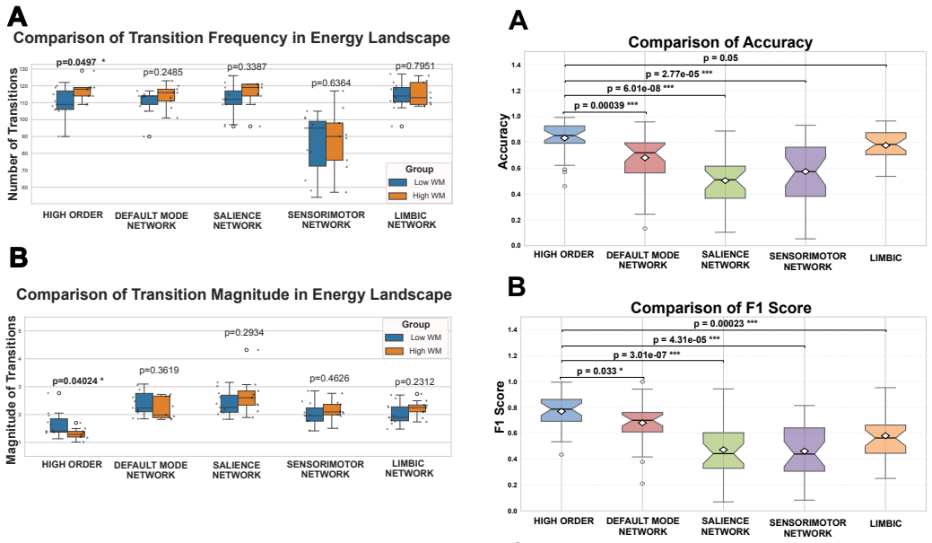
The authors claim that tumor-induced changes in high-order neural dynamics, quantified as energy landscapes before surgery, reliably predict whether a patient will have lower (2-5) or higher (6-9) postoperative Spatial Span scores, with the lower-scoring group showing fewer but more extreme transitions between local energy minima and maxima.
What carries the argument
Energy landscapes of high-order brain interactions, which describe the frequency and magnitude of transitions between local energy minima and maxima derived from presurgical fMRI.
If this is right
- Presurgical fMRI energy features could be used to estimate a patient's risk of working memory decline before choosing a surgical route.
- Patients exhibiting fewer extreme energy transitions may require extra precautions or postoperative support to preserve spatial memory.
- High-order interaction patterns carry outcome information beyond what standard pairwise connectivity measures provide.
Where Pith is reading between the lines
- The same energy-landscape approach might be tested for predicting other cognitive domains such as attention or language after resection.
- If the patterns prove stable, they could serve as a baseline for monitoring whether preoperative interventions alter energy transitions and improve outcomes.
- Similar energy metrics in non-tumor populations could reveal whether natural variation in these transitions tracks everyday working memory differences.
Load-bearing premise
The observed energy landscape differences reflect tumor effects on brain dynamics rather than being driven by tumor location, size, or other unmeasured clinical factors.
What would settle it
A replication study that matches patients on tumor location and size, then finds that energy features no longer separate low- and high-scoring groups above chance level, would falsify the predictive claim.
Figures


read the original abstract
Surgical resection is the primary treatment option for brain tumor patients, but it carries the risk of postoperative cognitive impairments. This study investigates how tumor-induced alterations in presurgical neural dynamics relate to postoperative working memory outcome assessed by Spatial Span (SSP) test. We analyzed functional magnetic resonance imaging (fMRI) of brain tumor patients before surgery and extracted energy landscapes of high-order brain interactions. We then examined the relation between these energy features and postoperative working memory performance using statistical and machine learning (random forest) models. Patients with lower postoperative SSP Scores (2 to 5) exhibited fewer but more extreme transitions between local energy minima and maxima, whereas patients with higher SSP Scores (6 to 9) showed more frequent but less extreme shifts. Furthermore, the presurgical high-order energy features were able to accurately predict postoperative working memory outcome with a mean accuracy of 90%, F1 score of 87.5%, and an AUC of 0.95. Our study suggests that the brain tumor-induced disruptions in high-order neural dynamics before surgery are predictive of postoperative working memory outcome. Our findings pave the path for personalized surgical planning and targeted interventions to mitigate cognitive risks associated with brain tumor resection.
Editorial analysis
A structured set of objections, weighed in public.
Referee Report
Summary. The manuscript analyzes presurgical fMRI data from brain tumor patients to extract high-order energy landscapes and relates these to postoperative working memory outcomes measured by the Spatial Span (SSP) test. It reports distinct transition patterns between energy minima and maxima: patients with lower postoperative SSP scores (2–5) exhibit fewer but more extreme transitions, while those with higher scores (6–9) show more frequent but less extreme shifts. A random forest classifier trained on these presurgical energy features is reported to predict postoperative SSP outcome with mean accuracy 90%, F1 score 87.5%, and AUC 0.95, suggesting utility for personalized surgical planning.
Significance. If the predictive relationship is shown to be robust under proper validation and confound control, the work could meaningfully advance preoperative risk assessment in neurosurgery by linking tumor-induced alterations in high-order neural dynamics to functional cognitive outcomes. The energy-landscape framing of fMRI interactions offers a potentially distinctive lens compared with conventional connectivity metrics. However, the absence of basic reporting on cohort size, validation, and baselines currently prevents assessment of whether the claimed performance reflects genuine predictive signal or methodological artifacts.
major comments (3)
- [Methods] Methods: No sample size (N), cross-validation scheme, feature dimensionality, or permutation baseline is reported for the random forest model that yields the central performance figures (90% accuracy, AUC 0.95). Without these, it is impossible to determine whether the quoted metrics are consistent with overfitting on a modest single-center cohort or reflect genuine generalization of the high-order energy features.
- [Results] Results: The manuscript provides no comparison of the energy-feature model against a baseline using only clinical variables (tumor location, size, grade, or laterality). Because tumor location can simultaneously influence both fMRI energy extrema and postoperative SSP deficit, the absence of this control leaves open the possibility that the reported accuracy is driven by location confounds rather than the claimed high-order dynamics.
- [Abstract] Abstract and Results: The claim that “presurgical high-order energy features were able to accurately predict” outcome rests on a fitted classifier whose robustness cannot be evaluated from the supplied text. Standard reporting elements required to support this claim—N, train/test split or CV folds, and any statistical test on the transition-pattern differences—are missing.
minor comments (2)
- [Abstract] Abstract: The SSP score ranges (2–5 vs. 6–9) are stated without reference to the normative range or maximum possible score on the Spatial Span test, which would aid interpretation of what constitutes “lower” versus “higher” performance.
- [Discussion] Discussion: A brief comparison to prior fMRI or connectivity-based predictors of post-resection cognitive outcome would help situate the incremental value of the energy-landscape approach.
Simulated Author's Rebuttal
We thank the referee for the constructive and detailed comments. We have reviewed each point carefully and will revise the manuscript to strengthen the reporting of methods, add necessary controls, and clarify the validation procedures supporting our claims. Point-by-point responses follow.
read point-by-point responses
-
Referee: [Methods] Methods: No sample size (N), cross-validation scheme, feature dimensionality, or permutation baseline is reported for the random forest model that yields the central performance figures (90% accuracy, AUC 0.95). Without these, it is impossible to determine whether the quoted metrics are consistent with overfitting on a modest single-center cohort or reflect genuine generalization of the high-order energy features.
Authors: We agree that these details are essential for readers to assess model robustness and rule out overfitting. The initial submission omitted explicit reporting of these elements. In the revised manuscript we will add the cohort size, a clear description of the cross-validation procedure, the dimensionality of the energy-landscape feature set, and results from a permutation test that establishes a chance-level baseline for the reported accuracy, F1, and AUC. revision: yes
-
Referee: [Results] Results: The manuscript provides no comparison of the energy-feature model against a baseline using only clinical variables (tumor location, size, grade, or laterality). Because tumor location can simultaneously influence both fMRI energy extrema and postoperative SSP deficit, the absence of this control leaves open the possibility that the reported accuracy is driven by location confounds rather than the claimed high-order dynamics.
Authors: We acknowledge the value of this control analysis. In the revised manuscript we will introduce a baseline random-forest model trained exclusively on clinical variables (tumor location, size, grade, and laterality) and directly compare its performance metrics with those obtained from the high-order energy features. This addition will allow readers to evaluate the incremental predictive contribution of the energy-landscape measures beyond standard clinical information. revision: yes
-
Referee: [Abstract] Abstract and Results: The claim that “presurgical high-order energy features were able to accurately predict” outcome rests on a fitted classifier whose robustness cannot be evaluated from the supplied text. Standard reporting elements required to support this claim—N, train/test split or CV folds, and any statistical test on the transition-pattern differences—are missing.
Authors: We agree that the abstract and results should contain the minimal information needed to evaluate the predictive claim. In the revision we will update the abstract to reference the sample size and validation approach, and we will expand the results section to report the cross-validation scheme together with appropriate statistical tests (e.g., permutation or non-parametric tests) comparing transition-pattern statistics between the low- and high-SSP-score groups. revision: yes
Circularity Check
Random forest accuracy on same-cohort energy features presented as presurgical prediction
specific steps
-
fitted input called prediction
[Abstract]
"Furthermore, the presurgical high-order energy features were able to accurately predict postoperative working memory outcome with a mean accuracy of 90%, F1 score of 87.5%, and an AUC of 0.95."
Energy features are computed from the identical patient cohort whose postoperative SSP scores are then used to train and evaluate the random forest; the reported accuracy is therefore the training-set performance of a model fitted to those same features and labels, not an out-of-sample prediction.
full rationale
The paper extracts high-order energy landscape features from presurgical fMRI of the studied patients, fits a random forest classifier to postoperative SSP scores within that cohort, and reports 90% accuracy / 0.95 AUC as evidence that the features 'predict' outcome. Because no cross-validation scheme, held-out test set, or external cohort is described, the quoted performance metrics are the in-sample fit rather than an independent prediction. This matches the fitted-input-called-prediction pattern and accounts for the moderate circularity burden noted in the reader's assessment.
Axiom & Free-Parameter Ledger
axioms (1)
- domain assumption fMRI BOLD signals can be used to construct high-order interaction energy landscapes whose local minima and maxima reflect functionally relevant brain states
Lean theorems connected to this paper
-
IndisputableMonolith/Cost/FunctionalEquation.leanwashburn_uniqueness_aczel echoes?
echoesECHOES: this paper passage has the same mathematical shape or conceptual pattern as the Recognition theorem, but is not a direct formal dependency.
the energy function E(xk) is given by: E(xk) = −∑i<j Wi j xkixkj − ∑i hi xki
-
IndisputableMonolith/Foundation/ArrowOfTime.leanz_monotone_absolute echoes?
echoesECHOES: this paper passage has the same mathematical shape or conceptual pattern as the Recognition theorem, but is not a direct formal dependency.
Patients with lower postoperative SSP Scores (2 to 5) exhibited fewer but more extreme transitions between local energy minima and maxima
What do these tags mean?
- matches
- The paper's claim is directly supported by a theorem in the formal canon.
- supports
- The theorem supports part of the paper's argument, but the paper may add assumptions or extra steps.
- extends
- The paper goes beyond the formal theorem; the theorem is a base layer rather than the whole result.
- uses
- The paper appears to rely on the theorem as machinery.
- contradicts
- The paper's claim conflicts with a theorem or certificate in the canon.
- unclear
- Pith found a possible connection, but the passage is too broad, indirect, or ambiguous to say the theorem truly supports the claim.
Reference graph
Works this paper leans on
-
[1]
T. K. Owonikoko, J. Arbiser, A. Zelnak, H.-K. G. Shu, H. Shim, A. M. Robin, S. N. Kalkanis, T. G. Whitsett, B. Salhia, N. L. Tran, et al., Current approaches to the treatment of metastatic brain tumours, Nat Rev Clin Oncol 11 (4) (2014) 203–222
work page 2014
-
[2]
B. Rodriguez, D. Rivera, J. Y . Zhang, C. Brown, T. Young, T. Williams, J. Kallos, S. Huq, C. Hadjpanayis, Innovations in intraoperative therapies in neurosurgical oncology: a narrative review, J Neurooncol (2024) 1–9
work page 2024
-
[3]
J. S. Young, R. A. Morshed, S. L. Hervey-Jumper, M. S. Berger, The surgical management of di ffuse gliomas: current state of neurosurgical management and future directions, Neuro Oncol 25 (12) (2023) 2117– 2133
work page 2023
- [4]
-
[5]
M. J. Taphoorn, M. Klein, Cognitive deficits in adult patients with brain tumours, Lancet Neurol 3 (3) (2004) 159–168
work page 2004
-
[6]
M. A. Kirkman, J. O. Ekert, B. H. Hunn, M. S. Thomas, A. K. Tolmie, A systematic review of cognitive interventions for adult patients with brain tumours, Cancer Med 12 (10) (2023) 11191–11210
work page 2023
-
[7]
M. Dallabona, S. Sarubbo, S. Merler, F. Corsini, G. Pulcrano, U. Roz- zanigo, M. Barbareschi, F. Chioffi, Impact of mass effect, tumor location, age, and surgery on the cognitive outcome of patients with high-grade gliomas: a longitudinal study, Neuro Oncol Pract 4 (4) (2017) 229–240
work page 2017
-
[8]
S. J. Rijnen, I. Meskal, M. Bakker, W. De Baene, G.-J. M. Rutten, K. Gehring, M. M. Sitskoorn, Cognitive outcomes in meningioma pa- tients undergoing surgery: individual changes over time and predictors of late cognitive functioning, Neuro Oncol 21 (7) (2019) 911–922
work page 2019
-
[9]
S. Schiavolin, A. Raggi, C. Scaratti, C. Toppo, F. Silvaggi, D. Sattin, M. Broggi, P. Ferroli, M. Leonardi, Outcome prediction in brain tumor surgery: A literature review on the influence of nonmedical factors, Neu- rosurg Rev 44 (2) (2021) 807–819
work page 2021
-
[10]
A. Zangrossi, E. Silvestri, M. Bisio, A. Bertoldo, S. De Pellegrin, A. Vallesi, A. Della Puppa, D. D’Avella, L. Denaro, R. Scienza, et al., Presurgical predictors of early cognitive outcome after brain tumor resec- tion in glioma patients, Neuroimage Clin 36 (2022) 103219
work page 2022
- [11]
-
[12]
N. B. Dadario, B. Brahimaj, J. Yeung, M. E. Sughrue, Reducing the cog- nitive footprint of brain tumor surgery, Front Neurol 12 (2021) 711646
work page 2021
-
[13]
D. A. Lakhani, D. S. Sabsevitz, K. L. Chaichana, A. Qui ˜nones-Hinojosa, E. H. Middlebrooks, Current state of functional mri in the presurgical planning of brain tumors, Radiol Imaging Cancer 5 (6) (2023) e230078
work page 2023
- [14]
-
[15]
D. J. Gri ffiths-King, C. Delivett, A. Peet, J. Waite, J. Novak, Limited research investigating the value of mri in predicting future cognitive mor- bidity in survivors of paediatric brain tumours: A systematic-review and call to action for clinical neuroimaging researchers, PLoS One 20 (1) (2025) e0314721
work page 2025
-
[16]
C. Wang, K. Van Dyk, N. Cho, C. Raymond, J. Choi, N. Salamon, W. B. Pope, A. Lai, T. F. Cloughesy, P. L. Nghiemphu, et al., Characterization of cognitive function in survivors of di ffuse gliomas using resting-state functional mri (rs-fmri), Brain Imaging Behav (2022) 1–13
work page 2022
- [17]
-
[18]
P. E. Tarapore, J. Martino, A. G. Guggisberg, J. Owen, S. M. Honma, A. Findlay, M. S. Berger, H. E. Kirsch, S. S. Nagarajan, Magnetoen- cephalographic imaging of resting-state functional connectivity predicts postsurgical neurological outcome in brain gliomas, Neurosurgery 71 (5) (2012) 1012
work page 2012
-
[19]
S. Lang, I. Gaxiola-Valdez, M. Opoku-Darko, L. A. Partlo, B. G. Goodyear, J. J. Kelly, P. Federico, Functional connectivity in frontopari- etal network: indicator of preoperative cognitive function and cognitive outcome following surgery in patients with glioma, World Neurosurg 105 (2017) 913–922
work page 2017
- [20]
-
[21]
D. van Nieuwenhuizen, L. Douw, M. Klein, S. M. Peerdeman, J. J. Heimans, J. C. Reijneveld, C. J. Stam, A. Hillebrand, Cognitive function- ing and functional brain networks in postoperative who grade i menin- gioma patients, J Neurooncol 140 (2018) 605–613
work page 2018
-
[22]
P. H. Luckett, M. O. Olufawo, K. Y . Park, B. Lamichhane, D. Dierker, G. T. Verastegui, J. J. Lee, P. Yang, A. Kim, O. H. Butt, et al., Predicting post-surgical functional status in high-grade glioma with resting state fmri and machine learning, J Neurooncol (2024) 1–11
work page 2024
-
[23]
A. M. Bastos, J.-M. Scho ffelen, A tutorial review of functional connec- tivity analysis methods and their interpretational pitfalls, Front Syst Neu- rosci 9 (2016) 175
work page 2016
-
[24]
M. E. Fox, T. Z. King, Functional connectivity in adult brain tumor pa- tients: A systematic review, Brain Connect 8 (7) (2018) 381–397
work page 2018
- [25]
-
[26]
A. Santoro, F. Battiston, M. Lucas, G. Petri, E. Amico, Higher-order con- nectomics of human brain function reveals local topological signatures of task decoding, individual identification, and behavior, Nat Commun 15 (1) (2024) 10244
work page 2024
-
[27]
T. M. Tran, T. T. Tran, S. Khanmohammadi, High-order resting-state functional connectivity is predictive of working memory decline after brain tumor resection, in: 2024 46th Annual International Conference of the IEEE Engineering in Medicine and Biology Society (EMBC), IEEE, 2024, pp. 1–5
work page 2024
- [28]
-
[29]
M. G. Preti, T. A. Bolton, D. Van De Ville, The dynamic functional con- nectome: State-of-the-art and perspectives, Neuroimage 160 (2017) 41– 54
work page 2017
-
[30]
H. Aerts, N. Colenbier, H. Almgren, T. Dhollander, J. R. Daparte, K. Clauw, A. Johri, J. Meier, J. Palmer, M. Schirner, et al., Pre-and post- surgery brain tumor multimodal magnetic resonance imaging data opti- mized for large scale computational modelling, Sci Data 9 (1) (2022) 676
work page 2022
-
[31]
J. Kang, C. Pae, H.-J. Park, Graph-theoretical analysis for energy land- scape reveals the organization of state transitions in the resting-state hu- man cerebral cortex, PLoS One 14 (9) (2019) e0222161
work page 2019
-
[32]
T. Watanabe, S. Hirose, H. Wada, Y . Imai, T. Machida, I. Shirouzu, S. Konishi, Y . Miyashita, N. Masuda, A pairwise maximum entropy model accurately describes resting-state human brain networks, Nat Com- mun 4 (1) (2013) 1370
work page 2013
-
[33]
T. Watanabe, N. Masuda, F. Megumi, R. Kanai, G. Rees, Energy land- 8 scape and dynamics of brain activity during human bistable perception, Nat Commun 5 (1) (2014) 4765
work page 2014
-
[34]
J. Kang, C. Pae, H.-J. Park, Energy landscape analysis of the subcortical brain network unravels system properties beneath resting state dynamics, Neuroimage 149 (2017) 153–164
work page 2017
- [35]
-
[36]
E. T. Jaynes, Information theory and statistical mechanics, Phys Rev 106 (4) (1957) 620
work page 1957
- [37]
-
[38]
N. Roy, S. P. Singleton, K. Jamison, P. Mukherjee, S. Shah, A. Kuceyeski, Brain activity dynamics after traumatic brain injury indicate increased state transition energy and preference of lower order states, Neuroimage Clin (2025) 103799
work page 2025
-
[39]
W. L. Yue, K. K. Ng, S. Liu, X. Qian, J. S. X. Chong, A. J. Koh, M. Q. W. Ong, S. K. S. Ting, A. S. L. Ng, N. Kandiah, et al., Di fferential spa- tial working memory–related functional network reconfiguration in young and older adults, Netw Neurosci 8 (2) (2024) 395–417
work page 2024
-
[40]
A. Corriveau, K. Yoo, Y . H. Kwon, M. M. Chun, M. D. Rosenberg, Func- tional connectome stability and optimality are markers of cognitive per- formance, Cereb Cortex 33 (8) (2023) 5025–5041
work page 2023
-
[41]
J. O. Ekert, A. Goyal, J. S. Young, S. L. Hervey-Jumper, M. S. Berger, In- terventional neurorehabilitation for glioma patients: A systematic review, Neuro Oncol Pract 11 (6) (2024) 679–690
work page 2024
-
[42]
A. Poologaindran, C. Profyris, I. M. Young, N. B. Dadario, S. A. Ahsan, K. Chendeb, R. G. Briggs, C. Teo, R. Romero-Garcia, J. Suckling, et al., Interventional neurorehabilitation for promoting functional recovery post- craniotomy: a proof-of-concept, Sci Rep 12 (1) (2022) 3039
work page 2022
-
[43]
T. F. Boerger, P. Pahapill, A. M. Butts, E. Arocho-Quinones, M. Ragha- van, M. O. Kruco ff, Large-scale brain networks and intra-axial tumor surgery: a narrative review of functional mapping techniques, criti- cal needs, and scientific opportunities, Front Hum Neurosci 17 (2023) 1170419
work page 2023
-
[44]
Breakspear, Dynamic models of large-scale brain activity, Nat Neu- rosci 20 (3) (2017) 340–352
M. Breakspear, Dynamic models of large-scale brain activity, Nat Neu- rosci 20 (3) (2017) 340–352
work page 2017
- [45]
-
[46]
G. Deco, J. Cruzat, J. Cabral, E. Tagliazucchi, H. Laufs, N. K. Logo- thetis, M. L. Kringelbach, Awakening: Predicting external stimulation to force transitions between different brain states, Proc Natl Acad Sci U S A 116 (36) (2019) 18088–18097
work page 2019
-
[47]
B. A. Seitzman, C. Gratton, T. O. Laumann, E. M. Gordon, B. Adeyemo, A. Dworetsky, B. T. Kraus, A. W. Gilmore, J. J. Berg, M. Ortega, et al., Trait-like variants in human functional brain networks, Proc Natl Acad Sci U S A 116 (45) (2019) 22851–22861
work page 2019
-
[48]
A. I. Luppi, D. Golkowski, A. Ranft, R. Ilg, D. Jordan, D. Bzdok, A. M. Owen, L. Naci, E. A. Stamatakis, E. Amico, et al., General anaesthesia decreases the uniqueness of brain functional connectivity across individ- uals and species, Nat Hum Behav (2025) 1–18. 9
work page 2025
discussion (0)
Sign in with ORCID, Apple, or X to comment. Anyone can read and Pith papers without signing in.