REVIEW 4 major objections 6 minor 32 references
Learning Semantic Directions for Feature Augmentation in Domain-Generalized Medical Segmentation
T0 review · 4 major / 6 minor · reviewed 2026-08-06 · deepseek-v4-flash
Pith's one-line read Learning which feature channels to perturb, and how strongly, improves medical segmentation on unseen domains.
desk verdict Solid feature-augmentation DG paper with a genuinely interesting selective consistency loss, but the core SDS mechanism is underspecified to the point that the main claim is not reproducible as written. read the letter →
The pith
A machine-rendered reading of the paper's core claim, the machinery that carries it, and where it could break.
The reading
What carries the argument
The load-bearing object is the semantic direction mask $d \in \{0,1\}^C$, a per-channel on/off choice that turns blind feature noise into selective perturbation through $\tilde z_i = z_i + d \odot s$. The selector $f_{\mathrm{cs}}$ is described as two convolutional layers, global average pooling, and a fully connected layer, and the intensity $s$ is sampled as $\mu + \sigma \odot \xi$ with $\xi \sim N(0,\Sigma_k)$ where $\Sigma_k$ is the inter-domain covariance of a randomly chosen domain pair. The selective consistency loss is the stabilizer: by penalizing only augmentations that increase the segmentation loss, it gives the selector a training signal that favors channels whose perturbation is safe. The paper does not specify how the binary mask in Equation (2) is produced from continuous network outputs, so the learnability of the selector rests on an unstated binarization step.
What would settle it
Train the same framework with the learned direction selector replaced by a random binary mask of matched channel count; if the prostate average Dice stays at 86.34, the learned direction is not carrying the gain.
Extended reading notes
Core claim
The central claim is that channel-selective, statistics-guided feature perturbation generalizes better to unseen clinical domains than image-level augmentation or uniform feature-level augmentation. The paper defines the augmented feature as $\tilde z_i = z_i + d \odot s$, with $d \in \{0,1\}^C$ the output of a learnable semantic direction selector built from convolutions, global average pooling, and a fully connected layer, and $s$ an instance-adaptive intensity produced by shifting and scaling noise drawn from inter-domain covariance matrices. The supervised loss supervises segmentation from both the original and augmented features, and the selective consistency loss $\mathcal{L}_{\mathrm{cons}}$ applies only when the augmented feature gives a higher segmentation loss than the original, so harmful perturbations are penalized while helpful ones are not. On the three benchmark tasks the paper reports average Dice of 86.34 over 85.53 on prostate, 79.62 over 77.99 on optic cup, and 92.00 over 91.35 on optic disc.
Load-bearing premise
The method depends on the semantic direction selector being able to learn a useful binary channel mask by gradient descent, but the paper never specifies how the binary output in Equation (2) is obtained from continuous network outputs or how gradients flow through it.
Editorial extensions
If this is right
- If the reported results are correct, SDFA becomes the top-scoring method on all three leave-one-domain-out tasks among the compared approaches, with the largest margin on optic cup segmentation.
- The ablation results imply that the direction selector and intensity sampler each contribute independently: SDS alone improves average Dice to 85.63 and SIS alone to 85.51 from 85.29, and together they reach 86.34.
- The selective consistency loss stabilizes selection: with it, the average Dice rises to 86.34 from 85.94 and the number of selected channels fluctuates less during training.
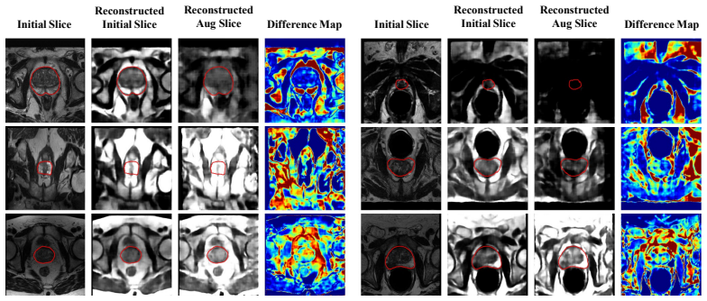
- Reconstruction visualizations suggest the augmentation edits contextual regions while sparing target anatomy, which would be why segmentation Dice can improve without distorting the region of interest.
- Because the method tunes feature noise by domain statistics rather than target data, it applies directly to the standard multi-source DG setting where only source centers are available at training time.
Reading between the lines
- Editorial extension: if the gain comes from channel selectivity, then the number of selected channels per sample should correlate with domain gap; one could test this by checking whether low-selection samples come from source-like domains within the training set.
- Editorial extension: the covariance-sampled intensity could be recycled for test-time adaptation by re-estimating $\Sigma_k$ on a small unlabeled target batch, a use the paper does not consider.
- Editorial extension: the selective consistency loss is a 'do no harm' regularizer; replacing the hard indicator in Eq. (9) with a margin-based soft penalty could smooth optimization and may improve stability on smaller datasets, but this is not tested in the paper.
Editorial analysis
A structured set of objections, weighed in public.
Referee Report
Summary. The paper proposes a domain generalization framework (SDFA) for medical image segmentation, built around three components: a Semantic Direction Selector (SDS) that learns which feature channels to perturb, a covariance-based Semantic Intensity Sampler (SIS) that controls perturbation magnitudes via inter-domain statistics, and a Selective Consistency Loss (SCL) that only penalizes augmentations that degrade segmentation. The method is evaluated on the Prostate and Fundus benchmarks under a leave-one-domain-out protocol, reporting the best average DSC among compared methods in Tables II-IV.
Significance. If fully specified and reproducible, the method is a plausible structured feature-augmentation approach for domain generalization, and the use of public multi-center benchmarks with a fixed leave-one-domain-out protocol is a strength. However, the core technical details needed to implement and train the proposed SDS and SIS are missing, and the empirical claim of 'consistently outperforming' is not supported by the per-domain results. The paper would be a useful contribution only after filling these gaps and providing statistical evidence for the reported improvements.
major comments (4)
- [III-B1, Eq. (2)] Equation (2) defines the semantic direction mask as d = f_cs(z) ∈ {0,1}^C, but f_cs is specified only as 'two convolutional layers, followed by a global average pooling and a fully connected layer,' which outputs continuous logits. The paper never states how the binary mask is produced or how gradients flow through it during training. If a hard threshold is applied, the gradient of the total loss with respect to the SDS parameters is zero almost everywhere, so the selector cannot be learned by standard backpropagation; if a soft relaxation or straight-through estimator is used, Eq. (2) is inaccurate and the actual training procedure is unspecified. Since SDS is the core novel component, this omission makes the method irreproducible and undermines the empirical results in Tables II-IV.
- [III-B2, Eqs. (3)-(4)] The construction of the inter-domain covariance matrices Σ_k is not operationally defined. The text says 'we compute a set of covariance matrices {Σ_k}_{k=1}^N' where each Σ_k captures 'the inter-domain covariance between a pair of domains (D_si, D_sj)', but it never states which feature maps enter the covariance (per-sample features, per-batch means, or per-domain statistics), how the domain pair is chosen for a given feature z, or how the index k is enumerated (with K=4 or 6 source domains, the number of pairs is not N as in a batch). Also, because z = Φ(x_i) depends on the input, it is unclear whether Σ_k is a fixed precomputed statistic or a running batch estimate; the two interpretations lead to different training dynamics and different gradients. This needs to be specified precisely for reproduction.
- [III-C, Eq. (9)] The Selective Consistency Loss uses an indicator function I(Laug > Lori), which is non-differentiable with respect to the SDS parameters. The paper claims SCL 'stabilizes the learning of the SDS,' but without a differentiable surrogate or a gradient approximation, the loss as written cannot provide learning signal to the SDS. The absolute value in Eq. (9) is also non-smooth, though that is secondary. Please specify how gradients are computed through the indicator, or reformulate SCL as a smooth loss.
- [IV-C, Tables II-IV] The abstract claims the framework 'consistently outperforms existing domain generalization approaches,' but the per-domain results do not support 'consistently': in Table III, SDFA is below CSU on Domain-1 (80.40 vs 80.48) and below TriD on Domain-4 (81.94 vs 84.21); in Table IV, SDFA is below EFDMixStyle on Domain-3 (92.16 vs 93.19). In addition, no standard deviations or significance tests are reported, and the average DSC gaps are small (0.81, 1.63, and 0.65 points). Please report multiple seeds with variance and statistical tests, or revise the claim to 'best average performance in most settings.'
minor comments (6)
- [Table II caption] The caption says 'Average indicates the mean DSC and ASD across all four domains,' but the Prostate benchmark has six domains (Domain-1 through Domain-6); this should read 'six domains.'
- [IV-A] There is a stray subsection header '1) Quantitative Results on the Fundus Dataset:' immediately after the evaluation metrics paragraph, with no content following it; this appears to be a leftover from an earlier draft and should be removed.
- [Figure 3] The caption reads 'Segmentation results with and without consistency loss,' but the surrounding text describes the number of selected channels over training steps; the caption should be updated to match the actual content of the figure.
- [Tables VI and VII] The word 'Efficay' in the captions 'Efficay of Each Module' and 'Efficay of SCL' is misspelled; it should be 'Efficacy.'
- [Table V and surrounding text] The text states 'the DSC increases as λ grows,' but the reported average DSC at λ=0.2 is 85.51 and at λ=0.4 is 85.49, so the trend is not monotonic; the claim should be stated as 'DSC tends to increase with λ, with the best value at λ=1.0.'
- [Notation, Eqs. (3) and (9)] The symbol N is overloaded: in Eq. (3) it denotes the number of covariance matrices/domain pairs, while in Eq. (9) it denotes the batch size; please use distinct symbols such as N_pairs and N_batch.
Circularity Check
No circularity: SDFA's claim rests on held-out-domain evaluation; the one self-citation is non-load-bearing.
full rationale
The derivation chain is self-contained. The augmented feature z̃_i = z_i + d⊙s (Eq. 1) is produced by the learnable selector f_cs (Eq. 2) and the covariance-based sampler (Eqs. 3-5), and the segmentation losses (Eqs. 6-8) plus the selective consistency term (Eq. 9) are all defined on source-domain training examples. No parameter is fitted to a target domain, and the leave-one-domain-out protocol in Sec. IV-A tests on held-out centers, so the average DSC gains in Tables II-IV are not forced by construction. The only self-citation, Ref. [25], is used to motivate a conceptual decomposition of semantic augmentation into direction and intensity; the paper adds its own learnable components and the empirical claim is evaluated independently. The unspecified binarization of d in Eq. (2) and the non-differentiable indicator in Eq. (9) are reproducibility/correctness concerns, but they do not make any prediction equivalent to an input. Hence no circular step is present.
Assumptions & free parameters
free parameters (3)
- mu (semantic intensity shift) =
learned, in R^C, no final values reported
- sigma (semantic intensity scale) =
learned, in R^C, no final values reported
- lambda (augmented-loss weight) =
1.0 (best in Table V)
assumptions (4)
- domain assumption Medical images have consistent anatomy, and domain shift mainly changes imaging conditions.
- domain assumption Semantic augmentation in feature space, z~ = z + d ⊙ s, preserves segmentation labels when d is correctly selected.
- domain assumption Covariance-sampled noise xi ~ N(0, Sigma_k) from paired domains is a valid model of domain-variant feature directions.
- ad hoc to paper The binary mask d in Eq. (2) can be optimized by gradient descent despite unspecified binarization.
Cite this review
Pith. "Pith review of Learning Semantic Directions for Feature Augmentation in Domain-Generalized Medical Segmentation." pith.science (2026). https://pith.science/paper/6JCNGFYU
@misc{pith2026250723326,
  author       = {Pith},
  title        = {Pith review of: Learning Semantic Directions for Feature Augmentation in Domain-Generalized Medical Segmentation},
  year         = {2026},
  howpublished = {\url{https://pith.science/paper/6JCNGFYU}},
  note         = {Machine review of arXiv:2507.23326}
}
read the original abstract
Medical image segmentation plays a crucial role in clinical workflows, but domain shift often leads to performance degradation when models are applied to unseen clinical domains. This challenge arises due to variations in imaging conditions, scanner types, and acquisition protocols, limiting the practical deployment of segmentation models. Unlike natural images, medical images typically exhibit consistent anatomical structures across patients, with domain-specific variations mainly caused by imaging conditions. This unique characteristic makes medical image segmentation particularly challenging. To address this challenge, we propose a domain generalization framework tailored for medical image segmentation. Our approach improves robustness to domain-specific variations by introducing implicit feature perturbations guided by domain statistics. Specifically, we employ a learnable semantic direction selector and a covariance-based semantic intensity sampler to modulate domain-variant features while preserving task-relevant anatomical consistency. Furthermore, we design an adaptive consistency constraint that is selectively applied only when feature adjustment leads to degraded segmentation performance. This constraint encourages the adjusted features to align with the original predictions, thereby stabilizing feature selection and improving the reliability of the segmentation. Extensive experiments on two public multi-center benchmarks show that our framework consistently outperforms existing domain generalization approaches, achieving robust and generalizable segmentation performance across diverse clinical domains.
Figures


Figures from the paper (1 more)
Reference graph
Works this paper leans on
-
[1]
A survey on deep learning in medical image analysis,
G. Litjens, T. Kooi, B. E. Bejnordi, A. A. A. Setio, F. Ciompi, M. Ghafoorian, J. A. Van Der Laak, B. Van Ginneken, and C. I. S´anchez, “A survey on deep learning in medical image analysis,” Medical image analysis, vol. 42, pp. 60–88, 2017
2017
-
[2]
U-net: Convolutional networks for biomedical image segmentation,
O. Ronneberger, P. Fischer, and T. Brox, “U-net: Convolutional networks for biomedical image segmentation,” in Medical image computing and computer-assisted intervention–MICCAI 2015: 18th international con- ference, Munich, Germany, October 5-9, 2015, proceedings, part III 18 . Springer, 2015, pp. 234–241
2015
-
[3]
nnu-net: a self-configuring method for deep learning-based biomedical image segmentation,
F. Isensee, P. F. Jaeger, S. A. Kohl, J. Petersen, and K. H. Maier-Hein, “nnu-net: a self-configuring method for deep learning-based biomedical image segmentation,” Nature methods, vol. 18, no. 2, pp. 203–211, 2021
2021
-
[4]
Unetr: Transformers for 3d medical image segmentation,
A. Hatamizadeh, Y . Tang, V . Nath, D. Yang, A. Myronenko, B. Land- man, H. R. Roth, and D. Xu, “Unetr: Transformers for 3d medical image segmentation,” in Proceedings of the IEEE/CVF winter conference on applications of computer vision , 2022, pp. 574–584
work page 2022
-
[5]
Swin unetr: Swin transformers for semantic segmentation of brain tumors in mri images,
A. Hatamizadeh, V . Nath, Y . Tang, D. Yang, H. R. Roth, and D. Xu, “Swin unetr: Swin transformers for semantic segmentation of brain tumors in mri images,” in International MICCAI brainlesion workshop . Springer, 2021, pp. 272–284
2021
-
[6]
Machine learning with multi-site imaging data: An empirical study on the impact of scanner effects,
B. Glocker, R. Robinson, D. C. Castro, Q. Dou, and E. Konukoglu, “Machine learning with multi-site imaging data: An empirical study on the impact of scanner effects,” arXiv preprint arXiv:1910.04597 , 2019
arXiv 1910
-
[7]
Domain generalization for medical image analysis: A review,
J. S. Yoon, K. Oh, Y . Shin, M. A. Mazurowski, and H.-I. Suk, “Domain generalization for medical image analysis: A review,” Proceedings of the IEEE, 2024
2024
-
[8]
P. Zhang, B. Zhang, T. Zhang, D. Chen, Y . Wang, and F. Wen, “Proto- typical pseudo label denoising and target structure learning for domain adaptive semantic segmentation,” in Proceedings of the IEEE/CVF con- ference on computer vision and pattern recognition , 2021, pp. 12 414– 12 424
work page 2021
Show all 32 references
-
[9]
Self-attentive spatial adaptive normalization for cross-modality domain adaptation,
D. Tomar, M. Lortkipanidze, G. Vray, B. Bozorgtabar, and J.-P. Thiran, “Self-attentive spatial adaptive normalization for cross-modality domain adaptation,” IEEE transactions on medical imaging , vol. 40, no. 10, pp. 2926–2938, 2021
2021
-
[10]
Where and how to transfer: Knowledge aggregation-induced transferability perception for unsupervised domain adaptation,
J. Dong, Y . Cong, G. Sun, Z. Fang, and Z. Ding, “Where and how to transfer: Knowledge aggregation-induced transferability perception for unsupervised domain adaptation,” IEEE Transactions on Pattern Analysis and Machine Intelligence, vol. 46, no. 3, pp. 1664–1681, 2021
2021
-
[11]
Shape-aware meta-learning for generalizing prostate mri segmentation to unseen domains,
Q. Liu, Q. Dou, and P.-A. Heng, “Shape-aware meta-learning for generalizing prostate mri segmentation to unseen domains,” in Interna- tional conference on medical image computing and computer-assisted intervention. Springer, 2020, pp. 475–485
2020
-
[12]
Causality-inspired single-source domain generalization for medical image segmentation,
C. Ouyang, C. Chen, S. Li, Z. Li, C. Qin, W. Bai, and D. Rueckert, “Causality-inspired single-source domain generalization for medical image segmentation,” IEEE Transactions on Medical Imaging , vol. 42, no. 4, pp. 1095–1106, 2022
2022
-
[13]
Bayeseg: Bayesian modeling for medical image segmentation with interpretable generalizability,
S. Gao, H. Zhou, Y . Gao, and X. Zhuang, “Bayeseg: Bayesian modeling for medical image segmentation with interpretable generalizability,” Medical Image Analysis , vol. 89, p. 102889, 2023
2023
-
[14]
Improving cross- domain generalizability of medical image segmentation using uncer- tainty and shape-aware continual test-time domain adaptation,
J. Zhu, B. Bolsterlee, Y . Song, and E. Meijering, “Improving cross- domain generalizability of medical image segmentation using uncer- tainty and shape-aware continual test-time domain adaptation,” Medical Image Analysis, vol. 101, p. 103422, 2025
2025
-
[15]
Synthseg: Segmentation of brain mri scans of any contrast and resolution without retraining,
B. Billot, D. N. Greve, O. Puonti, A. Thielscher, K. Van Leemput, B. Fischl, A. V . Dalca, J. E. Iglesias et al. , “Synthseg: Segmentation of brain mri scans of any contrast and resolution without retraining,” Medical image analysis , vol. 86, p. 102789, 2023
2023
-
[16]
Randstainna: Learning stain- agnostic features from histology slides by bridging stain augmentation and normalization,
Y . Shen, Y . Luo, D. Shen, and J. Ke, “Randstainna: Learning stain- agnostic features from histology slides by bridging stain augmentation and normalization,” in International Conference on Medical Image Computing and Computer-Assisted Intervention . Springer, 2022, pp. 212–221
2022
-
[17]
Test-time image- to-image translation ensembling improves out-of-distribution generaliza- tion in histopathology,
M. Scalbert, M. Vakalopoulou, and F. Couzini ´e-Devy, “Test-time image- to-image translation ensembling improves out-of-distribution generaliza- tion in histopathology,” in International Conference on Medical Image Computing and Computer-Assisted Intervention . Springer, 2022,...
2022
-
[18]
Mutual information-based disentangled neural net- works for classifying unseen categories in different domains: Application to fetal ultrasound imaging,
Q. Meng, J. Matthew, V . A. Zimmer, A. Gomez, D. F. Lloyd, D. Rueck- ert, and B. Kainz, “Mutual information-based disentangled neural net- works for classifying unseen categories in different domains: Application to fetal ultrasound imaging,” IEEE transactions on medical imagi...
2020
-
[19]
Mi- segnet: Mutual information-based us segmentation for unseen domain generalization,
Y . Bi, Z. Jiang, R. Clarenbach, R. Ghotbi, A. Karlas, and N. Navab, “Mi- segnet: Mutual information-based us segmentation for unseen domain generalization,” in International Conference on Medical Image Comput- ing and Computer-Assisted Intervention . Springer, 2023, pp. 130–140
2023
-
[20]
Generalizable cross- modality medical image segmentation via style augmentation and dual normalization,
Z. Zhou, L. Qi, X. Yang, D. Ni, and Y . Shi, “Generalizable cross- modality medical image segmentation via style augmentation and dual normalization,” in Proceedings of the IEEE/CVF conference on com- puter vision and pattern recognition , 2022, pp. 20 856–20 865
2022
-
[21]
Treasure in distribution: A domain randomization based multi-source domain generalization for 2d medical image segmentation,
Z. Chen, Y . Pan, Y . Ye, H. Cui, and Y . Xia, “Treasure in distribution: A domain randomization based multi-source domain generalization for 2d medical image segmentation,” in International Conference on Medical Image Computing and Computer-Assisted Intervention. Springer, 20...
2023
-
[22]
Domain generalization with correlated style uncertainty,
Z. Zhang, B. Wang, D. Jha, U. Demir, and U. Bagci, “Domain generalization with correlated style uncertainty,” in Proceedings of the IEEE/CVF Winter Conference on Applications of Computer Vision , 2024, pp. 2000–2009
2024
-
[23]
Deep feature interpolation for image content changes,
P. Upchurch, J. Gardner, G. Pleiss, R. Pless, N. Snavely, K. Bala, and K. Weinberger, “Deep feature interpolation for image content changes,” in Proceedings of the IEEE conference on computer vision and pattern recognition, 2017, pp. 7064–7073
2017
-
[24]
Regularizing deep networks with semantic data augmentation,
Y . Wang, G. Huang, S. Song, X. Pan, Y . Xia, and C. Wu, “Regularizing deep networks with semantic data augmentation,” IEEE Transactions on Pattern Analysis and Machine Intelligence , vol. 44, no. 7, pp. 3733– 3748, 2021
2021
-
[25]
Bsda: Bayesian random semantic data augmentation for medical image classification,
Y . Zhu, X. Cai, X. Wang, X. Chen, Z. Fu, and Y . Yao, “Bsda: Bayesian random semantic data augmentation for medical image classification,” Sensors, vol. 24, no. 23, p. 7511, 2024
2024
-
[26]
Dofe: Domain-oriented feature embedding for generalizable fundus image seg- mentation on unseen datasets,
S. Wang, L. Yu, K. Li, X. Yang, C.-W. Fu, and P.-A. Heng, “Dofe: Domain-oriented feature embedding for generalizable fundus image seg- mentation on unseen datasets,” IEEE Transactions on Medical Imaging , vol. 39, no. 12, pp. 4237–4248, 2020
2020
-
[27]
Learning generalized medical image segmentation from decoupled feature queries,
Q. Bi, J. Yi, H. Zheng, W. Ji, Y . Huang, Y . Li, and Y . Zheng, “Learning generalized medical image segmentation from decoupled feature queries,” in Proceedings of the AAAI conference on artificial intelligence, vol. 38, no. 2, 2024, pp. 810–818
2024
-
[28]
A comprehensive retinal image dataset for the assessment of glaucoma from the optic nerve head analysis,
J. Sivaswamy, S. Krishnadas, A. Chakravarty, G. Joshi, A. S. Tabish et al. , “A comprehensive retinal image dataset for the assessment of glaucoma from the optic nerve head analysis,” JSM Biomedical Imaging Data Papers, vol. 2, no. 1, p. 1004, 2015
2015
-
[29]
Rim-one: An open retinal image database for optic nerve evaluation,
F. Fumero, S. Alay ´on, J. L. Sanchez, J. Sigut, and M. Gonzalez- Hernandez, “Rim-one: An open retinal image database for optic nerve evaluation,” in 2011 24th international symposium on computer-based medical systems (CBMS) . IEEE, 2011, pp. 1–6
2011
-
[30]
Refuge challenge: A unified framework for evaluating automated methods for glaucoma assessment from fundus photographs,
J. I. Orlando, H. Fu, J. B. Breda, K. Van Keer, D. R. Bathula, A. Diaz- Pinto, R. Fang, P.-A. Heng, J. Kim, J. Lee et al. , “Refuge challenge: A unified framework for evaluating automated methods for glaucoma assessment from fundus photographs,” Medical image analysis, vol. 59...
2020
-
[31]
Domain generalization with mixstyle,
K. Zhou, Y . Yang, Y . Qiao, and T. Xiang, “Domain generalization with mixstyle,” arXiv preprint arXiv:2104.02008 , 2021
2021 arXiv
-
[32]
Exact feature distribution matching for arbitrary style transfer and domain generalization,
Y . Zhang, M. Li, R. Li, K. Jia, and L. Zhang, “Exact feature distribution matching for arbitrary style transfer and domain generalization,” in Proceedings of the IEEE/CVF conference on computer vision and pattern recognition, 2022, pp. 8035–8045
2022
Reviewed August 6, 2026 · model on record in the stance chip above.
Discussion (0). Sign in to comment.