REVIEW 4 major objections 5 minor 3 references
LV-Net: Anatomy-aware lateral ventricle shape modeling with a case study on Alzheimer's disease
T0 review · 4 major / 5 minor · reviewed 2026-08-05 · deepseek-v4-flash
Pith's one-line read LV-Net reconstructs whole lateral ventricle surfaces from brain MRI by deforming a joint ventricle-hippocampus template, and uses the stable vertex correspondence to localize Alzheimer's-associated ventricle subregions.
desk verdict Solid anatomical prior, under-specified core module, and no error bars; worth a serious referee but with mandatory revisions. read the letter →
The pith
A machine-rendered reading of the paper's core claim, the machinery that carries it, and where it could break.
The reading
What carries the argument
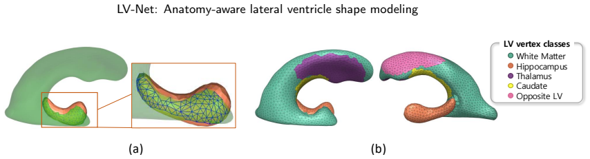
The anatomy-aware joint LV-hippocampus template mesh is a deformable mesh pair that shares a strip of boundary vertices between the ventricle and the hippocampus, with each ventricle vertex labeled by one of five surrounding structures. This template is the load-bearing object: its fixed topology supplies the parameterization, its shared boundary gives the hippocampus leverage to repair missing inferior ventricle segments, and its per-vertex labels define what correspondence means. The fitting mechanism is an iterative loop in which a neural vertex-deformation module predicts displacements, a distance loss (Chamfer distance plus point-to-mesh and mesh-to-point terms) pulls the mesh to the ta
What would settle it
Run the paper's own ablation quantitatively: take a set of low-resolution segmentations with inferior-LV holes, reconstruct with the full joint template and with the paper's LV-Net* variant, and compare both to a high-resolution reference using ASSD restricted to the inferior horn. If the joint template does not beat the LV-Net* variant by more than its internal variability, the anatomical-correction claim has no support. A sharper version: rerun the 5,000-iteration optimization with a randomly initialized vertex-deformation module; since all deformation comes from that module's predicted disp
Extended reading notes
Core claim
The central claim is that anatomical prior knowledge, encoded in the template rather than applied afterward, is what makes LV shape reconstruction robust. LV-Net begins with a joint LV-hippocampus template mesh whose boundary vertices belong to both structures; during iterative deformation, the hippocampus mesh is fitted to its own target point cloud, and the shared vertices pull the inferior ventricle into anatomically correct position even where the ventricle mask has holes. Each LV vertex is assigned one of five periventricular labels, and a per-region distance loss keeps labeled vertices near same-labeled target points, giving point correspondence that follows anatomy rather than raw geo
Load-bearing premise
The whole optimization depends on a neural network that predicts vertex displacements at every step, but the paper never states how that network is trained, what data it uses, or how it is initialized; if that module is unavailable or under-trained, the iterative template deformation cannot be reproduced as written.
Editorial extensions
If this is right
- Whole-ventricle analysis becomes feasible in routine datasets: studies no longer need to discard the inferior horn because its mask is disconnected or absent.
- Automatic segmentation outputs with holes or fragments are still usable, because the hippocampus-anchored template restores the missing boundary anatomy.
- Vertex-level shape statistics are interpretable across subjects, since corresponding vertices are tied to the same periventricular structure rather than to geometric nearest neighbors.
- The Alzheimer's case study pinpoints candidate local biomarkers: ventricle subregions adjacent to the hippocampus, thalamus, and part of the caudate are where shape distributions differ between AD and cognitively normal groups.
Reading between the lines
- Beyond the paper, the shared-boundary repair mechanism should transfer to other anatomical pairs where one structure is harder to segment than a neighboring structure; each pair would need its own template, but the repair logic is generic.
- A direct test the paper does not run: feed LV-Net target point clouds from a second segmentation method and see whether the reported accuracy gap over the baselines persists; the current experiments hinge on point clouds from a single automatic segmenter.
- The AD map is built from per-vertex Wilcoxon tests at $p \le 0.1$ with no stated multiple-comparison correction; a permutation test with false-discovery-rate control would show which parts of the map, especially the caudate-adjacent region, are robust.
- Because the template fixes topology, the fitted meshes could be subdivided anatomically (frontal horn, body, atrium, temporal horn) for subregional shape analysis, a level of localization the current vertex-wise map only hints at.
Signed reviews
Editorial analysis
A structured set of objections, weighed in public.
Referee Report
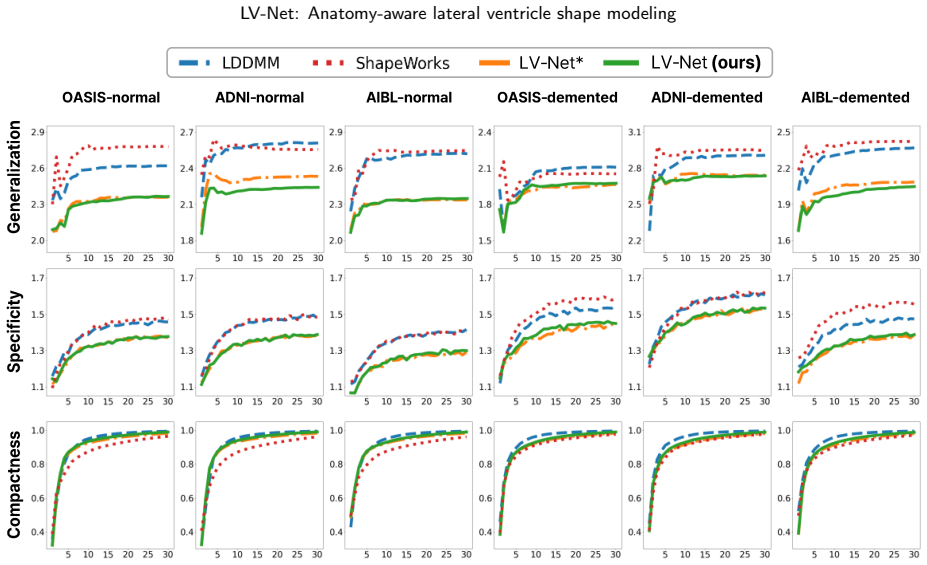
Summary. LV-Net proposes an automated pipeline for reconstructing subject-specific lateral ventricle (LV) meshes from brain MRI. The method deforms an anatomy-aware LV–hippocampus joint template mesh toward a target point cloud extracted from SynthSeg segmentations. The template includes shared boundary vertices between the LV and hippocampus and per-vertex labels for adjacent subcortical structures. Reconstruction is cast as 5,000 AdamW iterations of mesh deformation driven by the distance and regularization losses in Eqs. (1) and (2), with a PointNet-based vertex deformation module mentioned as the displacement predictor. Experiments compare LV-Net with LDDMM and ShapeWorks on OASIS, ADNI, and AIBL using DSC, ASSD, and HD95, and also evaluate statistical shape model quality (generalization, specificity, compactness). A case study applies the method to detect LV subregions associated with Alzheimer's disease using vertex-wise Wilcoxon tests.
Significance. The core idea of jointly modeling the LV and hippocampus to compensate for segmentation loss in the inferior LV, and of embedding periventricular anatomical labels into the template for correspondence, is original and clinically plausible. The code release is a clear strength. If the implementation were fully specified and the quantitative claims properly supported, LV-Net could be a useful contribution to anatomical shape modeling. As it stands, however, the central quantitative results rest on an underspecified deformation module and on aggregate statistics without variance or significance testing, and the AD analysis uses a lenient uncorrected threshold. These issues currently limit the reliability of the stated conclusions.
major comments (4)
- [§3.2.2] The core deformation step is not implementable as written. The text states only that 'a PointNet-based vertex deformation module (Qi et al., 2017) predicts vertex displacements' but gives no architecture, input features, layer sizes, parameter count, training data, or initialization. It is also unclear whether the module is pretrained, frozen, or optimized jointly during the 5,000 AdamW iterations. Since every subject-specific reconstruction in Table 1 is produced by this module, the claimed superiority over LDDMM and ShapeWorks cannot be reproduced or falsified from the manuscript. Please provide a complete specification, or correct the description if the module is not a learned network.
- [Table 1 / §4.2] The central comparison reports only three aggregate numbers (DSC 0.893, ASSD 0.715 mm, HD95 2.064 mm for LV-Net), with no per-dataset breakdown, no subject counts, no standard deviations/confidence intervals, and no significance tests. The statement that 'LV-Net outperforms all methods' is therefore supported only by point estimates. The table title also promises 'ASSD for each subcortical region' but the table has no such columns. Please provide per-dataset results with error bars and appropriate statistical tests, and repair the table/hierarchy mismatch.
- [§5, Figure 7] The AD subregion analysis uses a vertex-wise Wilcoxon signed-rank test at p≤0.1 with no multiple-comparison correction. With 2,490 LV vertices, even under a global null one would expect roughly 249 vertices to fall below this threshold by chance, so the reported 'significant' regions are not a reliable basis for clinical claims. In addition, the signed-rank test is normally for paired observations, whereas the comparison is between independent CN and AD groups; a Mann-Whitney U test or a model adjusted for age/sex would be more appropriate. Please apply a corrected threshold or permutation-based FDR control and use a standard significance level.
- [§4.3, Figure 4(a)] The error-correction validation compares LV-Net reconstruction from a downsampled mask with LV-Net reconstruction from the high-resolution mask. This is a self-consistency check: it shows the method is stable under resolution changes, but it does not establish that the restored inferior LV is anatomically correct, because both reconstructions come from the same method and share the same template bias. The claim that the joint template 'effectively restores the inferior LV' needs an independent reference, such as the original high-resolution segmentation or manual ground truth, and should be quantified rather than shown with a single qualitative example.
minor comments (5)
- [§3.1 and §3.2.2] Section 3.1 lists five peripheral brain structures, including the hippocampus, but Section 3.2.2 defines only four peripheral regions (m=4) and omits the hippocampus from the peripheral loss. Please clarify this taxonomy and state explicitly how the hippocampal label is handled.
- [Table 1] LV-Net* is discussed in the text as an important ablation for the joint template, but it does not appear in the quantitative table. Adding it to Table 1 (or a companion table) would make the ablation quantitative.
- [Figure 5] The generalization/specificity/compactness plots are described as supporting 'higher or comparable' performance, but no error bars or subject-level variability are shown and the legend is not described. Please add variability information.
- [General] Typos and small errors: 'Housdroff' should be 'Hausdorff'; 'significantly associations' should be 'significant associations'; the reference 'Shaphiro, S.' should be 'Shapiro, S.'; and 'The codes' is nonstandard ('The code').
- [§3.1] The paper does not describe how the joint LV-hippocampus template mesh is constructed (e.g., which atlas, manual labeling, or prior method). Since the template is central to the method, please provide this information.
Circularity Check
One secondary validation is circular: 'error correction' is measured against LV-Net's own high-resolution reconstruction; the central reconstruction comparison is not circular.
-
self definitional
[Section 4.3, 'Anatomy correction with the template mesh' (Figure 4a)]
"We then reconstructed the low-resolution masks with and without the joint template mesh and compared them to the high-resolution reconstruction. As illustrated in Figure 4(a), the joint template mesh effectively restores the inferior LV to a shape nearly identical to the high-resolution reconstruction, demonstrating its anatomical correction capability."
The 'high-resolution reconstruction' used as the reference for evaluating error correction is itself produced by LV-Net. Therefore, the experiment compares two outputs of the same model: one fitted to a low-resolution mask and one fitted to a high-resolution mask. Agreement between these two outputs shows self-consistency of LV-Net, not that the restored inferior LV matches true anatomy. The conclusion 'anatomical correction capability' is thus supported by a reference that is generated by the method under evaluation, rather than by an independent ground truth. This makes the validation circular for the specific claim of anatomical correction, although it does not undermine the main reconstruction comparison against LDDMM and ShapeWorks.
full rationale
The central derivation of LV-Net is a per-subject fitting procedure: a fixed joint template is deformed to target point clouds extracted from segmentation maps, with losses that directly minimize geometric distance to those targets. The headline reconstruction metrics (DSC, ASSD, HD95) evaluate how well each method fits the same target geometry, so the comparison against LDDMM and ShapeWorks is a fair benchmark rather than a circular prediction. The peripheral-region loss is an explicit design term that enforces correspondence by anatomical label; it is a construction choice, not a circular derivation. The only circular element found is the error-correction validation in Section 4.3, which uses LV-Net's own high-resolution reconstruction as the reference for 'anatomical correction.' This does not affect the main claim's independence but does weaken the secondary claim. The under-specification of the PointNet-based vertex deformation module is a reproducibility concern, not a circularity, since no equation is defined in terms of the claimed result and no fitted parameter is renamed as a prediction.
Assumptions & free parameters
free parameters (6)
- Distance loss weights λ_cf, λ_pm, λ_mp =
2, 1.4, 0.6
- Regularization weights λ_vert, λ_norm, λ_edge, λ_cons, λ_lap =
1, 1, 1000, 100, 300
- Template resolution =
2 mm edge, 2,490 LV vertices, 96 shared vertices
- Optimization schedule =
5000 iterations, lr 5e-4 halved every 1000 iterations
- Peripheral region count m =
4
- AD significance threshold =
p <= 0.1
assumptions (4)
- domain assumption The PointNet-based vertex deformation module is available and predicts useful vertex displacements.
- domain assumption The LV-hippocampus joint template mesh and its periventricular vertex labels are anatomically correct.
- domain assumption SynthSeg segmentation maps provide sufficiently accurate target point clouds and peripheral labels.
- ad hoc to paper Shared vertices along the LV-hippocampus boundary enforce correct inferior LV structure when LV masks are incomplete.
Cite this review
Pith. "Pith review of LV-Net: Anatomy-aware lateral ventricle shape modeling with a case study on Alzheimer's disease." pith.science (2026). https://pith.science/paper/6HJFDEJR
@misc{pith2026250806055,
  author       = {Pith},
  title        = {Pith review of: LV-Net: Anatomy-aware lateral ventricle shape modeling with a case study on Alzheimer's disease},
  year         = {2026},
  howpublished = {\url{https://pith.science/paper/6HJFDEJR}},
  note         = {Machine review of arXiv:2508.06055}
}
read the original abstract
Lateral ventricle (LV) shape analysis holds promise as a biomarker for neurological diseases; however, challenges remain due to substantial shape variability across individuals and segmentation difficulties arising from limited MRI resolution. We introduce LV-Net, a novel framework for producing individualized 3D LV meshes from brain MRI by deforming an anatomy-aware joint LV-hippocampus template mesh. By incorporating anatomical relationships embedded within the joint template, LV-Net reduces boundary segmentation artifacts and improves reconstruction robustness. In addition, by classifying the vertices of the template mesh based on their anatomical adjacency, our method enhances point correspondence across subjects, leading to more accurate LV shape statistics. We demonstrate that LV-Net achieves superior reconstruction accuracy, even in the presence of segmentation imperfections, and delivers more reliable shape descriptors across diverse datasets. Finally, we apply LV-Net to Alzheimer's disease analysis, identifying LV subregions that show significantly associations with the disease relative to cognitively normal controls. The codes for LV shape modeling are available at https://github.com/PWonjung/LV_Shape_Modeling.
Figures


Figures from the paper (4 more)



Reference graph
Works this paper leans on
-
[2008]
The alzheimer’s disease neuroimaging initiative (adni): Mri methods. Journal of Magnetic Resonance Imaging: An Official Journal of the International Society for Magnetic Resonance in Medicine 27, 685–691. Johnson, S.L., Wang, L., Alpert, K.I., Greenstein, D., Clasen, L., Lalonde, F., Miller, R., Rapoport, J., Gogtay, N., 2013. Hippocampal shape abnormalit...
arXiv 2013
-
[2021]
Deepcsr: A 3d deep learning approach for cortical surface reconstruction,in:ProceedingsoftheIEEE/CVFWinterConferenceon Applications of Computer Vision, pp. 806–815. Desbrun, M., Meyer, M., Schröder, P., Barr, A.H., 1999. Implicit fairing of irregular meshes using diffusion and curvature flow, in: Proceedings of the 26th annual conference on Computer graph...
arXiv 1999
-
[2022]
IEEE Transactions on Medical Imaging 42, 430–443
Cortexode: Learning cortical surface reconstruction by neural odes. IEEE Transactions on Medical Imaging 42, 430–443. Marcus, D.S., Fotenos, A.F., Csernansky, J.G., Morris, J.C., Buckner, R.L., 2010. Open access series of imaging studies: longitudinal mri data in nondemented and demented older adults. Journal of cognitive neuroscience 22, 2677–2684. Olive...
work page 2010
Reviewed August 5, 2026 · model on record in the stance chip above.
Discussion (0). Continue with ORCID to comment.