REVIEW 4 major objections 6 minor 62 references
CardioMorphNet: Cardiac Motion Prediction Using a Shape-Guided Bayesian Recurrent Deep Network
T0 review · 4 major / 6 minor · reviewed 2026-08-05 · deepseek-v4-flash
Pith's one-line read CardioMorphNet claims cardiac motion is learned more accurately by recursively registering segmentation maps than by matching image intensities, and reports superior shape-registration metrics and lower DVF uncertainty on a large population
desk verdict Plausible shape-guided registration method, but the headline comparison is biased: the model trains on the same automated masks it is then scored against. read the letter →
The pith
A machine-rendered reading of the paper's core claim, the machinery that carries it, and where it could break.
The reading
What carries the argument
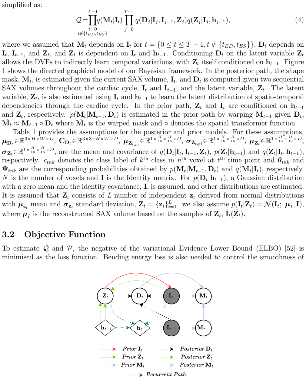
The load-bearing machinery is a recurrent variational autoencoder with two posterior networks and one warping prior. SegNet computes q(Mt|It), the per-voxel distribution over cardiac labels for the current frame; DeformNet computes q(Dt|It, It−1, Zt), a Gaussian displacement field with mean and covariance sigma-squared-I plus V-transpose-V; and the prior p(Mt|Mt−1, Dt) is implemented by warping the previous segmentation map with the current DVF. A ConvLSTM hidden state carries spatio-temporal context into the latent variable Zt. The ELBO loss combines a supervised and semi-supervised shape cross-entropy term that replaces intensity similarity, KL divergences for the displacement field and th
What would settle it
On a held-out set with expert manual contours (or a digital phantom with known ground-truth deformation), compare CardioMorphNet's warped masks and DVFs against independent ground truth rather than the automated tool's masks. If its advantage over intensity-based baselines disappears, the claim that shape-guided mask registration yields superior cardiac motion estimation is refuted.
Extended reading notes
Core claim
The central claim, stated on the paper's own terms, is that a recurrent variational Bayesian framework can learn cardiac motion by recursively registering segmentation maps, and doing so is enough to beat methods that optimize intensity-based image similarity. In the proposed generative model, the joint probability of the sequence factorizes so that the current mask depends on the previous mask and the current displacement field; the posterior estimates masks, displacement fields, and latent variables from the current and previous SAX volumes and a recurrent hidden state. Minimizing the resulting ELBO loss aligns the warped previous mask with the current segmentation at every time step, supe
Load-bearing premise
The automated tool that supplies the end-diastolic and end-systolic masks is accurate enough to serve as both training labels and the evaluation standard; if those masks are biased, the reported scores mainly measure agreement with the tool, not true cardiac motion.
Editorial extensions
If this is right
- DVFs are trained by mask overlap across the whole cardiac cycle, so only the ED and ES frames need manual or automated labels; intermediate frames are held together by recursive shape consistency.
- The framework produces per-voxel DVF uncertainty directly from the learned covariance, without dropout or test-time augmentation.
- On the reported cohort, shape-guided registration gives higher DSC/JAC and lower HD95/MSD than intensity-based and joint registration baselines in the LV, LV myocardium, and RV.
- The DVF uncertainty in the cardiac region is lower than in DragNet and SegMorph, which the paper interprets as higher confidence in the estimated heart motion.
Reading between the lines
- The evaluation uses the same automated contour tool for training supervision and for scoring; a reader should treat the reported accuracy as agreement with that tool's segmentations. An expert-annotation or synthetic-deformation benchmark would test whether the shape-guided loss truly captures cardiac motion rather than fitting the tool's contours.
- The abstract asserts more accurate clinical-index estimation, but the body reports only mask-overlap, uncertainty, and diffeomorphism evaluations; that assertion lacks a demonstrated result in the presented text.
- The recursive mask-registration loss is not cardiac-specific in principle; the same two-frame-supervision scheme could transfer to other organs or imaging modalities where reliable contours exist at two timepoints and intensity matching is unreliable.
- Because the model uses only short-axis volumes and discrete masks, through-plane motion and rotation that do not change mask overlap may be under-constrained; combining with long-axis views or mesh/tagging evidence would be a natural stress test.
Editorial analysis
A structured set of objections, weighed in public.
Referee Report
Summary. The paper proposes CardioMorphNet, a recurrent Bayesian deep network for 3D cardiac motion estimation from cine short-axis CMR sequences. The model combines a recurrent variational autoencoder, a segmentation network (SegNet), and a registration network (DeformNet). It estimates segmentation masks, DVFs, and latent variables jointly, with a loss derived from an ELBO plus a bending-energy regularizer. The key design feature is that registration is guided by warping segmentation maps rather than by an intensity-based image similarity loss. The method is evaluated on UK Biobank data, comparing mask overlap (DSC, JAC, HD95, MSD) with VoxelMorph, DragNet, SegMorph, JMS, SyN, and LCC-Demons, and also reports uncertainty maps and an ablation study.
Significance. If the claims are substantiated, the paper makes a useful contribution by demonstrating that shape-guided, recurrent Bayesian registration can produce accurate cardiac DVFs with explicit uncertainty estimates, without intensity-based similarity losses. The code is made available, and the method is compared against several strong baselines. The uncertainty maps are a valuable addition over deterministic registration methods. However, the evaluation currently has a serious circularity issue: the same automated masks are used both as training supervision at ED/ES and as the evaluation target, and the abstract claims M&M validation that does not appear in the experiments. These issues must be resolved before the central claim of superior performance can be accepted.
major comments (4)
- [§4.1, §4.3, Eq. (7), Table 2] The evaluation is circular with respect to the supervision. Section 4.1 states that ED/ES time steps and ground-truth masks are obtained with the automated tool of [54]. These same masks are used in Eq. (7) as supervised training labels for the warped masks, and in Table 2 as the evaluation target for DSC/JAC/HD95/MSD. CardioMorphNet is therefore explicitly trained to maximize the evaluation metric, while the comparison methods (VoxelMorph, DragNet, SegMorph, etc.) are not trained with this mask-overlap supervision. The reported improvements may reflect optimization alignment rather than genuine motion-estimation accuracy. The paper provides no independent check, such as manual expert contours, an alternative motion benchmark, or M&M results. This is the load-bearing weakness of the paper; please add an independent evaluation or retrain baselines with the same mask supervision.
- [§3.2, Eqs. (3)–(6)] The claim that the loss is 'derived' from the Bayesian formulation is not supported by the derivation. The factorization in Eq. (3) is simplified to Eq. (4) by strong conditional-independence assumptions that are neither justified nor derived. Then the ELBO in Eq. (6) introduces hand-set weights λ1, λ2, λ3, and ρ, with values chosen empirically (Section 3.2). These ad hoc weights mean the objective is not a pure variational bound but a weighted sum of terms. This weakens the paper's central methodological claim. Please provide the full ELBO derivation, state the exact independence assumptions, and clarify how the λ weights are justified.
- [§3.3, Eq. (8)] The semi-supervised shape loss is self-referential. Lsemi-shape minimizes the cross-entropy between Ψ, the SegNet posterior q(Mt|It), and θ, the warped previous mask p(Mt|Mt−1, Dt). At non-ED/ES frames, Ψ is the model's own output and is also being optimized. Therefore the DVF is trained against a moving target produced by the same network, which can trivially reduce this term by making SegNet copy its own predictions. This is not a fixed supervisory signal. Please clarify whether Ψ is detached or fixed, and discuss the implications for the registration objective.
- [Abstract, §4] The abstract states that the method is 'Validated on the UK Biobank and M&M datasets', but the experiments in Section 4 only report UK Biobank. There is no M&M dataset description, preprocessing, results table, or analysis anywhere in the paper. Either add the M&M experiments or remove the claim from the abstract. This is a factual inconsistency in the headline claim.
minor comments (6)
- [§4.3] The text states that a paired t-test was employed, but no p-values or confidence intervals are reported in Table 2 or the surrounding text. Please report statistical significance for the main comparisons.
- [§4.1] Since the automated tool of [54] is used for ground-truth masks, it would be helpful to report the tool's segmentation accuracy (e.g., Dice vs. manual contours) on the UK Biobank subset used here.
- [§5] There are typos: 'DrageNet' should be 'DragNet', and 'SegMorh' should be 'SegMorph'.
- [Eq. (9)] In Eq. (9), the notation Tr(CDj) is unclear because CD is a voxel-wise 3×3 covariance, not a matrix. Please define the trace operation on this tensor.
- [Table 3] The column header 'NoDeformNet' is confusing; it appears to mean that DeformNet is always enabled. Consider renaming to 'DeformNet' with ✓/✗ like the other columns.
- [§4.4] The uncertainty maps in Figure 6 use a color scale but no numerical colorbar; adding one would make the visualization more interpretable.
Circularity Check
No significant circularity: the ELBO-based loss is a genuine variational derivation, evaluation uses a held-out split, and the mask-supervised training/evaluation overlap is a standard supervised design rather than a definitional reduction.
full rationale
CardioMorphNet's derivation chain is self-contained. The variational bound (Eqs. 5–11) follows algebraically from the stated generative and posterior factorisations in Eqs. (1)–(4) and Table 1; no term is defined as the quantity it is claimed to predict. The central quantitative claim is evaluated on a held-out 10% test split of UK Biobank (Section 4.1), with DSC/JAC/HD95/MSD computed between warped and ground-truth masks at ED/ES. The fact that L_sup-shape (Eq. 7) supervises the same ED/ES masks used for evaluation is a standard supervised-training/evaluation design, not a definitional equivalence: cross-entropy is not identical to the reported overlap/surface metrics, and the test subjects are unseen during training. The semi-supervised term (Eq. 8) does use SegNet's own q(Mt|It) as a target for the warped mask, which creates a self-supervised consistency objective rather than a hidden identity; the paper explicitly acknowledges that segmentation errors can propagate into registration (Section 5, 'Another restriction is the segmentation error of the segmentation component in the proposed framework, which may affect cardiac shape registration'). No load-bearing self-citation or imported uniqueness theorem appears: [49] and [51] are baseline methods from overlapping authors and are compared empirically, not used to rule out alternatives. The automated-mask source [54] is an external tool, and while sharing a label source between training and evaluation and giving unequal mask supervision to baselines is a legitimate fairness/benchmark concern, it does not make the reported results equivalent to the paper's inputs by construction. Separately, the abstract mentions M&M validation and clinical-index assessment that are not reported in the experiments, but missing support is a correctness concern, not a circularity step.
Assumptions & free parameters
free parameters (4)
- Loss weights lambda_1, lambda_2, lambda_3 =
1e-4, 2e-4, 0.3
- Bending energy weight rho =
not specified
- Latent dimension L =
not specified
- Downsampled sequence length T =
6 frames
assumptions (5)
- domain assumption Automated tool [54] masks are reliable ground truth.
- ad hoc to paper Conditional independence assumptions in Eqs. (2) and (4).
- domain assumption Mask overlap is a sufficient proxy for cardiac motion.
- domain assumption Cyclic cardiac sequence assumption.
- ad hoc to paper Gaussian prior and posterior forms.
Cite this review
Pith. "Pith review of CardioMorphNet: Cardiac Motion Prediction Using a Shape-Guided Bayesian Recurrent Deep Network." pith.science (2026). https://pith.science/paper/LEK6Y37G
@misc{pith2026250820734,
  author       = {Pith},
  title        = {Pith review of: CardioMorphNet: Cardiac Motion Prediction Using a Shape-Guided Bayesian Recurrent Deep Network},
  year         = {2026},
  howpublished = {\url{https://pith.science/paper/LEK6Y37G}},
  note         = {Machine review of arXiv:2508.20734}
}
read the original abstract
Accurate cardiac motion estimation from cine cardiac magnetic resonance (CMR) images is vital for assessing cardiac function and detecting its abnormalities. Existing methods often struggle to accurately capture heart motion because they rely on intensity-based image registration similarity losses that may overlook cardiac anatomical regions. To address this, we propose CardioMorphNet, a recurrent Bayesian deep learning framework for 3D cardiac shape-guided deformable registration using short-axis (SAX) CMR images. It employs a recurrent variational autoencoder to model spatio-temporal dependencies across the cardiac cycle, along with two posterior models for bi-ventricular segmentation and motion estimation. The derived loss function from the Bayesian formulation guides the framework to focus on anatomical regions by recursively registering segmentation maps without using intensity-based image registration similarity loss, while leveraging sequential SAX volumes and spatio-temporal features. The Bayesian modelling also enables the computation of uncertainty maps for the estimated motion fields. Validated on the UK Biobank and M&M datasets by comparing warped mask shapes with ground-truth masks, CardioMorphNet demonstrates superior performance in cardiac motion estimation, outperforming state-of-the-art methods. Uncertainty assessment shows that it also yields lower uncertainty values for estimated motion fields in the cardiac region compared with other probabilistic-based cardiac registration methods, indicating higher confidence in its predictions. In addition, the clinical indices extraction assessment shows that CardioMorphNet estimates the clinical indices more accurately than other approaches.
Figures


Figures from the paper (5 more)


Reference graph
Works this paper leans on
-
[54]
Automated car- diovascular magnetic resonance image analysis with fully convolutional networks
Wenjia Bai, Matthew Sinclair, Giacomo Tarroni, Ozan Oktay, Martin Rajchl, Ghislain Vail- lant, Aaron M Lee, Nay Aung, Elena Lukaschuk, Mihir M Sanghvi, et al. Automated car- diovascular magnetic resonance image analysis with fully convolutional networks. Journal of cardiovascular magnetic resonance, 20(1):65, 2018
work page 2018
-
[1]
Global atlas on cardiovascular disease prevention and control
Shanthi Mendis, Pekka Puska, B editors Norrving, World Health Organization, et al. Global atlas on cardiovascular disease prevention and control . World Health Organization, 2011
work page 2011
-
[2]
European cardiovascular disease statistics 2017
Elizabeth Wilkins, L Wilson, Kremlin Wickramasinghe, Prachi Bhatnagar, Jose Leal, Ramon Luengo-Fernandez, R Burns, Mike Rayner, and Nick Townsend. European cardiovascular disease statistics 2017. 2017
work page 2017
-
[3]
Gregory A Roth, Degu Abate, Kalkidan Hassen Abate, Solomon M Abay, Cristiana Abbafati, Nooshin Abbasi, Hedayat Abbastabar, Foad Abd-Allah, Jemal Abdela, Ahmed Abdelalim, et al. Global, regional, and national age-sex-specific mortality for 282 causes of death in 195 countries and territories, 1980–2017: a systematic analysis for the global burden of diseas...
work page 1980
-
[4]
BHF CVD Statistics UK Factsheet, 2022
British Heart Foundation. BHF CVD Statistics UK Factsheet, 2022. [Online; accessed March 26, 2024]. 19
work page 2022
-
[5]
Manuel Franco, Richard S Cooper, Usama Bilal, and Valent ´ ın Fuster. Challenges and opportu- nities for cardiovascular disease prevention.The American journal of medicine, 124(2):95–102, 2011
work page 2011
-
[6]
Corrine Y Jurgens, Christopher S Lee, Dawn M Aycock, Ruth Masterson Creber, Quin E Denfeld, Holli A DeVon, Linda R Evers, Miyeon Jung, Gianluca Pucciarelli, Megan M Streur, et al. State of the science: the relevance of symptoms in cardiovascular disease and research: a scientific statement from the american heart association. Circulation, 146(12):e173–e184, 2022
work page 2022
-
[7]
Binu Koirala, Ruth-Alma Turkson-Ocran, Diana Baptiste, Bibhu Koirala, Lucine Francis, Patricia Davidson, Cheryl Dennison Himmelfarb, and Yvonne Commodore-Mensah. Het- erogeneity of cardiovascular disease risk factors among asian immigrants: insights from the 2010 to 2018 national health interview survey. Journal of the American Heart Association , 10(13):...
work page 2010
Show all 62 references
-
[8]
Cvd screening in low-risk, asymptomatic adults: clinical trials needed
Tamar S Polonsky and Philip Greenland. Cvd screening in low-risk, asymptomatic adults: clinical trials needed. Nature reviews Cardiology, 9(10):599–604, 2012
2012
-
[9]
The association of abnormal ventricular wall motion and increased dispersion of repolarization in humans is independent of the presence of myocardial infarction
Tobias Opthof, Peter Sutton, Ruben Coronel, Susan Wright, Panny Kallis, and Peter Taggart. The association of abnormal ventricular wall motion and increased dispersion of repolarization in humans is independent of the presence of myocardial infarction. Frontiers in physiology ...
2012
-
[10]
Usefulness of the severity and extent of wall motion abnormalities as prognostic markers of an adverse outcome after a first myocardial infarction treated with thrombolytic therapy
Erberto Carluccio, Stefano Tommasi, Maurizio Bentivoglio, Massimo Buccolieri, Lucio Prosci- utti, and Luigi Corea. Usefulness of the severity and extent of wall motion abnormalities as prognostic markers of an adverse outcome after a first myocardial infarction treated with th...
2000
-
[11]
Prevalence and prognostic significance of wall-motion abnormalities in adults without clinically recognized cardiovascular disease: the strong heart study
Silvana Cicala, Giovanni de Simone, Mary J Roman, Lyle G Best, Elisa T Lee, Wenyu Wang, Thomas K Welty, James M Galloway, Barbara V Howard, and Richard B Devereux. Prevalence and prognostic significance of wall-motion abnormalities in adults without clinically recognized cardi...
2007
-
[12]
Recent advances in cardiovascular magnetic resonance: techniques and applications
Michael Salerno, Behzad Sharif, H ˚ akan Arheden, Andreas Kumar, Leon Axel, Debiao Li, and Stefan Neubauer. Recent advances in cardiovascular magnetic resonance: techniques and applications. Circulation: Cardiovascular Imaging , 10(6):e003951, 2017
2017
-
[13]
Deep-learning cardiac motion analysis for human survival prediction
Ghalib A Bello, Timothy JW Dawes, Jinming Duan, Carlo Biffi, Antonio De Marvao, Luke SGE Howard, J Simon R Gibbs, Martin R Wilkins, Stuart A Cook, Daniel Rueckert, et al. Deep-learning cardiac motion analysis for human survival prediction. Nature machine intelligence, 1(2):95–...
2019
-
[14]
Cardiac mr: from theory to practice
Tevfik F Ismail, Wendy Strugnell, Chiara Coletti, Maˇ sa Boˇ zi´ c-Iven, Sebastian Weingaertner, Kerstin Hammernik, Teresa Correia, and Thomas Kuestner. Cardiac mr: from theory to practice. Frontiers in cardiovascular medicine, 9:826283, 2022
2022
-
[15]
Intra-observer and interobserver variability of biven- tricular function, volumes and mass in patients with congenital heart disease measured by cmr imaging
Saskia E Luijnenburg, Dani¨ elle Robbers-Visser, Adriaan Moelker, Hubert W Vliegen, Bar- bara JM Mulder, and Willem A Helbing. Intra-observer and interobserver variability of biven- tricular function, volumes and mass in patients with congenital heart disease measured by cmr i...
2010
-
[16]
Cardiac motion tracking using cine harmonic phase (harp) magnetic resonance imaging
Nael F Osman, William S Kerwin, Elliot R McVeigh, and Jerry L Prince. Cardiac motion tracking using cine harmonic phase (harp) magnetic resonance imaging. Magnetic Resonance in Medicine: An Official Journal of the International Society for Magnetic Resonance in Medicine, 42(6)...
1999
-
[17]
Automated 3d motion tracking using gabor filter bank, robust point matching, and deformable models
Ting Chen, Xiaoxu Wang, Sohae Chung, Dimitris Metaxas, and Leon Axel. Automated 3d motion tracking using gabor filter bank, robust point matching, and deformable models. IEEE Transactions on Medical Imaging, 29(1):1–11, 2009
2009
-
[18]
Incompressible deformation estimation algorithm (idea) from tagged mr images
Xiaofeng Liu, Khaled Z Abd-Elmoniem, Maureen Stone, Emi Z Murano, Jiachen Zhuo, Rao P Gullapalli, and Jerry L Prince. Incompressible deformation estimation algorithm (idea) from tagged mr images. IEEE transactions on medical imaging , 31(2):326–340, 2011
2011
-
[19]
Fast lv motion estimation using subspace approximation techniques
Y-P Wang, Yasheng Chen, and Amir A Amini. Fast lv motion estimation using subspace approximation techniques. IEEE transactions on medical imaging , 20(6):499–513, 2001
2001
-
[20]
Estimation of 3d left ventricular deformation from echocardiography
Xenophon Papademetris, Albert J Sinusas, Donald P Dione, and James S Duncan. Estimation of 3d left ventricular deformation from echocardiography. Medical image analysis, 5(1):17–28, 2001
2001
-
[21]
Nonrigid registration using free-form deformations: application to breast mr images
Daniel Rueckert, Luke I Sonoda, Carmel Hayes, Derek LG Hill, Martin O Leach, and David J Hawkes. Nonrigid registration using free-form deformations: application to breast mr images. IEEE transactions on medical imaging , 18(8):712–721, 1999
1999
-
[22]
Tem- poral diffeomorphic free-form deformation: Application to motion and strain estimation from 3d echocardiography
Mathieu De Craene, Gemma Piella, Oscar Camara, Nicolas Duchateau, Etelvino Silva, Adelina Doltra, Jan D’hooge, Josep Brugada, Marta Sitges, and Alejandro F Frangi. Tem- poral diffeomorphic free-form deformation: Application to motion and strain estimation from 3d echocardiogra...
2012
-
[23]
Consistent estimation of cardiac motions by 4d image registration
Dinggang Shen, Hari Sundar, Zhong Xue, Yong Fan, and Harold Litt. Consistent estimation of cardiac motions by 4d image registration. InMedical Image Computing and Computer-Assisted Intervention–MICCAI 2005: 8th International Conference, Palm Springs, CA, USA, October 26-29, 20...
2005
-
[24]
Analysis of 3-d myocardial motion in tagged mr images using nonrigid image registration
Raghavendra Chandrashekara, Raad H Mohiaddin, and Daniel Rueckert. Analysis of 3-d myocardial motion in tagged mr images using nonrigid image registration. IEEE Transactions on Medical Imaging , 23(10):1245–1250, 2004
2004
-
[25]
A compre- hensive cardiac motion estimation framework using both untagged and 3-d tagged mr images based on nonrigid registration
Wenzhe Shi, Xiahai Zhuang, Haiyan Wang, Simon Duckett, Duy VN Luong, Catalina Tobon- Gomez, KaiPin Tung, Philip J Edwards, Kawal S Rhode, Reza S Razavi, et al. A compre- hensive cardiac motion estimation framework using both untagged and 3-d tagged mr images based on nonrigid ...
2012
-
[26]
Benchmarking framework for myocardial tracking and deformation algorithms: An open access database
Catalina Tobon-Gomez, Mathieu De Craene, Kristin Mcleod, Lennart Tautz, Wenzhe Shi, Anja Hennemuth, Adityo Prakosa, Hengui Wang, Gerry Carr-White, Stam Kapetanakis, et al. Benchmarking framework for myocardial tracking and deformation algorithms: An open access database. Medic...
2013
-
[27]
Image matching as a diffusion process: an analogy with maxwell’s demons
J-P Thirion. Image matching as a diffusion process: an analogy with maxwell’s demons. Medical image analysis , 2(3):243–260, 1998. 21
1998
-
[28]
Non-parametric diffeomorphic image registration with the demons algorithm
Tom Vercauteren, Xavier Pennec, Aymeric Perchant, and Nicholas Ayache. Non-parametric diffeomorphic image registration with the demons algorithm. In International conference on medical image computing and computer-assisted intervention , pages 319–326. Springer, 2007
2007
-
[29]
An incompressible log-domain demons algorithm for tracking heart tissue
Kristin McLeod, Adityo Prakosa, Tommaso Mansi, Maxime Sermesant, and Xavier Pen- nec. An incompressible log-domain demons algorithm for tracking heart tissue. In Interna- tional Workshop on Statistical Atlases and Computational Models of the Heart , pages 55–67. Springer, 2011
2011
-
[30]
Deep learning in medical image registration
Xiang Chen, Andres Diaz-Pinto, Nishant Ravikumar, and Alejandro F Frangi. Deep learning in medical image registration. Progress in Biomedical Engineering, 3(1):012003, 2021
2021
-
[31]
Real- time deep pose estimation with geodesic loss for image-to-template rigid registration
Seyed Sadegh Mohseni Salehi, Shadab Khan, Deniz Erdogmus, and Ali Gholipour. Real- time deep pose estimation with geodesic loss for image-to-template rigid registration. IEEE Transactions on Medical Imaging, 38(2):470–481, 2019
2019
-
[32]
Pulmonary ct registration through supervised learn- ing with convolutional neural networks
Koen AJ Eppenhof and Josien PW Pluim. Pulmonary ct registration through supervised learn- ing with convolutional neural networks. IEEE transactions on medical imaging , 38(5):1097– 1105, 2018
2018
-
[33]
Deformable image registration using convolutional neural networks
Koen AJ Eppenhof, Maxime W Lafarge, Pim Moeskops, Mitko Veta, and Josien PW Pluim. Deformable image registration using convolutional neural networks. In Medical Imaging 2018: Image Processing, volume 10574, pages 192–197. SPIE, 2018
2018
-
[34]
Gdl-fire: Deep learning-based fast 4d ct image registration
Thilo Sentker, Frederic Madesta, and Ren´ e Werner. Gdl-fire: Deep learning-based fast 4d ct image registration. In International Conference on Medical Image Computing and Computer- Assisted Intervention, pages 765–773. Springer, 2018
2018
-
[35]
Birnet: Brain image reg- istration using dual-supervised fully convolutional networks
Jingfan Fan, Xiaohuan Cao, Pew-Thian Yap, and Dinggang Shen. Birnet: Brain image reg- istration using dual-supervised fully convolutional networks. Medical image analysis, 54:193– 206, 2019
2019
-
[36]
Training cnns for image registration from few samples with model-based data augmentation
Hristina Uzunova, Matthias Wilms, Heinz Handels, and Jan Ehrhardt. Training cnns for image registration from few samples with model-based data augmentation. In Medical Image Com- puting and Computer Assisted Intervention- MICCAI 2017: 20th International Conference, Quebec City...
2017
-
[37]
3d convolutional neural net- works image registration based on efficient supervised learning from artificial deformations
Hessam Sokooti, Bob de Vos, Floris Berendsen, Mohsen Ghafoorian, Sahar Yousefi, Boudewijn PF Lelieveldt, Ivana Isgum, and Marius Staring. 3d convolutional neural net- works image registration based on efficient supervised learning from artificial deformations. arXiv preprint a...
1908 arXiv
-
[38]
Flownet: Learning optical flow with convolutional networks
Alexey Dosovitskiy, Philipp Fischer, Eddy Ilg, Philip Hausser, Caner Hazirbas, Vladimir Golkov, Patrick Van Der Smagt, Daniel Cremers, and Thomas Brox. Flownet: Learning optical flow with convolutional networks. In Proceedings of the IEEE international conference on computer v...
2015
-
[39]
Voxel- morph: a learning framework for deformable medical image registration
Guha Balakrishnan, Amy Zhao, Mert R Sabuncu, John Guttag, and Adrian V Dalca. Voxel- morph: a learning framework for deformable medical image registration. IEEE transactions on medical imaging , 38(8):1788–1800, 2019. 22
2019
-
[40]
Symmetric diffeo- morphic image registration with cross-correlation: evaluating automated labeling of elderly and neurodegenerative brain
Brian B Avants, Charles L Epstein, Murray Grossman, and James C Gee. Symmetric diffeo- morphic image registration with cross-correlation: evaluating automated labeling of elderly and neurodegenerative brain. Medical image analysis , 12(1):26–41, 2008
2008
-
[41]
A reproducible evaluation of ants similarity metric performance in brain image registra- tion
Brian B Avants, Nicholas J Tustison, Gang Song, Philip A Cook, Arno Klein, and James C Gee. A reproducible evaluation of ants similarity metric performance in brain image registra- tion. Neuroimage, 54(3):2033–2044, 2011
-
[42]
Global image registration using a symmetric block-matching approach
Marc Modat, David M Cash, Pankaj Daga, Gavin P Winston, John S Duncan, and S´ ebastien Ourselin. Global image registration using a symmetric block-matching approach. Journal of medical imaging, 1(2):024003–024003, 2014
2014
-
[43]
Fast free-form deformation using graphics processing units
Marc Modat, Gerard R Ridgway, Zeike A Taylor, Manja Lehmann, Josephine Barnes, David J Hawkes, Nick C Fox, and S´ ebastien Ourselin. Fast free-form deformation using graphics processing units. Computer methods and programs in biomedicine , 98(3):278–284, 2010
2010
-
[44]
Cnn-based cardiac motion extraction to generate deformable geometric left ventricle myocardial models from cine mri
Roshan Reddy Upendra, Brian Jamison Wentz, Richard Simon, Suzanne M Shontz, and Cristian A Linte. Cnn-based cardiac motion extraction to generate deformable geometric left ventricle myocardial models from cine mri. In International Conference on Functional Imaging and Modeling...
2021
-
[45]
A deep learning framework for unsupervised affine and deformable image registration
Bob D De Vos, Floris F Berendsen, Max A Viergever, Hessam Sokooti, Marius Staring, and Ivana Iˇ sgum. A deep learning framework for unsupervised affine and deformable image registration. Medical image analysis , 52:128–143, 2019
2019
-
[46]
Mulvimotion: Shape-aware 3d myocardial motion tracking from multi- view cardiac mri
Qingjie Meng, Chen Qin, Wenjia Bai, Tianrui Liu, Antonio De Marvao, Declan P O’Regan, and Daniel Rueckert. Mulvimotion: Shape-aware 3d myocardial motion tracking from multi- view cardiac mri. IEEE transactions on medical imaging , 41(8):1961–1974, 2022
1961
-
[47]
Deepmesh: Mesh-based cardiac motion tracking using deep learning
Qingjie Meng, Wenjia Bai, Declan P O’Regan, and Daniel Rueckert. Deepmesh: Mesh-based cardiac motion tracking using deep learning. IEEE Transactions on Medical Imaging , 2023
2023
-
[48]
Learn- ing a probabilistic model for diffeomorphic registration
Julian Krebs, Herv´ e Delingette, Boris Mailh´ e, Nicholas Ayache, and Tommaso Mansi. Learn- ing a probabilistic model for diffeomorphic registration. IEEE transactions on medical imag- ing, 38(9):2165–2176, 2019
2019
-
[49]
Dragnet: Learning-based de- formable registration for realistic cardiac mr sequence generation from a single frame
Arezoo Zakeri, Alireza Hokmabadi, Ning Bi, Isuru Wijesinghe, Michael G Nix, Steffen E Petersen, Alejandro F Frangi, Zeike A Taylor, and Ali Gooya. Dragnet: Learning-based de- formable registration for realistic cardiac mr sequence generation from a single frame. Medical Image ...
2023
-
[50]
Joint learning of motion estimation and segmentation for cardiac mr image sequences
Chen Qin, Wenjia Bai, Jo Schlemper, Steffen E Petersen, Stefan K Piechnik, Stefan Neubauer, and Daniel Rueckert. Joint learning of motion estimation and segmentation for cardiac mr image sequences. In Medical Image Computing and Computer Assisted Intervention–MICCAI 2018: 21st...
2018
-
[51]
Segmorph: Concurrent motion estimation and segmentation for cardiac mri sequences
Ning Bi, Arezoo Zakeri, Yan Xia, Nina Cheng, Zeike A Taylor, Alejandro F Frangi, and Ali Gooya. Segmorph: Concurrent motion estimation and segmentation for cardiac mri sequences. IEEE transactions on medical imaging , 2024. 23
2024
-
[52]
Auto-encoding variational bayes
Diederik P Kingma. Auto-encoding variational bayes. arXiv preprint arXiv:1312.6114 , 2013
2013 arXiv
-
[53]
Back to basics: Unsuper- vised learning of optical flow via brightness constancy and motion smoothness
Jason J Yu, Adam W Harley, and Konstantinos G Derpanis. Back to basics: Unsuper- vised learning of optical flow via brightness constancy and motion smoothness. In Computer Vision–ECCV 2016 Workshops: Amsterdam, The Netherlands, October 8-10 and 15-16, 2016, Proceedings, Part I...
2016
-
[55]
Mesh-based 3d motion tracking in cardiac mri using deep learning
Qingjie Meng, Wenjia Bai, Tianrui Liu, Declan P O’regan, and Daniel Rueckert. Mesh-based 3d motion tracking in cardiac mri using deep learning. In International conference on medical image computing and computer-assisted intervention , pages 248–258. Springer, 2022
2022
-
[56]
Monai: An open-source framework for deep learning in healthcare
Richard Brown Nic Ma Eric Kerfoot Yiheng Wang Benjamin Murrey Andriy Myronenko Can Zhao Dong Yang Vishwesh Nath Yufan He Ziyue Xu Ali Hatamizadeh Andriy Myronenko Wentao Zhu Yun Liu Mingxin Zheng Yucheng Tang Isaac Yang Michael Zephyr Behrooz Hashemian Sachidanand Alle Mohamma...
2022 arXiv
-
[57]
Lcc-demons: a robust and accurate symmetric diffeo- morphic registration algorithm
Marco Lorenzi, Nicholas Ayache, Giovanni B Frisoni, Xavier Pennec, Alzheimer’s Disease Neuroimaging Initiative (ADNI, et al. Lcc-demons: a robust and accurate symmetric diffeo- morphic registration algorithm. NeuroImage, 81:470–483, 2013
2013
-
[58]
A fast diffeomorphic image registration algorithm
John Ashburner. A fast diffeomorphic image registration algorithm. Neuroimage, 38(1):95– 113, 2007
2007
-
[59]
Joint segmentation and discontinuity-preserving deformable registration: Application to cardiac cine-mr images
Xiang Chen, Yan Xia, Nishant Ravikumar, and Alejandro F Frangi. Joint segmentation and discontinuity-preserving deformable registration: Application to cardiac cine-mr images. arXiv preprint arXiv:2211.13828 , 2022
2022 arXiv
-
[60]
A locally adaptive regularization based on anisotropic diffusion for deformable image registration of sliding organs
Danielle F Pace, Stephen R Aylward, and Marc Niethammer. A locally adaptive regularization based on anisotropic diffusion for deformable image registration of sliding organs. IEEE transactions on medical imaging , 32(11):2114–2126, 2013
2013
-
[61]
Multiresolution extended free-form deformations (xffd) for non-rigid registration with discontinuous transforms.Medical image analysis , 36:113–122, 2017
Rui Hua, Jose M Pozo, Zeike A Taylor, and Alejandro F Frangi. Multiresolution extended free-form deformations (xffd) for non-rigid registration with discontinuous transforms.Medical image analysis , 36:113–122, 2017
2017
-
[62]
A bi-ventricular cardiac atlas built from 1000+ high resolution mr images of healthy subjects and an analysis of shape and motion
Wenjia Bai, Wenzhe Shi, Antonio de Marvao, Timothy JW Dawes, Declan P O’Regan, Stu- art A Cook, and Daniel Rueckert. A bi-ventricular cardiac atlas built from 1000+ high resolution mr images of healthy subjects and an analysis of shape and motion. Medical image analysis, 26(1)...
2015
Reviewed August 5, 2026 · model on record in the stance chip above.
Discussion (0). Continue with ORCID to comment.