REVIEW 4 major objections 4 minor 63 references
A Modality-agnostic Multi-task Foundation Model for Human Brain Imaging
T0 review · 4 major / 4 minor · reviewed 2026-08-05 · deepseek-v4-flash
Pith's one-line read One model handles five brain-imaging tasks with no fine-tuning
desk verdict Solid multi-task engineering with a real usability payoff, but the SOTA claim is unverifiable as written because the external comparisons never appear. read the letter →
The pith
A machine-rendered reading of the paper's core claim, the machinery that carries it, and where it could break.
The reading
What carries the argument
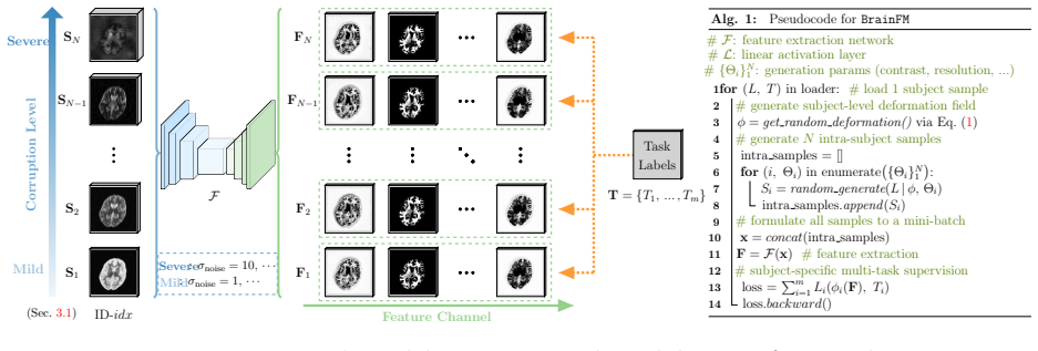
The engine is the on-the-fly intra-subject data generator: given a subject's segmentation labels, it samples a deformation (affine plus stationary velocity field), paints each label with Gaussian-distributed intensities, applies corruption and resolution degradation, and optionally mixes intensities with a real same-subject scan. Training with several such samples per subject in one mini-batch forces the 3D U-Net backbone to encode stable subject identity across appearance changes. Multi-task supervision with linear heads—L1 and gradient-L1 for synthesis, soft dice plus cross-entropy for segmentation, L1 for distance and registration, L2 for bias field—makes the features carry the informatio
What would settle it
Take the released BrainFM, run it on a held-out cohort collected with an acquisition protocol not represented in its corruption range (for example low-field portable MRI or a CT scanner with a different Hounsfield calibration), and compare segmentation Dice and synthesis L1 against a task-specific model fine-tuned on that cohort. If the zero-fine-tuning model's error on that cohort exceeds the fine-tuned model's by more than the paper's reported margins, the modality-agnostic generalization claim fails exactly where clinical utility would be tested.
Extended reading notes
Core claim
BrainFM's central discovery is that contrast and resolution robustness can be learned by generating, for each subject, a mini-batch of intra-subject images deformed and painted with random Gaussian intensities per anatomical label, then corrupted at mild-to-severe levels and mixed with real same-subject scans. The network is supervised simultaneously on five tasks with per-task linear heads, and the paper reports that this yields a representation that is both robust—features transfer to unseen contrasts and resolutions—and expressive, so that one linear head per task suffices. The empirical claim is that BrainFM outperforms the earlier single-task model, a from-scratch baseline with identica
Load-bearing premise
BrainFM's central promise stands or falls on whether the synthetic data generator—random Gaussian intensities painted per anatomical label, deformed and corrupted, then mixed with real same-subject scans—produces images whose appearance statistics match real clinical T1w/T2w/FLAIR and CT closely enough that the model transfers without fine-tuning; CT is the most fragile case because its intensities are physically calibrated in Hounsfield units, not label-conditional Gaussians
Editorial extensions
If this is right
- If the claim holds, one pretrained BrainFM can be dropped into clinical pipelines for T1w/T2w/FLAIR/CT without per-dataset retraining, covering synthesis, segmentation, distance maps, bias-field correction and registration.
- The modality-agnostic representation would also serve as a general feature extractor: the paper shows adaptation to hemisphere and low-field domains, super-resolution and brain-age prediction with minimal extra training.
- Hospitals that acquire only one or two contrasts could still obtain CT-like or T1w-like images through synthesis, because the model is trained to output all four modalities from any input.
- Because training relies on anatomy labels rather than curated paired multi-modal datasets, the approach avoids the cross-institution data collection bottleneck that limits most medical foundation models.
- The mild-to-severe corruption schedule and real-synth mix-up are presented as the reason larger intra-subject batches improve feature robustness, making batch size a practical lever for future models of this kind.
Reading between the lines
- The 'modality-agnostic' claim is only as strong as the synthetic generator's coverage of real acquisition physics; a natural next test would be to probe BrainFM on contrasts and resolutions not in the generator's corruption range, such as low-field portable MRI or non-Cartesian acquisitions.
- Because CT intensity is calibrated in Hounsfield units, the label-conditional Gaussian painting used for MR may be the least faithful simulation; the paper's own tables show CT synthesis has the largest L1 errors, hinting that a physics-informed CT generator could tighten the gap.
- A single multi-task model with linear heads suggests a path toward 'zero-shot' neuroimaging toolkits: if the representation is truly anatomy-anchored, new tasks could be added by training only a new linear head on a small labeled set while keeping the backbone frozen.
- The paper's stated limitation—poor handling of extensive pathology regions—means the strongest version of the claim applies to near-normal anatomy; extending the generator with pathology priors would be the direct next step.
Editorial analysis
A structured set of objections, weighed in public.
Referee Report
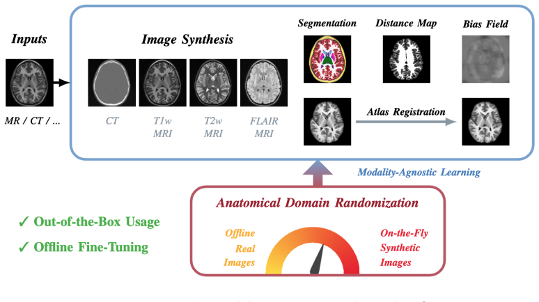
Summary. BrainFM is a proposed modality-agnostic, multi-task 3D vision model for human brain imaging. The model is trained on a mixture of synthetic images generated from anatomical labels via random deformations, label-conditional Gaussian contrast painting, corruption/resolution simulation, and a 'real-synth' mix-up with same-subject real scans. A single U-Net backbone with linear task heads is trained jointly on five tasks: T1w/T2w/FLAIR/CT synthesis, anatomy segmentation, scalp-to-cortical distance map regression, atlas registration, and bias field estimation. The authors report experiments on eleven public datasets under three train/test setups, comparing against two internal baselines (SCRATCH and Brain-ID) and claiming state-of-the-art performance on all tasks and modalities.
Significance. If the claims are substantiated, this would be a valuable contribution: one model that generalizes across MR contrasts and CT without fine-tuning, trained largely from synthetic data, with code released. The internal comparisons against SCRATCH and Brain-ID and the ablations in Table 4 are consistently favorable and support the central architecture choices (multi-task training, real-synth mix-up, mild-to-severe corruption schedule, intra-subject batches). However, the headline 'state-of-the-art' claim is not verifiable from the provided manuscript because the external baselines named in Sec. 4.1.3 never appear in the results, and the segmentation evaluation is anchored to SynthSeg, the same tool used to generate training labels. The lack of error bars or significance tests further weakens the strength of the reported gains, some of which are numerically small.
major comments (4)
- [Abstract, Sec. 1, Sec. 4.1.3, Tables 2-3] The abstract and introduction claim 'state-of-the-art performance in all tasks and input image modalities.' Sec. 4.1.3 names SAMSEG, FastSurfer, SynthSR, and SynthSR-FT as comparison models, but no results against any of these appear in Tables 2-3 or anywhere else in the text; the tables report only SCRATCH and Brain-ID, which are the authors' own baselines. The SOTA claim therefore rests on missing evidence. Either these external comparisons must be added, or the claims must be reduced to comparisons against the presented baselines.
- [Sec. 3.2.2 and Sec. 4.1.4, Table 3 (Seg)] The segmentation evaluation is circular with respect to the training data. Sec. 3.2.2 states that SynthSeg [18] provides the gold-standard segmentation labels used as training targets, and Sec. 4.1.4 states that the gold-standard test labels are also obtained by running SynthSeg on T1-weighted images. Dice scores in Table 3 therefore measure agreement with SynthSeg, not independent anatomical accuracy. This is especially problematic because SynthSeg is from the same lab lineage and defines the training label distribution. An independent reference (e.g., manual annotations or a different segmentation method) is needed, or the results should be framed specifically as 'agreement with SynthSeg.'
- [Tables 2-3] All results are reported as single numbers without error bars, confidence intervals, or significance tests. Some differences between BrainFM and Brain-ID are very small (e.g., Setup I bias-field norm-L2 for T1w: 0.268 vs 0.270; Setup III T2w bias-field: 0.283 vs 0.282), so the claim of 'consistent improvements' is not statistically supported. At minimum, results over multiple training runs or a paired significance test should be reported for the main comparisons.
- [Eq. (2), Sec. 3.1.2, Tables 2-3 (CT rows)] The modality-agnostic claim is strongest for CT, yet the synthetic generator paints each label with a Gaussian intensity (Eq. 2), which does not model the physically calibrated Hounsfield-unit scale of CT. Unsurprisingly, CT synthesis has the highest L1 errors in Tables 2-3 (e.g., Setup I BrainFM CT L1 = 0.050 vs 0.015 for T1w). The paper does not compare against any external CT synthesis method or evaluate errors in HU units. This is a correctness-risk concern, not a circularity one: the CT transfer is the most likely point of failure for the 'modality-agnostic' claim, and it needs either a dedicated external baseline or a clear statement of the CT-specific limitation.
minor comments (4)
- [Sec. 3.1, Sec. 4.1.4, Sec. 2.2, Sec. 4.1.5] Typos: 'generatation' (Sec. 3.1), 'datesets' (Sec. 4.1.4), 'sites sites' (Sec. 2.2), 'T raining' (Sec. 4.1.5 heading).
- [Sec. 4.1.1 (OASIS3)] The dataset description says 'We used T1-weighted MRI, T1-weighted MRI, and CT pairs'; this should presumably read 'T1-weighted MRI, T2-weighted MRI, and CT pairs.'
- [Table 2] The table layout is hard to parse: each row appears to contain four numbers per condition, but the caption says entries report L1/PSNR/SSIM. A legend or sub-table structure would clarify which metric corresponds to which column.
- [Sec. 4.3.3] The limitation that BrainFM is not adept at images with extensive pathology is acknowledged, which is fine, but it should be reconciled with the abstract's unqualified 'all tasks and input modalities' claim.
Circularity Check
Segmentation and distance-map evaluations reduce to agreement with the same SynthSeg labels used for supervision; the promised external SOTA comparisons are absent.
-
self definitional
[Sec. 3.2.2, 'Multi-task Supervision / Anatomy Segmentation' and Sec. 4.1.4, 'Data Preprocessing']
"For all training subjects, we use SynthSeg [18] to obtain the segmentation labels with 30 brain anatomical regions [40], as the gold standard segmentation target. ... The gold-standard brain segmentation maps are obtained by performing SynthSeg [18] on the T1-weighted MR images of all the subjects."
The labels used as supervision for BrainFM's segmentation head are the same SynthSeg outputs used as the evaluation reference. The Dice scores in Table 3 therefore measure agreement between BrainFM and SynthSeg, not independent anatomical accuracy. The 'anatomy segmentation' prediction is trained with soft Dice and cross-entropy against SynthSeg labels, so the reported performance is a self-agreement score by construction. The claim of state-of-the-art segmentation is thus a consistency check with the training label source, not an external validation.
-
self definitional
[Sec. 3.2.2, 'Multi-task Supervision / Distance Maps Prediction']
"We predict distance maps to represent extracted surfaces, where each voxel encodes the shortest distance to the segmented boundary [21]. All the distance values are trained with L1 loss."
The distance-map task is defined as distance to the segmented boundary. The segmented boundary used for supervision (and for the evaluation target) is the same SynthSeg label map used for the segmentation task. Consequently, the L1 errors for distance prediction are a deterministic transform of the SynthSeg agreement already scored in the segmentation output. No independent geometric ground truth is introduced, so this task does not provide separate evidence for BrainFM's anatomical accuracy.
full rationale
BrainFM's image synthesis tasks are evaluated against real same-subject acquisitions, which is an independent reference and not circular. The multi-task architecture and the 'real-synth' mix-up are internally coherent and could be validated by those synthesis results. However, the anatomy segmentation and distance-map evaluations are circular in a specific, quotable way: the gold-standard segmentation maps are produced by SynthSeg, and the same SynthSeg labels serve as the training supervision. Reporting Dice or distance L1 against that source measures self-agreement with the label generator, not independent anatomical truth. In addition, Sec. 4.1.3 promises comparisons with SAMSEG, FastSurfer, SynthSR, and SynthSR-FT, but Tables 2 and 3 report only SCRATCH and Brain-ID, so the 'state-of-the-art in all tasks' claim is unverifiable from the manuscript as written. This missing support is a correctness gap rather than a circularity, but it compounds the self-referential evaluation. Overall, the central multi-task model has substantial independent content in the synthesis tasks, but the segmentation and distance 'predictions' reduce by construction to agreement with the same SynthSeg-derived labels, yielding a partial circularity score of 6.
Assumptions & free parameters
free parameters (5)
- Contrast synthesis hyperparameters (theta_l, theta_mu, theta_sigma) =
not stated in text; inherited from SynthSeg-style label-conditional Gaussian painting
- Deformation range parameters theta_phi =
not stated
- Mild-to-severe corruption schedule =
noise sigma from 1 to 10 (Tab 4), plus resolution/artifact levels from [6]
- Intra-subject mini-batch size N =
4
- Multi-task loss weights (L1, grad-L1, dice, CE, L2) =
not stated
assumptions (6)
- standard math Stationary velocity field integration yields smooth, invertible deformations that preserve brain topology
- domain assumption Label-conditional Gaussian intensities cover the real MR/CT appearance space
- domain assumption Real-synth mix-up bridges sim-to-real gap without corrupting anatomy
- ad hoc to paper SynthSeg labels are a valid gold standard for segmentation training and evaluation
- domain assumption R2R atlas coordinates are a valid registration supervision target
- domain assumption SynthStrip skull-stripping plus NiftyReg rigid alignment is sufficient preprocessing for all modalities
Cite this review
Pith. "Pith review of A Modality-agnostic Multi-task Foundation Model for Human Brain Imaging." pith.science (2026). https://pith.science/paper/BQOWGG3D
@misc{pith2026250900549,
  author       = {Pith},
  title        = {Pith review of: A Modality-agnostic Multi-task Foundation Model for Human Brain Imaging},
  year         = {2026},
  howpublished = {\url{https://pith.science/paper/BQOWGG3D}},
  note         = {Machine review of arXiv:2509.00549}
}
read the original abstract
Recent learning-based approaches have made astonishing advances in calibrated medical imaging like computerized tomography (CT), yet they struggle to generalize in uncalibrated modalities -- notably magnetic resonance (MR) imaging, where performance is highly sensitive to the differences in MR contrast, resolution, and orientation. This prevents broad applicability to diverse real-world clinical protocols. Here we introduce BrainFM, a modality-agnostic, multi-task vision foundation model for human brain imaging. With the proposed "mild-to-severe" intra-subject generation and "real-synth" mix-up training strategy, BrainFM is resilient to the appearance of acquired images (e.g., modality, contrast, deformation, resolution, artifacts), and can be directly applied to five fundamental brain imaging tasks, including image synthesis for CT and T1w/T2w/FLAIR MRI, anatomy segmentation, scalp-to-cortical distance, bias field estimation, and registration. We evaluate the efficacy of BrainFM on eleven public datasets, and demonstrate its robustness and effectiveness across all tasks and input modalities. Code is available at https://github.com/jhuldr/BrainFM.
Figures


Figures from the paper (1 more)
Reference graph
Works this paper leans on
-
[18]
SynthSeg: Segmentation of brain MRI scans of any contrast and resolution without retraining,
B. Billot, D. N. Greve, O. Puonti, A. Thielscher, K. V. Leemput, B. R. Fischl et al. , “SynthSeg: Segmentation of brain MRI scans of any contrast and resolution without retraining,” Medical Image Analysis, 2021. 2, 3, 5, 8
work page 2021
-
[1]
MP RAGE: A three-dimensional, T1-weighted, gradient-echo sequence–initial experience in the brain
M. N. Brant-Zawadzki, G. D. Gillan, and W. R. Nitz, “MP RAGE: A three-dimensional, T1-weighted, gradient-echo sequence–initial experience in the brain.” Radiology, 1992. 1
work page 1992
-
[2]
U-Net: Convolutional networks for biomedical image seg- mentation,
O. Ronneberger, P. Fischer, and T. Brox, “U-Net: Convolutional networks for biomedical image seg- mentation,” in MICCAI, 2015. 1, 8
work page 2015
-
[3]
V-Net: Fully convolutional neural networks for volumetric medical image segmentation,
F. Milletar ` ı, N. Navab, and S.-A. Ahmadi, “V-Net: Fully convolutional neural networks for volumetric medical image segmentation,” 3DV, 2016. 1
work page 2016
-
[4]
Efficient multi-scale 3D CNN with fully connected CRF for accurate brain lesion segmentation,
K. Kamnitsas, C. Ledig, V. F. J. Newcombe, J. P. Simpson, A. D. Kane, D. K. Menon et al., “Efficient multi-scale 3D CNN with fully connected CRF for accurate brain lesion segmentation,” Medical Image Analysis, 2016. 1
work page 2016
-
[5]
Local temperature scaling for probability calibration,
Z. Ding, X. Han, P. Liu, and M. Niethammer, “Local temperature scaling for probability calibration,” in ICCV, 2021. 1
work page 2021
-
[6]
J. E. Iglesias, B. Billot, Y. Balbastre, C. G. Magdamo, S. Arnold, S. Das et al. , “SynthSR: A pub- lic AI tool to turn heterogeneous clinical brain scans into high-resolution T1-weighted images for 3D morphometry,” Science Advances, 2023. 1, 2, 3, 4, 8
work page 2023
-
[7]
VoxelMorph: A learning framework for deformable medical image registration,
G. Balakrishnan, A. Zhao, M. R. Sabuncu, J. V. Guttag, and A. V. Dalca, “VoxelMorph: A learning framework for deformable medical image registration,” IEEE Transactions on Medical Imaging , 2018. 1
work page 2018
Show all 63 references
-
[8]
Quicksilver: Fast predictive image registration – a deep learning approach,
X. Yang, R. Kwitt, and M. Niethammer, “Quicksilver: Fast predictive image registration – a deep learning approach,” NeuroImage, 2017. 1
2017
-
[9]
A deep learning framework for unsupervised affine and deformable image registration,
B. D. de Vos, F. F. Berendsen, M. A. Viergever, H. Sokooti, M. Staring, and I. Iˇ sgum, “A deep learning framework for unsupervised affine and deformable image registration,” Medical Image Analysis , 2019. 1
2019
-
[10]
Accurate point cloud registration with robust optimal transport,
Z. Shen, J. Feydy, P. Liu, A. H. Curiale, R. San Jose Estepar, R. San Jose Estepar, and M. Niethammer, “Accurate point cloud registration with robust optimal transport,” in NeurIPS, 2021. 1
2021
-
[11]
Improving in vivo human cerebral cortical surface reconstruction using data-driven super-resolution
Q. Tian, B. Bilgi¸ c, Q. Fan, C. Ngamsombat, N. Zaretskaya, N. E. Fultzet al., “Improving in vivo human cerebral cortical surface reconstruction using data-driven super-resolution.” Cerebral Cortex, 2020. 1
2020
-
[12]
Uncertainty modelling in deep learning for safer neuroimage enhancement: Demonstration in diffusion MRI,
R. Tanno, D. E. Worrall, E. Kaden, and D. C. Alexander, “Uncertainty modelling in deep learning for safer neuroimage enhancement: Demonstration in diffusion MRI,” NeuroImage, 2020. 1
2020
-
[13]
Underconnected, but how? A survey of functional connectivity MRI studies in autism spectrum disorders
R.-A. M¨ uller, P. Shih, B. Keehn, J. R. Deyoe, K. M. Leyden, and D. Shukla, “Underconnected, but how? A survey of functional connectivity MRI studies in autism spectrum disorders.” Cerebral Cortex,
-
[14]
Deep visual domain adaptation: A survey,
M. Wang and W. Deng, “Deep visual domain adaptation: A survey,” Neurocomputing, 2018. 2
2018
-
[15]
Joint super- resolution and synthesis of 1 mm isotropic MP-RAGE volumes from clinical MRI exams with scans of different orientation, resolution and contrast,
J. E. Iglesias, B. Billot, Y. Balbastre, A. Tabari, J. Conklin, D. C. Alexander et al. , “Joint super- resolution and synthesis of 1 mm isotropic MP-RAGE volumes from clinical MRI exams with scans of different orientation, resolution and contrast,” NeuroImage, 2020. 2, 3, 4 10
2020
-
[16]
Discovering hidden physics behind transport dynamics,
P. Liu, L. Tian, Y. Zhang, S. Aylward, Y. Lee, and M. Niethammer, “Discovering hidden physics behind transport dynamics,” in CVPR, 2021. 2
2021
-
[17]
Deep decomposition for stochastic normal-abnormal transport,
P. Liu, Y. Lee, S. Aylward, and M. Niethammer, “Deep decomposition for stochastic normal-abnormal transport,” in CVPR, 2022. 2
2022
-
[19]
SynthMorph: Learn- ing contrast-invariant registration without acquired images,
M. Hoffmann, B. Billot, D. N. Greve, J. E. Iglesias, B. R. Fischl, and A. V. Dalca, “SynthMorph: Learn- ing contrast-invariant registration without acquired images,” IEEE Transactions on Medical Imaging ,
-
[20]
Quantifying white matter hyperintensity and brain volumes in heterogeneous clinical and low-field portable MRI,
P. Laso, S. Cerri, A. Sorby-Adams, J. Guo, F. Mateen, P. Goebl et al. , “Quantifying white matter hyperintensity and brain volumes in heterogeneous clinical and low-field portable MRI,” arXiv, vol. abs/2312.05119, 2023. 2, 3
2023 arXiv
-
[21]
“Recon-all-clinical
K. Gopinath, D. N. Greve, C. Magdamo, S. Arnold, S. Das, O. Puonti, J. E. Iglesias, A. D. N. Initiative et al., ““Recon-all-clinical”: Cortical surface reconstruction and analysis of heterogeneous clinical brain MRI,” Medical Image Analysis, 2025. 2, 5
2025
-
[22]
Language models are few-shot learners,
T. Brown, B. Mann, N. Ryder, M. Subbiah, J. D. Kaplan, P. Dhariwal et al. , “Language models are few-shot learners,” in NeurIPS, 2020. 2
2020
-
[23]
PaLM: Scaling language modeling with pathways,
A. Chowdhery, S. Narang, J. Devlin, M. Bosma, G. Mishra, A. Roberts et al., “PaLM: Scaling language modeling with pathways,” JMLR, 2022. 2
2022
-
[24]
Segment anything,
A. Kirillov, E. Mintun, N. Ravi, H. Mao, C. Rolland, L. Gustafson et al. , “Segment anything,” arXiv, vol. abs/2304.02643, 2023. 2
2023 arXiv
-
[25]
On the opportunities and risks of foundation models,
R. Bommasani, D. A. Hudson, E. Adeli, R. Altman, S. Arora, S. von Arx et al., “On the opportunities and risks of foundation models,” arXiv, vol. abs/2108.07258, 2021. 2
2021 arXiv
-
[26]
Foundational models defining a new era in vision: A survey and outlook,
M. Awais, M. Naseer, S. S. Khan, R. M. Anwer, H. Cholakkal, M. Shah et al. , “Foundational models defining a new era in vision: A survey and outlook,” arXiv, vol. abs/2307.13721, 2023. 2
2023 arXiv
-
[27]
MONAI: An open-source framework for deep learning in healthcare,
M. J. Cardoso, W. Li, R. Brown, N. Ma, E. Kerfoot, Y. Wang et al. , “MONAI: An open-source framework for deep learning in healthcare,” arXiv, vol. abs/2211.02701, 2022. 2
2022 arXiv
-
[28]
A foundation model for generalizable disease detection from retinal images,
Y. Zhou, M. A. Chia, S. K. Wagner, M. S. Ayhan, D. J. Williamson, R. R. Struyvenet al., “A foundation model for generalizable disease detection from retinal images,” Nature, 2023. 2
2023
-
[29]
Foundation models for generalist medical artificial intelligence,
M. Moor, O. Banerjee, Z. F. H. Abad, H. M. Krumholz, J. Leskovec, E. J. Topol, and P. Rajpurkar, “Foundation models for generalist medical artificial intelligence,” Nature, 2023. 2
2023
-
[30]
Large language models encode clinical knowledge,
K. Singhal, S. Azizi, T. Tu, S. Mahdavi, J. Wei, H. W. Chung et al. , “Large language models encode clinical knowledge,” Nature, 2022. 2
2022
-
[31]
Towards generalist biomed- ical AI,
T. Tu, S. Azizi, D. Driess, M. Schaekermann, M. Amin, P.-C. Chang et al., “Towards generalist biomed- ical AI,” arXiv, vol. abs/2307.14334, 2023. 2
2023 arXiv
-
[32]
Brain-ID: Learning robust feature representations for brain imaging,
P. Liu, O. Puonti, X. Hu, D. C. Alexander, and J. E. Iglesias, “Brain-ID: Learning robust feature representations for brain imaging,” in ECCV, 2024. 2, 3, 9
2024
-
[33]
Deep learning of feature representation with multiple instance learning for medical image analysis,
Y. Xu, T. Mo, Q. Feng, P. Zhong, M. Lai, and E. I.-C. Chang, “Deep learning of feature representation with multiple instance learning for medical image analysis,” in ICASSP, 2014. 2
2014
-
[34]
Momentum contrastive voxel-wise representation learning for semi-supervised volumetric medical image segmentation,
C. You, R. Zhao, L. H. Staib, and J. S. Duncan, “Momentum contrastive voxel-wise representation learning for semi-supervised volumetric medical image segmentation,” in MICCAI, 2021. 2
2021
-
[35]
SAM: Self-supervised learning of pixel- wise anatomical embeddings in radiological images,
K. Yan, J. Cai, D. Jin, S. Miao, A. P. Harrison, D. Guo et al., “SAM: Self-supervised learning of pixel- wise anatomical embeddings in radiological images,” IEEE Transactions on Medical Imaging , 2020. 2 11
2020
-
[36]
SAMConvex: Fast discrete optimization for CT registration using self-supervised anatomical embedding and correlation pyramid,
Z. Li, L. Tian, T. C. W. Mok, X. Bai, P. Wang, J. Ge et al. , “SAMConvex: Fast discrete optimization for CT registration using self-supervised anatomical embedding and correlation pyramid,” in MICCAI,
-
[37]
BrainPrint: Identifying subjects by their brain,
C. Wachinger, P. Golland, and M. Reuter, “BrainPrint: Identifying subjects by their brain,” inMICCAI,
-
[38]
Contrast invariant feature representations for segmentation and registration of medical images,
Y. Z. R. Chua and A. V. Dalca, “Contrast invariant feature representations for segmentation and registration of medical images,” in MIDL, 2023. 3
2023
-
[39]
A unifying framework for partial volume segmentation of brain MR images,
K. V. Leemput, F. Maes, D. Vandermeulen, and P. Suetens, “A unifying framework for partial volume segmentation of brain MR images,” IEEE Transactions on Medical Imaging , 2003. 3
2003
-
[40]
Whole brain segmentation automated labeling of neuroanatomical structures in the human brain,
B. R. Fischl, D. H. Salat, E. Busa, M. S. Albert, M. Dieterich, C. Haselgrove et al. , “Whole brain segmentation automated labeling of neuroanatomical structures in the human brain,” Neuron, 2002. 3, 5
2002
-
[41]
Fast and sequence-adaptive whole-brain segmentation using parametric bayesian modeling,
O. Puonti, J. E. Iglesias, and K. V. Leemput, “Fast and sequence-adaptive whole-brain segmentation using parametric bayesian modeling,” NeuroImage, 2016. 3, 8
2016
-
[42]
SynthStrip: skull-stripping for any brain image,
A. Hoopes, J. S. Mora, A. V. Dalca, B. R. Fischl, and M. Hoffmann, “SynthStrip: skull-stripping for any brain image,” NeuroImage, 2022. 3, 8
2022
-
[43]
Federated learning in medicine: Facilitating multi-institutional collaborations without sharing patient data,
M. J. Sheller, B. Edwards, G. A. Reina, J. Martin, S. Pati, A. Kotrotsou et al. , “Federated learning in medicine: Facilitating multi-institutional collaborations without sharing patient data,” Scientific reports, 2020. 4
2020
-
[44]
Federated learning and differential privacy for medical image analysis,
M. Adnan, S. Kalra, J. C. Cresswell, G. W. Taylor, and H. R. Tizhoosh, “Federated learning and differential privacy for medical image analysis,” Scientific reports, 2022. 4
2022
-
[45]
Federated learning in medical imaging: Part I: Toward multicentral health care ecosystems,
E. Darzidehkalani, M. Ghasemi-Rad, and P. van Ooijen, “Federated learning in medical imaging: Part I: Toward multicentral health care ecosystems,” Journal of the American College of Radiology , 2022. 4
2022
-
[46]
Registration by regression (rbr): a framework for interpretable and flexible atlas registration,
K. Gopinath, X. Hu, M. Hoffmann, O. Puonti, and J. E. Iglesias, “Registration by regression (rbr): a framework for interpretable and flexible atlas registration,” arXiv, vol. abs/2404.16781, 2024. 5
2024 arXiv
-
[47]
Hierarchical uncertainty estimation for learning-based registration in neuroimaging,
X. Hu, K. Gopinath, P. Liu, M. Hoffmann, K. Van Leemput, O. Puonti, and J. E. Iglesias, “Hierarchical uncertainty estimation for learning-based registration in neuroimaging,” in ICLR, 2025. 5
2025
-
[48]
Bias field correction for MRI images,
J. Juntu, J. Sijbers, D. V. Dyck, and J. Gielen, “Bias field correction for MRI images,” in CORES,
-
[49]
Evaluation of performance metrics for bias field correction in MR brain images,
Z. Y. Chua, W. Zheng, M. W. L. Chee, and V. Zagorodnov, “Evaluation of performance metrics for bias field correction in MR brain images,” Journal of Magnetic Resonance Imaging , 2009. 5, 8
2009
-
[50]
The autism brain imaging data exchange: towards a large-scale evaluation of the intrinsic brain architecture in autism,
A. Di Martino, C.-G. Yan, Q. Li, E. Denio, F. X. Castellanos, K. Alaerts et al. , “The autism brain imaging data exchange: towards a large-scale evaluation of the intrinsic brain architecture in autism,” Molecular psychiatry, 2014. 6
2014
-
[51]
Enhancing studies of the connectome in autism using the autism brain imaging data exchange II,
A. Di Martino, D. O’connor, B. Chen, K. Alaerts, J. S. Anderson, M. Assaf et al. , “Enhancing studies of the connectome in autism using the autism brain imaging data exchange II,” Scientific data, 2017. 6
2017
-
[52]
ADHD- 200 global competition: diagnosing ADHD using personal characteristic data can outperform resting state fMRI measurements,
M. R. G. Brown, G. P. S. Sidhu, R. Greiner, N. Asgarian, M. Bastani, P. H. Silverstone et al., “ADHD- 200 global competition: diagnosing ADHD using personal characteristic data can outperform resting state fMRI measurements,” Frontiers in Systems Neuroscience, 2012. 6
2012
-
[53]
The Alzheimer’s disease neuroimaging initiative 3: Continued innovation for clinical trial improvement,
M. W. Weiner, D. P. Veitch, P. S. Aisen, L. A. Beckett, N. J. Cairns, R. C. Greenet al., “The Alzheimer’s disease neuroimaging initiative 3: Continued innovation for clinical trial improvement,” Alzheimer’s & Dementia, 2017. 6
2017
-
[54]
Fowler, S
C. Fowler, S. R. Rainey-Smith, S. Bird, J. Bomke, P. Bourgeat, B. M. Brown et al., “Fifteen years of the australian imaging, biomarkers and lifestyle (AIBL) study: Progress and observations from 2,359 older adults spanning the spectrum from cognitive normality to Alzheimer’s d...
2021
-
[55]
Exploring the psychosis functional connectome: aberrant intrinsic networks in schizophrenia and bipolar disorder,
V. D. Calhoun, J. Sui, K. Kiehl, J. Turner, E. Allen, and G. Pearlson, “Exploring the psychosis functional connectome: aberrant intrinsic networks in schizophrenia and bipolar disorder,” Frontiers in psychiatry,
-
[56]
Longitudinal multiple sclerosis lesion segmentation: resource and challenge,
A. Carass, S. Roy, A. Jog, J. L. Cuzzocreo, E. Magrath, A. Gherman et al. , “Longitudinal multiple sclerosis lesion segmentation: resource and challenge,” NeuroImage, 2017. 7
2017
-
[57]
The human connectome project: A data acquisition perspective,
D. C. V. Essen, K. U˘ gurbil, E. J. Auerbach, D. M. Barch, T. E. J. Behrens, R. D. Bucholz et al., “The human connectome project: A data acquisition perspective,” NeuroImage, 2012. 7
2012
-
[58]
The chinese human connectome project,
N. Vogt, “The chinese human connectome project,” Nature Methods, 2023. 7
2023
-
[59]
The MCIC collection: a shared repository of multi-modal, multi-site brain image data from a clinical investigation of schizophrenia,
R. L. Gollub, J. M. Shoemaker, M. D. King, T. White, S. Ehrlich, S. R. Sponheim, V. P. Clark, J. A. Turner, B. A. Mueller, V. Magnotta et al. , “The MCIC collection: a shared repository of multi-modal, multi-site brain image data from a clinical investigation of schizophrenia,...
2013
-
[60]
OASIS-3: Longitudinal neuroimaging, clinical, and cognitive dataset for normal aging and Alzheimer’s disease,
P. J. LaMontagne, S. J. Keefe, W. Lauren, C. Xiong, E. A. Grant, K. L. Moulder et al. , “OASIS-3: Longitudinal neuroimaging, clinical, and cognitive dataset for normal aging and Alzheimer’s disease,” Alzheimer’s & Dementia , 2018. 7
2018
-
[61]
A contrast-adaptive method for simultaneous whole-brain and lesion segmentation in multiple sclerosis,
S. Cerri, O. Puonti, D. S. Meier, J. Wuerfel, M. M¨ uhlau, H. R. Siebner, and K. V. Leemput, “A contrast-adaptive method for simultaneous whole-brain and lesion segmentation in multiple sclerosis,” NeuroImage, 2020. 8
2020
-
[62]
FastSurferVINN: Building resolution-independence into deep learning segmentation methods — A solution for highres brain MRI,
L. Henschel, D. K¨ ugler, and M. Reuter, “FastSurferVINN: Building resolution-independence into deep learning segmentation methods — A solution for highres brain MRI,” NeuroImage, 2022. 8
2022
-
[63]
Fast free-form deformation using graphics processing units,
M. Modat, G. R. Ridgway, Z. A. Taylor, M. Lehmann, J. Barnes, D. J. Hawkes et al. , “Fast free-form deformation using graphics processing units,” Computer Methods and Programs in Biomedicine , 2010. 8 13 Table 2: Comparisons of BrainFM with SCRATCH and Brain-ID, on image synth...
2010
Reviewed August 5, 2026 · model on record in the stance chip above.
Discussion (0). Sign in to comment.