REVIEW 5 major objections 5 minor 29 references
Systematic Integration of Attention Modules into CNNs for Accurate and Generalizable Medical Image Diagnosis
T0 review · 5 major / 5 minor · reviewed 2026-08-05 · deepseek-v4-flash
Pith's one-line read The paper claims that adding lightweight attention modules to five standard CNNs consistently improves medical image classification on two datasets, with EfficientNetB5 plus hybrid attention reaching 89.97 percent accuracy on the POC datase
desk verdict The paper's central claim that attention consistently helps is unsupported because the test set was used for early stopping and model selection; the comparison framework is useful but the numbers cannot be trusted as is. read the letter →
The pith
A machine-rendered reading of the paper's core claim, the machinery that carries it, and where it could break.
The reading
What carries the argument
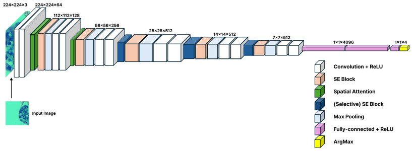
The load-bearing units are the Squeeze-and-Excitation block and a spatial-attention module of the CBAM family. SE compresses each feature map to a channel descriptor and then uses a small learned gate to rescale channels, emphasizing informative channels; spatial attention pools the feature map along the channel axis and applies a 7x7 convolution to produce a spatial importance map, emphasizing informative regions. The paper's controlled comparison of global, selective, and hybrid placements of these units is what carries the argument: it isolates the placement effect rather than just the attention mechanism itself.
What would settle it
Run the same five-backbone comparison with model selection and early stopping done on a separate validation set, leaving an untouched test set for the final numbers (or use nested cross-validation). If attention-augmented models no longer beat their baselines on that untouched test set, the paper's central claim fails.
Extended reading notes
Core claim
The paper's central claim is that attention-augmented CNNs consistently outperform their unaugmented counterparts, and that hybrid attention—channel recalibration combined with spatial attention—gives the largest gains. It further claims that selectively placing SE blocks in deeper layers beats inserting them everywhere, and that adding spatial attention to those selected blocks improves both accuracy and F1. The strongest configuration is EfficientNetB5 with SE blocks after blocks 2, 3, and 4 plus a spatial-attention module after block 3, which reaches 89.97% test accuracy on POC; on the brain-tumor dataset the best hybrid result is ResNet18 at 84.37% accuracy. The paper interprets these ga
Load-bearing premise
The claim assumes the test set used for final metrics did not influence training decisions, but the study used test accuracy for early stopping and model selection, so the reported gains may be inflated.
Editorial extensions
If this is right
- Pretrained CNN backbones can be upgraded with attention modules without redesigning the classifier or training from scratch.
- Selective placement of SE blocks in deeper layers gives the best accuracy-per-complexity trade-off, outperforming global insertion in the reported runs.
- Combining channel and spatial attention improves precision, recall, and F1, not just top-1 accuracy, so class-balanced performance benefits too.
- EfficientNetB5 with hybrid attention is the strongest single configuration identified for these two medical classification tasks.
- Lightweight backbones such as VGG16 and ResNet18 narrow the gap with heavier models when attention is added, which matters for deployment on limited hardware.
Reading between the lines
- The use of test accuracy for model selection and early stopping means the reported numbers are likely optimistic; a fair estimate of the attention gain would require a validation set for decisions and an untouched test set for the final report.
- Because the same attention modules helped in both MRI and histopathology, the intervention plausibly transfers to other medical imaging tasks where subtle localized features drive diagnosis, but the paper only demonstrates two datasets.
- The manually chosen attention positions leave open the possibility that automated placement search would find configurations that are even more accurate or more efficient.
- The paper's 'generalization' claim covers two held-out test sets from the same sources; true clinical generality across devices and institutions is not tested.
Editorial analysis
A structured set of objections, weighed in public.
Referee Report
Summary. The paper proposes to augment five pretrained CNNs (VGG16, ResNet18, InceptionV3, DenseNet121, EfficientNetB5) with Squeeze-and-Excitation (SE) blocks and/or spatial attention, in global, selective, and hybrid configurations, and evaluates the resulting variants on two medical image datasets: a POC histopathology dataset and a BT-Large-4c brain-tumor MRI dataset. The central claim is that attention-augmented CNNs consistently outperform their baselines across all metrics, with EfficientNetB5 plus hybrid attention achieving the highest overall performance. The results are reported in four stages: baseline fine-tuning, global SE insertion, selective SE insertion, and hybrid SE+spatial attention.
Significance. The comparative scope is useful in principle: five backbone families, two imaging modalities, and systematic placement variants are considered. However, the evaluation protocol uses the test set for both early stopping and model selection, so the reported test numbers are not unbiased estimates of generalization. The headline claim is also internally contradicted by the paper's own tables. Because the main contribution is empirical and the protocol is flawed, the current significance is low; a corrected re-run with a proper validation split and repeated seeds would be required before the claimed attention gains can be assessed.
major comments (5)
- [§3.4, Table 3] The training protocol uses the test set to make training decisions: Section 3.4 states that early stopping 'monitors test accuracy' and that 'The best-performing model was saved based on highest test accuracy,' and Table 3 lists 'Model Selection Criterion: Best test accuracy.' This is explicit test-set leakage. Every reported metric is the result of selection on the test set, so the reported improvements over baselines are not unbiased estimates of generalization. The paper must use a held-out validation set (or nested cross-validation) for early stopping, checkpoint selection, and comparing variants, and report final metrics only on a truly unseen test set.
- [§4.4 vs. abstract; Table 11; Table 6] The abstract claims that 'EfficientNetB5 with hybrid attention achieves the highest overall performance, delivering substantial gains on both datasets.' Table 11 (brain tumor) shows ResNet18+SE+SA at 84.37% accuracy versus EfficientNetB5+SE+SA at 83.41%, so EfficientNetB5 is not the best model on the brain-tumor dataset. In addition, Table 6 (POC) shows EfficientNetB5+SE at 85.74% accuracy and F1=0.8482, below the baseline EfficientNetB5 at 86.05% and F1=0.8519, directly contradicting the claim that attention-augmented CNNs 'consistently outperform' baselines. The claims and the experiments need to be reconciled.
- [§3.4, Table 3] Baseline and attention-augmented models are not trained under identical optimization settings. Table 3 and Section 3.4 specify a backbone learning rate of 1e-4 and an attention-module learning rate of 6e-4. Thus any performance difference between a baseline and its attention-augmented variant could be due to the higher learning rate applied to the added parameters rather than to the attention mechanism itself. A controlled comparison should hold the optimization protocol fixed across models, or include an ablation that trains the baseline with the same two-group learning-rate schedule.
- [§4, Tables 4–11] Each configuration appears to be run once. The text says experiments were 'repeated with consistent random seeds,' but this means the same seed was reused, not that multiple independent seeds were tried. No variance, confidence intervals, or statistical tests are reported. Differences as small as 0.5–1% in Tables 4–11 are treated as decisive, which is not justified, especially given the test-set selection described in §3.4. At minimum, the authors should report mean±std over several seeds on a proper validation split.
- [§5] The limitation statement admits that attention positions were 'fixed and manually chosen' and 'selected based on prior studies and empirical evaluation.' In the context of the test set being used for model selection and early stopping, this placement search is another channel through which test information enters the final configuration. An unbiased evaluation would fix the placement rules a priori or select them on a validation set, not on the test set.
minor comments (5)
- [§3.2.4 vs. §5.2, Table 2] The first EfficientNet variant is described inconsistently: Section 3.2.4 says SE is incorporated inside the MBConv blocks, while the text after Section 5.2 says a custom MBConv block is appended after the pretrained feature extractor. Please clarify which implementation was actually used.
- [§6] The conclusion refers to 'DenseNet12' instead of 'DenseNet121.'
- [§5.1] Section 5.1 mentions MobileNetV2 as one of the architectures used, but MobileNetV2 does not appear in the experiments. This should be removed or corrected.
- [Keywords/§3.3.4] The paper uses 'CBAM' in the keywords and introduction, but the implementation described in Section 3.3.4 is a sequential SE plus spatial-attention module, not the CBAM module of Woo et al. as a single integrated unit. Clarify the terminology.
- [Table 2] Table 2 uses block indices such as 'index 4,' 'index 9,' and 'index 13' without mapping them to PyTorch layer names or the original EfficientNet stages. A layer-name mapping would improve reproducibility.
Circularity Check
No circularity by construction; the work is an empirical benchmark study. The test-set leakage in Section 3.4 is a serious validity concern but not a circular derivation.
full rationale
The paper makes no first-principles or derivational claim: it reports fine-tuning experiments of standard pretrained CNN backbones with standard SE/CBAM attention modules on two datasets. The attention modules are cited to external prior work (Hu et al. 2018; Woo et al. 2018), and the backbones are standard ImageNet models; no result is derived from a self-imposed ansatz or from a theorem by the same authors. The only self-citations are the POC dataset [26] and two prior brain-tumor papers [9,10] in related work; these are used for data provenance and background, not to justify the central attention claim. Section 5 openly lists limitations (manually chosen positions, overfitting risk), which are soundness concerns rather than circularity. The most serious issue is that Section 3.4 and Table 3 set the model selection criterion to 'Best test accuracy' and use test accuracy for early stopping, so reported test metrics are selected on the test set and are not unbiased generalization estimates; Table 6 even shows EfficientNetB5 SE (0.8574) below its baseline (0.8605), contradicting 'consistently outperform.' This is test-set leakage/selection bias, a correctness and reproducibility problem, but it does not make the reported comparison equivalent to its inputs by construction: attention's superiority is not entailed by the selection rule. Hence no circular step meets the evidentiary bar, and the circularity score is 0.
Assumptions & free parameters
free parameters (4)
- SE reduction ratio =
16
- Backbone learning rate =
0.0001
- Attention learning rate =
0.0006
- Attention placement positions =
e.g., VGG16 blocks 3-5; EfficientNetB5 blocks 2,3,4
assumptions (3)
- domain assumption ImageNet-pretrained weights transfer to medical imaging domains
- domain assumption Test set is held out and not used for model selection
- domain assumption The two public datasets are representative of the clinical tasks
Cite this review
Pith. "Pith review of Systematic Integration of Attention Modules into CNNs for Accurate and Generalizable Medical Image Diagnosis." pith.science (2026). https://pith.science/paper/2PY7IZ3H
@misc{pith2026250905343,
  author       = {Pith},
  title        = {Pith review of: Systematic Integration of Attention Modules into CNNs for Accurate and Generalizable Medical Image Diagnosis},
  year         = {2026},
  howpublished = {\url{https://pith.science/paper/2PY7IZ3H}},
  note         = {Machine review of arXiv:2509.05343}
}
read the original abstract
Deep learning has become a powerful tool for medical image analysis; however, conventional Convolutional Neural Networks (CNNs) often fail to capture the fine-grained and complex features critical for accurate diagnosis. To address this limitation, we systematically integrate attention mechanisms into five widely adopted CNN architectures, namely, VGG16, ResNet18, InceptionV3, DenseNet121, and EfficientNetB5, to enhance their ability to focus on salient regions and improve discriminative performance. Specifically, each baseline model is augmented with either a Squeeze and Excitation block or a hybrid Convolutional Block Attention Module, allowing adaptive recalibration of channel and spatial feature representations. The proposed models are evaluated on two distinct medical imaging datasets, a brain tumor MRI dataset comprising multiple tumor subtypes, and a Products of Conception histopathological dataset containing four tissue categories. Experimental results demonstrate that attention augmented CNNs consistently outperform baseline architectures across all metrics. In particular, EfficientNetB5 with hybrid attention achieves the highest overall performance, delivering substantial gains on both datasets. Beyond improved classification accuracy, attention mechanisms enhance feature localization, leading to better generalization across heterogeneous imaging modalities. This work contributes a systematic comparative framework for embedding attention modules in diverse CNN architectures and rigorously assesses their impact across multiple medical imaging tasks. The findings provide practical insights for the development of robust, interpretable, and clinically applicable deep learning based decision support systems.
Figures


Figures from the paper (4 more)


Reference graph
Works this paper leans on
-
[1]
I. Abd El Kader, G. Xu, Z. Shuai, S. Saminu, I. Javaid, I. Salim Ahmad, Differential deep convolutional neural network model for brain tumor classification, Brain Sciences 11 (2021) 352
work page 2021
-
[2]
A. H. Khan, S. Abbas, M. A. Khan, U. Farooq, W. A. Khan, S. Y. Sid- diqui, A. Ahmad, Intelligent model for brain tumor identification using deep learning, Applied Computational Intelligence and Soft Computing 2022 (2022) 8104054
work page 2022
-
[3]
M. W. Nadeem, M. A. A. Ghamdi, M. Hussain, M. A. Khan, K. M. Khan, S. H. Almotiri, S. A. Butt, Brain tumor analysis empowered with deep learning: A review, taxonomy, and future challenges, Brain sciences 10 (2020) 118
work page 2020
-
[4]
J. Hu, L. Shen, G. Sun, Squeeze-and-excitation networks, in: Proceed- ings of the IEEE conference on computer vision and pattern recognition, 2018, pp. 7132–7141
2018
-
[5]
S. Woo, J. Park, J.-Y. Lee, I. S. Kweon, Cbam: Convolutional block attention module, in: Proceedings of the European conference on com- puter vision (ECCV), 2018, pp. 3–19
2018
-
[6]
Improved EATFormer: A Vision Transformer for Medical Image Classification
Y. Shisu, S. Mingwin, Y. Wanwag, Z. Chenso, S. Huing, Improved eatformer: A vision transformer for medical image classification, arXiv preprint arXiv:2403.13167 (2024). 34
work page Pith review arXiv 2024
-
[7]
D. Chung, Artificial intelligence in healthcare and medicine technology development review, Engineering Applications of Artificial Intelligence 143 (2025) 109801
work page 2025
-
[8]
H. Yu, L. T. Yang, Q. Zhang, D. Armstrong, M. J. Deen, Convolutional neural networks for medical image analysis: state-of-the-art, compar- isons, improvement and perspectives, Neurocomputing 444 (2021) 92– 110
work page 2021
Show all 29 references
-
[9]
Ullah, J
Z. Ullah, J. Kim, Hierarchical deep feature fusion and ensemble learning for enhanced brain tumor mri classification, arXiv preprint arXiv:2506.12363 (2025)
2025 arXiv
-
[10]
Ullah, D
Z. Ullah, D. Pamucar, J. Kim, Hybrid ensemble approaches: Optimal deep feature fusion and hyperparameter-tuned classifier ensembling for enhanced brain tumor classification, arXiv preprint arXiv:2507.12177 (2025)
2025 arXiv
-
[11]
R. N. Asif, M. T. Naseem, M. Ahmad, T. Mazhar, M. A. Khan, M. A. Khan, A. Al-Rasheed, H. Hamam, Brain tumor detection empowered with ensemble deep learning approaches from mri scan images, Scientific Reports 15 (2025) 15002
2025
-
[12]
Babayomi, O
M. Babayomi, O. A. Olagbaju, A. A. Kadiri, Convolutional xg- boost (c-xgboost) model for brain tumor detection, arXiv preprint arXiv:2301.02317 (2023)
2023 arXiv
-
[13]
Srinivasan, D
S. Srinivasan, D. Francis, S. K. Mathivanan, H. Rajadurai, B. D. Shiv- ahare, M. A. Shah, A hybrid deep cnn model for brain tumor image multi-classification, BMC Medical Imaging 24 (2024) 21
2024
-
[14]
Raffel, N
C. Raffel, N. Shazeer, A. Roberts, K. Lee, S. Narang, M. Matena, Y. Zhou, W. Li, P. J. Liu, Exploring the limits of transfer learning with a unified text-to-text transformer, Journal of machine learning research 21 (2020) 1–67
2020
-
[15]
H. E. Kim, A. Cosa-Linan, N. Santhanam, M. Jannesari, M. E. Maros, T. Ganslandt, Transfer learning for medical image classification: a lit- erature review, BMC medical imaging 22 (2022) 69. 35
2022
-
[16]
Y. Sun, C. Wang, Brain tumor detection based on a novel and high- quality prediction of the tumor pixel distributions, Computers in Biology and Medicine 172 (2024) 108196
2024
-
[17]
G. K. Thakur, A. Thakur, S. Kulkarni, N. Khan, S. Khan, Deep learning approaches for medical image analysis and diagnosis, Cureus 16 (2024)
2024
-
[18]
R. Ge, T. Shen, Y. Zhou, C. Liu, L. Zhang, B. Yang, Y. Yan, J.-L. Coatrieux, Y. Chen, Convolutional squeeze-and-excitation network for ecg arrhythmia detection, Artificial Intelligence in Medicine 121 (2021) 102181
2021
-
[19]
Y. Li, Y. Liu, W.-G. Cui, Y.-Z. Guo, H. Huang, Z.-Y. Hu, Epileptic seizure detection in eeg signals using a unified temporal-spectral squeeze- and-excitation network, IEEE Transactions on Neural Systems and Re- habilitation Engineering 28 (2020) 782–794
2020
-
[20]
Kitada, H
S. Kitada, H. Iyatomi, Skin lesion classification with ensemble of squeeze-and-excitation networks and semi-supervised learning, arXiv preprint arXiv:1809.02568 (2018)
2018 arXiv
-
[21]
Simonyan, A
K. Simonyan, A. Zisserman, Very deep convolutional networks for large- scale image recognition, arXiv preprint arXiv:1409.1556 (2014)
2014 arXiv
-
[22]
K. He, X. Zhang, S. Ren, J. Sun, Deep residual learning for image recognition, in: Proceedings of the IEEE conference on computer vision and pattern recognition, 2016, pp. 770–778
2016
-
[23]
Szegedy, W
C. Szegedy, W. Liu, Y. Jia, P. Sermanet, S. Reed, D. Anguelov, D. Er- han, V. Vanhoucke, A. Rabinovich, Going deeper with convolutions, in: Proceedings of the IEEE conference on computer vision and pattern recognition, 2015, pp. 1–9
2015
-
[24]
M. Tan, Q. E. Le, et al., Rethinking model scaling for convolutional neural networks, in: Proceedings of the International conference on machine learning, Long Beach, CA, USA, volume 15, 2019
2019
-
[25]
Huang, Z
G. Huang, Z. Liu, L. Van Der Maaten, K. Q. Weinberger, Densely con- nected convolutional networks, in: Proceedings of the IEEE conference on computer vision and pattern recognition, 2017, pp. 4700–4708. 36
2017
-
[26]
Mahmood, Z
T. Mahmood, Z. Ullah, A. Latif, B. A. Sultan, M. Zubair, Z. Ullah, A. Ansari, T. Zehra, S. Ahmed, N. Dilshad, Computer-aided diagnosis in spontaneous abortion: A histopathology dataset and benchmark for products of conception, Diagnostics 14 (2024) 2877
2024
-
[27]
Perez, J
L. Perez, J. Wang, The effectiveness of data augmentation in image clas- sification using deep learning, arXiv preprint arXiv:1712.04621 (2017)
2017 arXiv
-
[28]
S. Yang, W. Xiao, M. Zhang, S. Guo, J. Zhao, F. Shen, Image data aug- mentation for deep learning: A survey, arXiv preprint arXiv:2204.08610 (2022)
2022 arXiv
-
[29]
Szegedy, V
C. Szegedy, V. Vanhoucke, S. Ioffe, J. Shlens, Z. Wojna, Rethinking the inception architecture for computer vision, in: Proceedings of the IEEE conference on computer vision and pattern recognition, 2016, pp. 2818–2826. 37
2016
Reviewed August 5, 2026 · model on record in the stance chip above.
Discussion (0). Sign in to comment.