REVIEW 5 major objections 6 minor 36 references
The paper's central claim is that directly optimizing vertex displacements on a template mesh yields the most accurate and anatomically consistent aortic registrations from cardiac MRI, and that the resulting correspondences support meaning
Reviewed by Pith at T0; open to challenge. T0 means a machine referee read the full paper against a public rubric. the ladder, T0–T4 →
An MRI aortic pipeline with nnUNet segmentation, mesh reconstruction, and gradient-descent registration produces a 599-subject healthy shape model with reported PCA modes.
T0 review reviewed 2026-08-04 challenge →
load-bearing objection A competent pipeline whose registration superiority claim is undercut by self-referential evaluation; the healthy-cohort PCA is useful but needs independent anatomical validation. the 5 major comments →
A Comprehensive Pipeline for Aortic Segmentation and Shape Analysis
The pith
A machine-rendered reading of the paper's core claim, the machinery that carries it, and where it could break.
The reading
Core claim
The paper claims that mesh registration for aortic shape analysis is best done by direct optimization of vertex displacements on a fixed template, rather than by classical rigid alignment, coherent-point-drift deformation, or diffeomorphic registration. The deformed mesh is the template plus a learned displacement per vertex; the optimization minimizes a weighted sum of seven losses, with the weights themselves learned through softmax-normalized log parameters. On the paper's 744-scan cardiac MRI dataset, this method reports average surface distance 0.73 mm, RMSE 0.85 mm, IoU 0.99, and Dice 0.99, beating all compared methods on every metric. The registered meshes then feed a PCA analysis on
What carries the argument
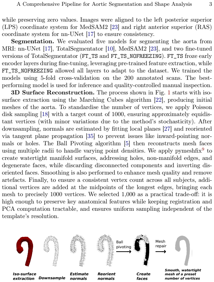
The engine is a template-to-target vertex displacement optimization. A population template is constructed by averaging 100 meshes aligned by an affine registration; each target mesh is then matched by deforming this template's 1000 vertices directly via gradient descent. The objective is a seven-term composite loss: Chamfer distance for point correspondence, edge-length loss for topology, normal-consistency and Laplacian terms for smoothness, mean-curvature Chamfer matching for intrinsic geometry, and IoU/Dice for volumetric overlap. Learnable softmax-normalized weights balance these terms, and a regularization penalty on the weight distribution prevents any single loss from dominating. This
Load-bearing premise
The evaluation assumes that surface distance and overlap metrics measure true anatomical correspondence, yet those exact metrics are also the objectives being optimized, and no independent landmark or clinical ground truth is used.
What would settle it
Take a held-out set of aortas with expert-placed anatomical landmarks (e.g., sinotubular junction, arch branch origins) and compare landmark-to-landmark distance after registration with this method versus the best classical baseline. If the 0.99 Dice / 0.73 mm advantage does not produce lower landmark error, the superiority claim is partly an artifact of evaluating with the same geometric losses being optimized.
If this is right
- If the registration claim holds, statistical shape models of the aorta can be built on dense vertex correspondences rather than sparse landmarks, making PCA modes directly interpretable as surface deformations.
- The healthy-subject PCA reference could serve as a normative baseline: new patients' aortas can be registered and projected onto the modes to quantify deviation from normal shape.
- The watertight, uniformly sampled meshes produced by the pipeline are ready inputs for computational fluid dynamics simulations, linking shape analysis to hemodynamic modeling.
- The finding that freezing early encoder layers during fine-tuning has minimal impact suggests small annotated datasets can leverage large pre-trained segmentation models, though training from scratch gave the best Dice here.
- The registration method is not tied to a particular segmentation model, so it could be applied to any aortic mesh with consistent topology, including meshes derived from CT.
Where Pith is reading between the lines
- Untested in the paper: because the same seven loss terms are not organ-specific, the registration recipe could transfer to other tubular structures (e.g., the pulmonary artery) with a matched template, but this is an editorial extension.
- Untested in the paper: the template was built from healthy aortas, so registering pathological shapes may bias results toward the healthy mean; a disease-aware template or coarse-to-fine scheme is a natural next step the authors do not explore.
- The paper does not treat this, but the reported 0.99 Dice values may be inflated by the fact that IoU and Dice are both optimization objectives and evaluation metrics; an independent landmark-based correspondence test would separate geometric accuracy from anatomical correctness.
Editorial analysis
A structured set of objections, weighed in public.
Referee Report
Summary. The manuscript describes an end-to-end pipeline for aortic shape analysis from cardiac MRI: it benchmarks five segmentation models, reconstructs watertight meshes with a fixed 1000-vertex count, registers all meshes to a template using a proposed per-mesh vertex-displacement optimization (termed “DL-based”), and performs PCA on 599 healthy subjects. The central empirical claim is that the proposed registration method “significantly outperforms” classical rigid and non-rigid methods in geometric accuracy and anatomical consistency, supported by Table 1 (e.g., avg. distance 0.73 vs. 1.00 for Deformetrica; Dice 0.99 vs. 0.98). The paper also reports nnU-Net as the best segmentation model, and identifies six PCA modes of aortic shape variation under rigid and similarity transformations.
Significance. The paper has several strengths: a large healthy cohort (n=599), a practical segmentation comparison with fine-tuning, a carefully described mesh post-processing chain, and a reproducible-looking pipeline. If the registration claim were independently validated, the work would be a useful contribution to aortic shape analysis. However, the headline result is currently not established: the evaluation metrics in Table 1 are largely the same quantities minimized in the proposed loss, no statistical significance tests are reported, no external landmarks or clinical measurements are used, and the “DL-based” label is inaccurate for a per-instance optimization. The PCA component is descriptive but could be a valuable normative reference if the correspondence quality is confirmed. With additional validation, reframing, and tighter reporting, the paper could become acceptable; in its present form the central claim overreaches the evidence.
major comments (5)
- [2. Proposed Learning-based Mesh Registration / Table 1] The headline comparison is not an independent test. The composite loss L_total explicitly includes Chamfer distance, IoU, Dice, curvature matching, edge length, and normal consistency, and Table 1 then reports average distance, RMSE, IoU, and Dice as evaluation metrics. Since the optimizer directly minimizes or maximizes these quantities on each target mesh, the reported advantage over Deformetrica (e.g., avg dist 0.73 vs 1.00, Dice 0.99 vs 0.98) is partly by construction. To support the claim of superior anatomical correspondence, the authors need an external criterion not present in the loss: manually annotated anatomical landmarks, clinical diameter measurements at defined locations, expert ratings of alignment, or a held-out geometric measure not optimized. Without this, the phrase “anatomical consistency” is not supported.
- [Table 1 / 3. Registration Performance] The word “significantly” is used without any statistical testing. Table 1 reports only means and standard deviations; no paired tests, confidence intervals, or effect sizes are given. Given the reported standard deviations (e.g., Dice 0.99±0.03 vs 0.98±0.03; Hausdorff 2.28±0.55 vs 2.63±1.91), some differences may not be significant. Please provide per-subject paired comparisons (e.g., Wilcoxon signed-rank or paired t-test), report the number of meshes used, and state the test chosen. This is required for any comparative claim of superiority.
- [3. Segmentation Performance / Fig. 3] Segmentation results are presented only as a bar chart (Fig. 3a) with no numerical values, error bars, or significance tests. The selection of nnU-Net as the best model, the claim that freezing layers had minimal impact, and the statement that MedSAM2 oversegments cannot be assessed from the current figure. Please report Dice, precision, and recall means and standard deviations for all five models across the 5 folds, and perform pairwise comparisons or at least provide the underlying numbers.
- [2. Proposed Learning-based Mesh Registration / Abstract] The method is labeled “DL-based” and “learning-based,” but no network is trained and no parameters are shared across subjects: vertex displacements are optimized directly for each target mesh via gradient descent, with per-mesh loss weights. This is a per-instance geometric optimization, not deep learning in the usual sense. The label overstates novelty and should be corrected throughout (e.g., “optimization-based mesh registration” or “gradient-based mesh registration”), unless the authors clarify what is learned and how it generalizes to unseen targets.
- [2. Baseline Mesh Registration Methods] The comparison with Deformetrica, the strongest baseline, depends on hyperparameters that are not justified or varied. The text reports one setting (Gaussian kernels with λV=10mm, λW=8mm, 1mm noise, 100 iterations, starting from a 2mm-remeshed template) without sensitivity analysis. Because the central claim rests on outperforming Deformetrica, please add a small sensitivity/ablation study for baseline hyperparameters, or a convergence criterion and a justification for the chosen settings. Otherwise the ranking in Table 1 may reflect unfavorable baseline tuning rather than method superiority.
minor comments (6)
- [Table 1 / Fig. 4] There is a second tabular block immediately after Table 1 with different numbers (e.g., RANSAC avg dist 4.29 vs 3.94; CPD deformable Dice 0.75 vs 0.85) and no caption. This appears to be a duplicate or leftover table. Please remove it or reconcile the values.
- [Throughout] “Haussdorff” is misspelled in Table 1 and in the text; it should be “Hausdorff.”
- [Fig. 3b] The segmentation sample panels are small and unlabeled. Please annotate which method produced each panel, and indicate the imaging plane or 3D rendering. The caption says “random sample” but the anatomical level is unclear.
- [Fig. 5] The three rows of the shape-mode visualization are described as “different viewing angles” but the angles are not specified. Please add view labels (e.g., anterior, lateral, superior) and state the number of standard deviations shown.
- [2. Proposed Learning-based Mesh Registration] The optimization details are incomplete: the maximum number of iterations is not given (only early stopping after 200 non-improving iterations), and the learning rate of 1.0 for vertex displacements is unusually high. Please report the convergence criterion, the median number of iterations, and the runtime per mesh for all methods, since Deformetrica is noted to be computationally expensive.
- [1. Introduction / 2. Materials and Methods] No code or data availability statement is provided. Given the reproducibility-oriented claims (“reproducible technical foundation”), please include a statement or an explicit reason for omission. Also, the manual annotations by two experts are mentioned without inter-observer variability metrics; adding this would strengthen the segmentation benchmark.
Circularity Check
The headline registration advantage is partly constructed: the proposed method directly optimizes IoU, Dice, and Chamfer-style losses, and Table 1 reports those same quantities as the evidence of superiority.
specific steps
-
fitted input called prediction
[Section 2, 'Proposed Learning-based Mesh Registration' (loss components) and 'Template Selection and Alignment' (evaluation metrics); Table 1]
"The optimisation minimises a composite loss function Ltotal ... – IoU and Dice Coefficients: Enforce volumetric consistency and spatial overlap between the deformed and target meshes. ... Subsequent registrations deform this template to match individual target meshes and are evaluated using root mean square error (RMSE), Haussdorff, IoU, Dice, and the average distance between the target and deformed source (template) mesh."
The proposed method optimizes IoU, Dice, and Chamfer distance (a pointwise distance similar to average distance/RMSE) directly on each target mesh, then Table 1 reports IoU, Dice, average distance, and RMSE as evidence that it 'significantly outperforms classical rigid and nonrigid methods in geometric accuracy and anatomical consistency.' Because the evaluation metrics are the very objectives being minimized, the reported advantage is substantially forced by construction. No independent landmarks, manually annotated correspondences, or clinical ground truth are used to show that the better overlap and lower distances correspond to anatomically correct alignment. Additionally, the per-mesh loss weights are fitted on the same meshes used for the evaluation, and early stopping is based on th
full rationale
The paper's central registration claim is self-referential in its evaluation: the proposed method is a per-mesh optimization of a composite loss that explicitly includes IoU, Dice, and Chamfer distance, and the same types of measures (IoU, Dice, average distance, RMSE) are then reported as the proof of superiority in Table 1. This does not make the whole paper circular—the segmentation comparison uses Dice on manually annotated ground truth and is a genuine benchmark, and the PCA shape analysis, while dependent on the registration, is at least a separate downstream use. But the headline 'outperforms ... in anatomical consistency' is not supported by an external or independent criterion; it largely reports how well the method minimized its own loss. The score of 6 reflects that one or more of the claimed 'predictions' (anatomical correspondence quality) reduce by construction to the optimized objective, while acknowledging that the technical work of the pipeline is substantial and not merely a renamed known result. No load-bearing self-citation chain or uniqueness-theorem import was found.
Axiom & Free-Parameter Ledger
free parameters (4)
- Per-mesh loss weights w_i =
7 weights optimized per registration (softmax normalized)
- Target vertex count =
1000
- Template construction sample K =
100
- Vertex displacement learning rate =
1.0
axioms (5)
- domain assumption Ground-truth annotations by two expert annotators on 200 scans are correct and sufficient for training and evaluation.
- domain assumption The CPD-affine average of 100 randomly chosen meshes is a neutral, unbiased template.
- domain assumption After post-processing, reconstructed meshes are watertight manifolds whose vertex correspondences are anatomically meaningful.
- domain assumption Excluding 119 symptomatic subjects and labeling 599 of 625 volunteers as healthy is sufficient to define a normal reference.
- domain assumption PCA on registered vertex positions yields clinically interpretable modes of aortic shape variation.
Cite this review
Pith. "Pith review of A Comprehensive Pipeline for Aortic Segmentation and Shape Analysis." pith.science (2026). https://pith.science/paper/K57GXQDM
@misc{pith2026250909718,
  author       = {Pith},
  title        = {Pith review of: A Comprehensive Pipeline for Aortic Segmentation and Shape Analysis},
  year         = {2026},
  howpublished = {\url{https://pith.science/paper/K57GXQDM}},
  note         = {Machine review of arXiv:2509.09718}
}
read the original abstract
Aortic shape analysis plays a key role in cardiovascular diagnostics, treatment planning, and understanding disease progression. We present a robust, fully automated pipeline for aortic shape analysis from cardiac MRI, combining deep learning and statistical techniques across segmentation, 3D surface reconstruction, and mesh registration. We benchmark leading segmentation models including nnUNet, TotalSegmentator, and MedSAM2 highlighting the effectiveness of domain specific training and transfer learning on a curated dataset. Following segmentation, we reconstruct high quality 3D meshes and introduce a DL based mesh registration method that directly optimises vertex displacements. This approach significantly outperforms classical rigid and nonrigid methods in geometric accuracy and anatomical consistency. Using the registered meshes, we perform statistical shape analysis on a cohort of 599 healthy subjects. Principal Component Analysis reveals dominant modes of aortic shape variation, capturing both global morphology and local structural differences under rigid and similarity transformations. Our findings demonstrate the advantages of integrating traditional geometry processing with learning based models for anatomically precise and scalable aortic analysis. This work lays the groundwork for future studies into pathological shape deviations and supports the development of personalised diagnostics in cardiovascular medicine.
Figures


Reference graph
Works this paper leans on
-
[1]
NPJ Genomic Medicine5(1), 46 (2020)
Aguib, Y., Allouba, M., Afify, A., Halawa, S., El-Khatib, M., Sous, M., Galal, A., Abdelrahman, E., Shehata, N., El Sawy, A., et al.: The egyptian collabora- tive cardiac genomics (ecco-gen) project: defining a healthy volunteer cohort. NPJ Genomic Medicine5(1), 46 (2020)
work page 2020
- [2]
-
[3]
In: 2007 IEEE Conference on Computer Vision and Pattern Recognition
Amberg, B., Romdhani, S., Vetter, T.: Optimal step nonrigid icp algorithms for surface registration. In: 2007 IEEE Conference on Computer Vision and Pattern Recognition. pp. 1–8. IEEE (2007).https://doi.org/10.1109/CVPR.2007.383165
-
[4]
Ayed, I.B., Wang, M., Miles, B., Garvin, G.J.: Tric: trust region for invariant compactnessanditsapplicationtoabdominalaortasegmentation.In:International Conference on Medical Image Computing and Computer-Assisted Intervention. pp. 381–388. Springer (2014)
work page 2014
-
[5]
IEEE transactions on visualization and computer graphics5(4), 349–359 (1999)
Bernardini, F., Mittleman, J., Rushmeier, H., Silva, C., Taubin, G.: The ball- pivoting algorithm for surface reconstruction. IEEE transactions on visualization and computer graphics5(4), 349–359 (1999)
work page 1999
-
[6]
In: Biomedical image processing and biomed- ical visualization
Boissonnat, J.D., Geiger, B.: Three-dimensional reconstruction of complex shapes based on the delaunay triangulation. In: Biomedical image processing and biomed- ical visualization. vol. 1905, pp. 964–975. SPIE (1993)
work page 1905
-
[7]
Bône, A., Louis, M., Martin, B., Durrleman, S.: Deformetrica 4: an open-source software for statistical shape analysis. In: Shape in medical imaging: international workshop, ShapeMI 2018, held in conjunction with MICCAI 2018, granada, Spain, september 20, 2018, proceedings. pp. 3–13. Springer (2018)
work page 2018
-
[8]
Bookstein, F.L.: Thin-plate splines and the atlas problem for biomedical images. In: Information Processing in Medical Imaging: 12th International Conference, IPMI’91 Wye, UK, July 7–12, 1991 Proceedings 12. pp. 326–342. Springer (1991)
work page 1991
-
[9]
Image and vision computing10(3), 145–155 (1992)
Chen, Y., Medioni, G.: Object modelling by registration of multiple range images. Image and vision computing10(3), 145–155 (1992)
work page 1992
-
[10]
arXiv preprint arXiv:2405.19492 (2024)
D’Antonoli, T.A., Berger, L.K., Indrakanti, A.K., Vishwanathan, N., Weiß, J., Jung, M., Berkarda, Z., Rau, A., Reisert, M., Küstner, T., et al.: Totalsegmentator mri: Sequence-independent segmentation of 59 anatomical structures in mr images. arXiv preprint arXiv:2405.19492 (2024)
Pith/arXiv arXiv 2024
-
[11]
Proceedings of the IEEE (2023)
Farshian, A., Götz, M., Cavallaro, G., Debus, C., Nießner, M., Benediktsson, J.A., Streit, A.: Deep-learning-based 3-d surface reconstruction—a survey. Proceedings of the IEEE (2023)
work page 2023
-
[12]
In: Handbook of dis- crete and computational geometry, pp
Fortune, S.: Voronoi diagrams and delaunay triangulations. In: Handbook of dis- crete and computational geometry, pp. 705–721. Chapman and Hall/CRC (2017)
work page 2017
-
[13]
Journal of Open Source Software7(80), 4681 (2022)
Gatti, A.A., Khallaghi, S.: Pycpd: Pure numpy implementation of the coherent point drift algorithm. Journal of Open Source Software7(80), 4681 (2022)
work page 2022
-
[14]
arXiv preprint arXiv:2005.11084 (2020)
Hanocka, R., Metzer, G., Giryes, R., Cohen-Or, D.: Point2mesh: A self-prior for deformable meshes. arXiv preprint arXiv:2005.11084 (2020)
Pith/arXiv arXiv 2005
-
[15]
In: International Workshop on Statistical Atlases and Computational Models of the Heart
Heinrich, M.P., Oster, J.: Mri whole heart segmentation using discrete nonlinear registration and fast non-local fusion. In: International Workshop on Statistical Atlases and Computational Models of the Heart. pp. 233–241. Springer (2017)
work page 2017
-
[16]
Hepp, T., Fischer, M., Winkelmann, M.T., Baldenhofer, S., Kuestner, T., Nikolaou, K., Yang, B., Gatidis, S.: Fully automated segmentation and shape analysis of the thoracic aorta in non–contrast-enhanced magnetic resonance images of the german national cohort study. Journal of thoracic imaging35(6), 389–398 (2020) A Comprehensive Pipeline for Aortic Segme...
work page 2020
-
[17]
Nature methods18(2), 203–211 (2021)
Isensee, F., Jaeger, P.F., Kohl, S.A., Petersen, J., Maier-Hein, K.H.: nnu-net: a self-configuring method for deep learning-based biomedical image segmentation. Nature methods18(2), 203–211 (2021)
2021
-
[18]
In: Proceed- ings of the fourth Eurographics symposium on Geometry processing
Kazhdan, M., Bolitho, M., Hoppe, H.: Poisson surface reconstruction. In: Proceed- ings of the fourth Eurographics symposium on Geometry processing. vol. 7 (2006)
work page 2006
-
[19]
Remote Sensing 12(10), 1643 (2020)
Kulawiak, M., Lubniewski, Z.: Improving the accuracy of automatic reconstruction of 3d complex buildings models from airborne lidar point clouds. Remote Sensing 12(10), 1643 (2020)
work page 2020
-
[20]
In: Proceedings of the IEEE conference on computer vision and pattern recognition
Liao, Y., Donne, S., Geiger, A.: Deep marching cubes: Learning explicit surface representations. In: Proceedings of the IEEE conference on computer vision and pattern recognition. pp. 2916–2925 (2018)
work page 2018
-
[21]
In: Proceedings of the IEEE international conference on computer vision
Litany, O., Remez, T., Rodola, E., Bronstein, A., Bronstein, M.: Deep functional maps: Structured prediction for dense shape correspondence. In: Proceedings of the IEEE international conference on computer vision. pp. 5659–5667 (2017)
work page 2017
-
[22]
ACM siggraph computer graphics21(4), 163–169 (1987)
Lorensen, W.E., Cline, H.E.: Marching cubes: A high resolution 3d surface con- struction algorithm. ACM siggraph computer graphics21(4), 163–169 (1987)
work page 1987
-
[23]
Ma, J., Kim, S., Li, F., Baharoon, M., Asakereh, R., Lyu, H., Wang, B.: Segment anythinginmedicalimagesandvideos:Benchmarkanddeployment.arXivpreprint arXiv:2408.03322 (2024)
Pith/arXiv arXiv 2024
-
[24]
Electronics13(24), 4919 (2024)
Mavridis, C., Vagenas, T.P., Economopoulos, T.L., Vezakis, I., Petropoulou, O., Kakkos, I., Matsopoulos, G.K.: Automatic segmentation in 3d ct images: A com- parative study of deep learning architectures for the automatic segmentation of the abdominal aorta. Electronics13(24), 4919 (2024)
work page 2024
-
[25]
IEEE trans- actions on pattern analysis and machine intelligence32(12), 2262–2275 (2010)
Myronenko, A., Song, X.: Point set registration: Coherent point drift. IEEE trans- actions on pattern analysis and machine intelligence32(12), 2262–2275 (2010)
2010
-
[26]
IEEE Trans- actions on pattern analysis and machine intelligence14(2), 239–256 (1992)
Paul, J.: Besl and neil mckay, a methode for registration of 3d shapes. IEEE Trans- actions on pattern analysis and machine intelligence14(2), 239–256 (1992)
work page 1992
-
[27]
Hartung-Gorre Süderdithmarschen, Germany (2003)
Pauly, M.: Point primitives for interactive modeling and processing of 3D geometry. Hartung-Gorre Süderdithmarschen, Germany (2003)
work page 2003
-
[28]
Ravi,N.,Reizenstein,J.,Novotny,D.,Gordon,T.,Lo,W.Y.,Johnson,J.,Gkioxari, G.: Accelerating 3d deep learning with pytorch3d. arXiv:2007.08501 (2020)
Pith/arXiv arXiv 2007
-
[29]
European radiology29, 4613–4623 (2019)
Sedghi Gamechi, Z., Bons, L.R., Giordano, M., Bos, D., Budde, R.P., Kofoed, K.F., Pedersen, J.H., Roos-Hesselink, J.W., de Bruijne, M.: Automated 3d segmentation and diameter measurement of the thoracic aorta on non-contrast enhanced ct. European radiology29, 4613–4623 (2019)
work page 2019
-
[30]
Sultan, A.A.: Image-Based Models for Left Heart Flow Morphodynamics in Hyper- trophic Obstructive Cardiomyopathy. Master’s thesis, The American University in Cairo (2025),https://fount.aucegypt.edu/etds/2500, master’s Thesis
work page 2025
-
[31]
IEEE Transactions on pattern analysis and machine intelligence 13(4), 376–380 (1991)
Umeyama, S.: Least-squares estimation of transformation parameters between two point patterns. IEEE Transactions on pattern analysis and machine intelligence 13(4), 376–380 (1991)
work page 1991
-
[32]
In: Proceedings of the IEEE/CVF Conference on Computer Vision and Pattern Recognition
Wang, W., Ceylan, D., Mech, R., Neumann, U.: 3dn: 3d deformation network. In: Proceedings of the IEEE/CVF Conference on Computer Vision and Pattern Recognition. pp. 1038–1046 (2019)
work page 2019
-
[33]
arXiv preprint arXiv:2404.13830 (2024)
Zhang, Y.X., Gui, J., Cong, X., Gong, X., Tao, W.: A comprehensive survey and taxonomy on point cloud registration based on deep learning. arXiv preprint arXiv:2404.13830 (2024)
Pith/arXiv arXiv 2024
-
[34]
In: European conference on computer vision
Zhou, Q.Y., Park, J., Koltun, V.: Fast global registration. In: European conference on computer vision. pp. 766–782. Springer (2016)
2016
-
[35]
arXiv preprint arXiv:1801.09847 (2018) 12 N
Zhou, Q.Y., Park, J., Koltun, V.: Open3d: A modern library for 3d data processing. arXiv preprint arXiv:1801.09847 (2018) 12 N. Shehata et al
Pith/arXiv arXiv 2018
-
[36]
Medical image analysis 58, 101537 (2019)
Zhuang, X., Li, L., Payer, C., Štern, D., Urschler, M., Heinrich, M.P., Oster, J., Wang, C., Smedby, Ö., Bian, C., et al.: Evaluation of algorithms for multi-modality whole heart segmentation: an open-access grand challenge. Medical image analysis 58, 101537 (2019)
2019
This paper was first reviewed by deepseek-v4-flash on August 4, 2026.
discussion (0)
Sign in with ORCID, Apple, or X to comment. Anyone can read and Pith papers without signing in.