REVIEW 4 major objections 5 minor 36 references
CORE -- A Cell-Level Coarse-to-Fine Image Registration Engine for Multi-stain Image Alignment
T0 review · 4 major / 5 minor · reviewed 2026-08-03 · deepseek-v4-flash
Pith's one-line read A two-stage pipeline aligns multi-stain tissue slides down to nuclei-level precision.
desk verdict Useful coarse-to-fine WSI registration platform with a genuinely fast coarse stage, but the nuclei-level claim outruns the evidence: all fine-stage numbers are TRE on sparse manual landmarks, not on nuclei. read the letter →
The pith
A machine-rendered reading of the paper's core claim, the machinery that carries it, and where it could break.
The reading
What carries the argument
The load-bearing object is the automatically detected nuclei centroid point set. The method's distinctive identity is a hybrid shape-aware distance metric that combines Euclidean distance between transformed source nuclei and target nuclei with the absolute difference of their normalised per-nucleus areas, balanced by a weight parameter set to 0.3. This metric drives a derivative-free Powell optimisation for rigid alignment, and the resulting correspondences feed Coherent Point Drift, a probabilistic non-rigid point-set registration algorithm, to produce the final dense displacement field. The paper also relies on a prompt-based tissue mask extraction step and an accelerated deep feature mat
What would settle it
Measure CORE's fine-stage target registration error on slide pairs with artificially withheld or poorly detected nuclei—for example, by running the watershed detector on patches with dense overlapping lymphocyte clusters and comparing against manual landmarks. If the median fine error rises sharply when detection completeness drops below roughly 90 percent, the central nuclei-correspondence premise is falsified.
Extended reading notes
Core claim
On the paper's own terms, the central discovery is that whole-slide registration across very different stains can be made both fast and nuclei-accurate by separating the problem into a morphology-first coarse stage and a nuclei-point-set fine stage. The coarse stage uses prompt-based tissue masks, centre-of-mass alignment, discrete rotation search, and accelerated dense feature matching at low magnification to produce a global alignment in about 15–20 seconds. The fine stage then detects nuclei centroids in the target and coarsely registered source, applies a shape-aware rigid point-set alignment that mixes spatial distance with normalised nuclear area, and finishes with Coherent Point Drift
Load-bearing premise
The fine stage assumes that automatically detected nuclei in one stain line up as corresponding landmarks with nuclei in the other stain; if detection fails or produces non-corresponding centroids in dense, overlapping, or artefact-heavy tissue, the point-set fit will target the wrong correspondences.
Editorial extensions
If this is right
- A fast, accurate coarse alignment becomes available as a standalone step, making interactive or high-throughput registration possible where nuclei-level precision is not required.
- The same untuned pipeline can align bright-field stains such as H&E, IHC, and PAS with multiplex immunofluorescence, a cross-modality combination that is difficult for stain-specific methods.
- Cell-level downstream analyses, such as matching single cells across stains or linking phenotype to morphology, can be run on the fine-stage output without manual landmark selection.
- Because nuclei detection is morphology-based rather than trained per stain, the method transfers to new stain protocols without needing annotated nuclei datasets.
- If the reported sub-micrometre errors on re-stained sections are reproducible, the fine stage reaches the precision needed to treat nuclei as corresponding biological units across staining rounds.
Reading between the lines
- The paper leaves implicit that the coarse stage's speed makes it suitable for interactive browsing or triage, but the reported runtime is for the alignment computation itself, not for full-resolution rendering or visualisation.
- A natural stress test the paper does not run is registration of slide pairs with large tissue folds or tears inside the tissue mask; such cases would exercise the rigid coarse estimate and the Jacobian-based folding correction simultaneously.
- The point-set machinery could be transferred to other intrinsic landmarks—glands, vessels, or immune-cell clusters—which would extend the method to tissues where nuclei are not the most reliable modality-invariant feature.
- The explicit dependence on nuclei detection suggests that a version of CORE using a learned detector, trained only where annotated nuclei exist, could improve the fine stage in dense tissue while keeping the coarse-to-fine structure intact.
Editorial analysis
A structured set of objections, weighed in public.
Referee Report
Summary. The paper proposes CORE, a two-stage whole-slide image (WSI) registration framework. The coarse stage combines prompt-based tissue-mask extraction (Florence-2-SAM), a morphology-based rigid alignment module (TriMorph), XFeat dense feature matching, and hierarchical non-rigid NCC optimization with smoothness regularization. The fine stage detects nuclei centroids via a morphology/watershed pipeline, performs a shape-aware point-set rigid alignment (Eqs. 4–7, Powell optimization), and then estimates local non-rigid deformations with Coherent Point Drift (CPD). The method is evaluated on ACROBAT, ANHIR, HYRECO, Multi-IHC CRC, and REACTIV AS, using TRE/rTRE-based landmarks and runtime comparisons, and the authors claim that CORE outperforms state-of-the-art methods in precision, robustness, and generalisability.
Significance. If the reported claims are substantiated, CORE would be a practically valuable contribution: the coarse stage is fast (e.g., 14.37 s on ACROBAT versus 50–120 s for published baselines), the method is tested on five cohorts spanning bright-field and immunofluorescence modalities, and open-source code plus a TiaViz demo are provided. The use of a classical, training-free nuclei detector is also attractive for data-scarce settings. However, the headline claim of 'accurate nuclei-level registration' is not directly supported by the current evaluation, which relies entirely on tissue-level landmark TRE/rTRE. The coarse-registration evidence is more credible than the fine-stage evidence; the fine-stage and 'generalises without manual tuning' claims need additional support before the paper can be accepted.
major comments (4)
- [§3.2, Tables 2–6] The abstract and §3.2 claim 'precise nuclei-level correspondence across modalities' and 'cellular-level precision.' But all quantitative results in Tables 2–6 use landmark-based TRE/rTRE (§4.2), and the landmarks (e.g., 11–19 per HYRECO section, ~43 per re-stained pair) are manually placed anatomical points, not shown to coincide with nuclei centroids. A low TRE after fine registration demonstrates tissue-level alignment, not nucleus-to-nucleus correspondence. Since the fine stage is a point-set registration on nuclei centroids, the evaluation should include a direct nuclei-level metric, such as mutual-nearest-neighbour centroid precision/recall, distances between corresponding nuclei, or a cell-matching F1 score. Without such a metric, the paper's central novelty is unverified. This is separate from the acknowledged detector-dependence limitation in §4.4.
- [Table 3] Table 3 reports 'Fine CORE' as having the lowest AArTRE and AMrTRE on ANHIR, but §4.3 states that fine-shape-aware registration was performed only on slides at 20×/40× magnification, while lower-resolution samples (1.25×–10×) were excluded. The baselines (DFBR, DeeperHistReg, HistokatFusion, etc.) are evaluated on the full ANHIR set. Thus the fine-stage comparison is not head-to-head: the improved global metrics could reflect case selection rather than method superiority. Please report per-subset results for CORE and all baselines on the same 20×/40× pairs, or provide a full-set fine-registration result if feasible. This is necessary to support the 'outperforms at fine registration' claim.
- [Tables 2–6] No confidence intervals, standard errors, or significance tests are reported for any metric. For example, in Table 2, CORE's AMTRE90 of 139.00 µm is only 2.6 µm below HistokatFusion (141.64) and 1.3 µm below DeeperHistReg (140.33). Median/mean differences of this size may be within case-level variability. Because the abstract claims 'outperforms current state-of-the-art methods' across five datasets, the paper should provide paired statistical comparisons (e.g., bootstrap confidence intervals or Wilcoxon signed-rank tests over WSI pairs) and report the number of pairs contributing to each aggregate. The same applies to runtime comparisons, where only single means are given.
- [§3.1.1–§3.1.2, Tables A.2–A.4] The introduction says the framework 'generalises across staining protocols without manual tuning,' but §3.1.1 states that gamma correction ranges (1.0–1.2 for H&E, 0.4–0.8 for IHC) were 'empirically determined based on evaluations conducted across the datasets,' and many other parameters are fixed values with no sensitivity analysis: Dice threshold 0.7, rotation step 10°, XFeat precision/iteration settings, Powell tolerance, CPD regularization parameters, and shape weight γ. Please clarify explicitly whether these were fixed a priori across all datasets or tuned per dataset. If they were tuned, the 'without manual tuning' claim should be softened; at minimum, provide a sensitivity study for the most influential parameters (e.g., γ in Eq. (5) and CPD α, β in Table A.4).
minor comments (5)
- [§4.1.5] The REACTIV AS dataset reference appears as '[citation]' in the text; it should be [33].
- [Table 1 vs §4.1.1] §4.1.1 states ACROBAT contains 3,406 WSIs, while Table 1 reports 4,212 WSIs. Please reconcile these numbers.
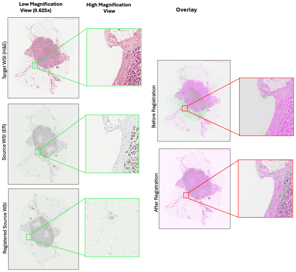
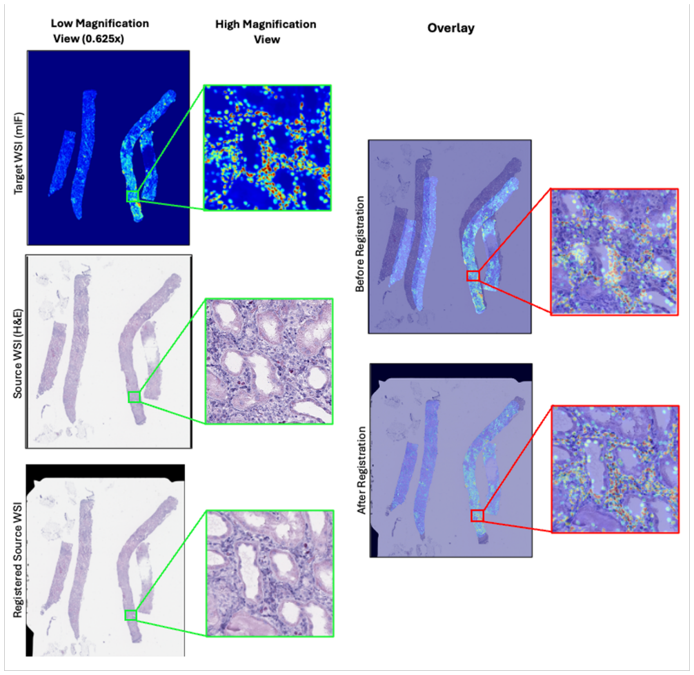
- [Figure 6 caption] Figure 6 caption says source (PAS), but HYRECO stain sets are H&E, PHH3, CD8, CD45; the text describes PHH3. Please correct the caption or clarify the stain.
- [Tables 4 and 6] Report the number of image pairs used for each metric, especially for the private REACTIV AS dataset (11 paired samples) and the HYRECO subsets, so that the aggregate values can be interpreted.
- [§2 Related Works] A few in-text citation numbers appear inconsistent with the reference list (e.g., 'Huang et al. [13]' while [13] lists Wei et al.). Please verify all citation labels.
Circularity Check
No significant circularity: accuracy claims are benchmarked externally; only minor non-load-bearing same-group citations.
full rationale
CORE is an empirical registration pipeline, not a derivation in which an output is defined as its input. The coarse stage uses externally pretrained models (Florence-2/SAM, XFeat) and standard metrics (NCC, NGF); no fitted parameter is relabeled as a prediction. The fine stage estimates a point-set transformation from detected nuclei centroids (Eqs. 4-8, CPD in Sec. 3.2.2), but the reported TRE/rTRE numbers come from manual landmarks in HYRECO, ANHIR, Multi-IHC CRC, and REACTIV AS, not from the nuclei set used in the objective, so the central evaluation is not the optimized quantity by construction. Same-group references exist ([1] review, [5] DFBR baseline and Multi-IHC CRC dataset, [20] sparse-matching efficiency, [36] TIA Viz viewer), but none is load-bearing for the headline claim; [20] only supports a computational-efficiency choice. The paper itself acknowledges the main evidence limitation in Sec. 4.4: 'the fine registration stage depends on accurate nuclei detection, which can be hindered by overlapping cells, staining artefacts, or suboptimal segmentation in dense tissue regions.' The absence of a nuclei-level quantitative metric and the fact that fine registration was not run on ACROBAT (10x slides) are evaluation gaps, not circular reductions. No equation or claimed prediction reduces by construction to the paper's own inputs.
Assumptions & free parameters
free parameters (9)
- Gamma correction range (H&E vs IHC) =
1.0–1.2 (H&E), 0.4–0.8 (IHC)
- Dice threshold for TriMorph acceptance =
0.7
- Rotation search step =
10 degrees
- XFeat matching settings =
precision 3–5 px; 1000–2000 iterations; 99.9% confidence
- Shape weight gamma =
0.3
- Powell optimization tolerance / max iterations =
ε=1e-8, i_max=100
- Coarse non-rigid parameters =
lr=0.001, NCC window=7, iterations 200–500, smoothness γ_s, 6 pyramid levels
- CPD parameters =
alpha=0.01, beta=0.5, max_iter=200, tol=1e-9, w=0.1, sigma_init=1.0, smoothing=10, max displacement=10
- MNN cap / progressive sample sizes =
~5000 MNNs; 500→200,000 points
assumptions (6)
- domain assumption Landmark annotations used for TRE/rTRE are accurate and correspond across stains.
- domain assumption Florence-2+SAM prompt-based segmentation yields tissue masks that exclude artifacts and include tissue.
- domain assumption Nuclei centroids are reliable cross-modality landmarks for cell-level alignment.
- domain assumption XFeat features are modality-invariant enough to match H&E to IHC/PAS/mIF.
- standard math CPD Gaussian-mixture model and smoothness regularization model tissue deformation plausibly.
- standard math Macenko stain normalization preserves structural information needed for mask extraction.
Cite this review
Pith. "Pith review of CORE -- A Cell-Level Coarse-to-Fine Image Registration Engine for Multi-stain Image Alignment." pith.science (2026). https://pith.science/paper/XIS7KXHV
@misc{pith2026251103826,
  author       = {Pith},
  title        = {Pith review of: CORE -- A Cell-Level Coarse-to-Fine Image Registration Engine for Multi-stain Image Alignment},
  year         = {2026},
  howpublished = {\url{https://pith.science/paper/XIS7KXHV}},
  note         = {Machine review of arXiv:2511.03826}
}
read the original abstract
Accurate and efficient registration of whole slide images (WSIs) is essential for high-resolution, nuclei-level analysis in multi-stained tissue slides. We propose a novel coarse-to-fine framework CORE for accurate nuclei-level registration across diverse multimodal whole-slide image (WSI) datasets. The coarse registration stage leverages prompt-based tissue mask extraction to effectively filter out artefacts and non-tissue regions, followed by global alignment using tissue morphology and accelerated dense feature matching with a pre-trained feature extractor. From the coarsely aligned slides, nuclei centroids are detected and subjected to fine-grained rigid registration using a custom, shape-aware point-set registration model. Finally, non-rigid alignment at the cellular level is achieved by estimating a non-linear displacement field using Coherent Point Drift (CPD). Our approach benefits from automatically generated nuclei that enhance the accuracy of deformable registration and ensure precise nuclei-level correspondence across modalities. The proposed model is evaluated on three publicly available WSI registration datasets, and two private datasets. We show that CORE outperforms current state-of-the-art methods in terms of generalisability, precision, and robustness in bright-field and immunofluorescence microscopy WSIs
Figures


Figures from the paper (7 more)




Reference graph
Works this paper leans on
-
[1]
Young, Nasir M
Behnaz Elhaminia, Abdullah Alsalemi, Esha Nasir, Mostafa Jahanifar, Ruqayya Awan, Lawrence S. Young, Nasir M. Rajpoot, Fayyaz Minhas, and Shan E Ahmed Raza. From Traditional to Deep Learning Approaches in Whole Slide Image Regis- tration: A Methodological Review, 2025. Version Number: 1
2025
-
[2]
Comparison of Consecutive and Re-stained Sections for Image Registration in Histopathology
Johannes Lotz, Nick Weiss, Jeroen van der Laak, and Stefan Heldmann. Comparison of Consecutive and Re-stained Sections for Image Registration in Histopathology. Journal of Medical Imaging, 10(06), November 2023. arXiv:2106.13150 [eess]
arXiv 2023
-
[3]
Gatenbee, Ann-Marie Baker, Sandhya Prabhakaran, Ottilie Swinyard, Robbert J
Chandler D. Gatenbee, Ann-Marie Baker, Sandhya Prabhakaran, Ottilie Swinyard, Robbert J. C. Slebos, Gunjan Mandal, Eoghan Mulholland, Noemi Andor, An- driy Marusyk, Simon Leedham, Jose R. Conejo-Garcia, Christine H. Chung, Mark Robertson-Tessi, Trevor A. Graham, and Alexander R. A. Anderson. Virtual align- ment of pathology image series for multi-gigapixe...
2023
-
[4]
Deeper- HistReg: Robust Whole Slide Images Registration Framework, 2024
Marek Wodzinski, Niccol` o Marini, Manfredo Atzori, and Henning M¨ uller. Deeper- HistReg: Robust Whole Slide Images Registration Framework, 2024. Version Num- ber: 1
2024
-
[5]
Ahmed Raza, Johannes Lotz, Nick Weiss, and Nasir Ra- jpoot
Ruqayya Awan, Shan E. Ahmed Raza, Johannes Lotz, Nick Weiss, and Nasir Ra- jpoot. Deep feature based cross-slide registration.Computerized Medical Imaging and Graphics, 104:102162, March 2023
2023
-
[6]
J. Lotz, J. Olesch, B. Muller, T. Polzin, P. Galuschka, J. M. Lotz, S. Heldmann, H. Laue, M. Gonzalez-Vallinas, A. Warth, B. Lahrmann, N. Grabe, O. Sedlaczek, K. Breuhahn, and J. Modersitzki. Patch-Based Nonlinear Image Registration for Gigapixel Whole Slide Images.IEEE Transactions on Biomedical Engineering, 63(9):1812–1819, September 2016
2016
-
[7]
Robust, fast and accurate: a 3-step method for automatic histological image registration, 2019
Johannes Lotz, Nick Weiss, and Stefan Heldmann. Robust, fast and accurate: a 3-step method for automatic histological image registration, 2019. Version Number: 2
2019
-
[8]
Sorokin, Gloria Bueno, Alexander V
Jiri Borovec, Jan Kybic, Ignacio Arganda-Carreras, Dmitry V. Sorokin, Gloria Bueno, Alexander V. Khvostikov, Spyridon Bakas, Eric I-Chao Chang, Stefan Held- mann, Kimmo Kartasalo, Leena Latonen, Johannes Lotz, Michelle Noga, Sarthak Pati, Kumaradevan Punithakumar, Pekka Ruusuvuori, Andrzej Skalski, Nazanin Tahmasebi, Masi Valkonen, Ludovic Venet, Yizhe Wa...
2020
Show all 36 references
-
[9]
Gatenbee, Alexander R.A
Philippe Weitz, Masi Valkonen, Leslie Solorzano, Circe Carr, Kimmo Kartasalo, Constance Boissin, Sonja Koivukoski, Aino Kuusela, Dusan Rasic, Yanbo Feng, San- dra Sinius Pouplier, Abhinav Sharma, Kajsa Ledesma Eriksson, Stephanie Robert- son, Christian Marzahl, Chandler D. Gat...
2022
-
[10]
Siegwart
Stefan Leutenegger, Margarita Chli, and Roland Y. Siegwart. BRISK: Binary Robust invariant scalable keypoints. In2011 International Conference on Computer Vision, pages 2548–2555, Barcelona, Spain, November 2011. IEEE
2011
-
[11]
A robust method for inter-marker whole slide registration of digital pathology images using lines based features
Anindya Sarkar, Quan Yuan, and Chukka Srinivas. A robust method for inter-marker whole slide registration of digital pathology images using lines based features. In2014 IEEE 11th International Symposium on Biomedical Imaging (ISBI), pages 762–765, Beijing, April 2014. IEEE
2014
-
[12]
Fischler and Robert C
Martin A. Fischler and Robert C. Bolles. Random sample consensus: a paradigm for model fitting with applications to image analysis and automated cartography. Communications of the ACM, 24(6):381–395, June 1981
1981
-
[13]
Xingyue Wei, Lin Ge, Lijie Huang, Jianwen Luo, and Yan Xu. Unsupervised Non- Rigid Histological Image Registration Guided by Keypoint Correspondences Based on Learnable Deep Features With Iterative Training.IEEE Transactions on Medical Imaging, 44(1):447–461, January 2025
2025
-
[14]
Pyatov and Dmitry V
Vladislav A. Pyatov and Dmitry V. Sorokin. Unsupervised Feature Matching for Affine Histological Image Registration. In Apostolos Antonacopoulos, Subhasis Chaudhuri, Rama Chellappa, Cheng-Lin Liu, Saumik Bhattacharya, and Uma- pada Pal, editors,Pattern Recognition, volume 1531...
2025
-
[15]
Unsupervised His- tological Image Registration Using Structural Feature Guided Convolutional Neural Network.IEEE Transactions on Medical Imaging, 41(9):2414–2431, September 2022
Lin Ge, Xingyue Wei, Yayu Hao, Jianwen Luo, and Yan Xu. Unsupervised His- tological Image Registration Using Structural Feature Guided Convolutional Neural Network.IEEE Transactions on Medical Imaging, 41(9):2414–2431, September 2022. Publisher: Institute of Electrical and Ele...
2022
-
[16]
SuperGlue: Learning Feature Matching With Graph Neural Networks
Paul-Edouard Sarlin, Daniel DeTone, Tomasz Malisiewicz, and Andrew Rabinovich. SuperGlue: Learning Feature Matching With Graph Neural Networks. In2020 IEEE/CVF Conference on Computer Vision and Pattern Recognition (CVPR), pages 4937–4946, Seattle, W A, USA, June 2020. IEEE
2020
-
[17]
Deep autoencoder features for registration of histology images
Awan R and Rajpoot N. Deep autoencoder features for registration of histology images. InAnnual Conference on Medical Image Understanding and Analysis, pages 371–378. Springer, 2018
2018
-
[18]
Flownet: Learning optical flow with convolutional networks
Dosovitskiy A, Fischer P, Ilg E, Hausser P, Hazirbas C, Golkov V, Van Der Smagt P, Cremers D, and Brox T. Flownet: Learning optical flow with convolutional networks. InProceedings of the IEEE international conference on computer vision, pages 2758–2766, 2015
2015
-
[19]
Registration of Histopathogy Images Using Structural Information From Fine Grained Feature Maps, 2020
Dwarikanath Mahapatra. Registration of Histopathogy Images Using Structural Information From Fine Grained Feature Maps, 2020. Version Number: 1. 29
2020
-
[20]
Ahmed Raza
Adith Jeyasangar, Abdullah Alsalemi, and Shan E. Ahmed Raza. Nuclei-Location Based Point Set Registration of Multi-stained Whole Slide Images. In Moi Hoon Yap, Connah Kendrick, Ardhendu Behera, Timothy Cootes, and Reyer Zwiggelaar, editors,Medical Image Understanding and Analy...
2024
-
[21]
HyReCo - Hybrid re-stained and consecutive histological serial sections, July 2021
Jeroen van der Laak, Johannes Lotz, Nick Weiss, and Stefan Heldmann. HyReCo - Hybrid re-stained and consecutive histological serial sections, July 2021
2021
-
[22]
Multimodal Alignment of Histopathological Images Using Cell Segmentation and Point Set Matching for Integrative Cancer Analysis, 2024
Jun Jiang, Raymond Moore, Brenna Novotny, Leo Liu, Zachary Fogarty, Ray Guo, Markovic Svetomir, and Chen Wang. Multimodal Alignment of Histopathological Images Using Cell Segmentation and Point Set Matching for Integrative Cancer Analysis, 2024. Version Number: 1
2024
-
[23]
Source data for V ALIS: Virtual Alignment of pathoLogy Image Series for multi-gigapixel whole slide images publication, May 2023
Chandler Gatenbee. Source data for V ALIS: Virtual Alignment of pathoLogy Image Series for multi-gigapixel whole slide images publication, May 2023
2023
-
[24]
Green, Margaret Eminizer, Daniel Jimenez-Sanchez, Steve Lu, Elizabeth L
Joshua Doyle, Benjamin F. Green, Margaret Eminizer, Daniel Jimenez-Sanchez, Steve Lu, Elizabeth L. Engle, Haiying Xu, Aleksandra Ogurtsova, Jonathan Lai, Sigfredo Soto-Diaz, Jeffrey S. Roskes, Julie S. Deutsch, Janis M. Taube, Joel C. Sunshine, and Alexander S. Szalay. Whole-S...
2023
-
[25]
Marc Macenko, Marc Niethammer, J. S. Marron, David Borland, John T. Woosley, Xiaojun Guan, Charles Schmitt, and Nancy E. Thomas. A method for normalizing histology slides for quantitative analysis. In2009 IEEE International Symposium on Biomedical Imaging: From Nano to Macro, ...
2009
-
[26]
Florence-2: Advancing a Unified Representation for a Variety of Vision Tasks, 2023
Bin Xiao, Haiping Wu, Weijian Xu, Xiyang Dai, Houdong Hu, Yumao Lu, Michael Zeng, Ce Liu, and Lu Yuan. Florence-2: Advancing a Unified Representation for a Variety of Vision Tasks, 2023. Version Number: 1
2023
-
[27]
Berg, Wan-Yen Lo, Piotr Doll´ ar, and Ross Girshick
Alexander Kirillov, Eric Mintun, Nikhila Ravi, Hanzi Mao, Chloe Rolland, Laura Gustafson, Tete Xiao, Spencer Whitehead, Alexander C. Berg, Wan-Yen Lo, Piotr Doll´ ar, and Ross Girshick. Segment Anything, April 2023. arXiv:2304.02643 [cs]
2023 arXiv
-
[28]
Nascimento
Guilherme Potje, Felipe Cadar, Andre Araujo, Renato Martins, and Erickson R. Nascimento. XFeat: Accelerated Features for Lightweight Image Matching, 2024. Version Number: 1
2024
-
[29]
Avants, Nicholas J
Brian B. Avants, Nicholas J. Tustison, Gang Song, Philip A. Cook, Arno Klein, and James C. Gee. A reproducible evaluation of ANTs similarity metric performance in brain image registration.NeuroImage, 54(3):2033–2044, February 2011. Publisher: Elsevier BV
-
[30]
Sabuncu, John Guttag, and Adrian V
Guha Balakrishnan, Amy Zhao, Mert R. Sabuncu, John Guttag, and Adrian V. Dalca. VoxelMorph: A Learning Framework for Deformable Medical Image Regis- tration. 2018. Publisher: arXiv Version Number: 3. 30
2018
-
[31]
Fessler, Michael Roberson, James Balter, and Marc Kessler
Dan Ruan, Jeffrey A. Fessler, Michael Roberson, James Balter, and Marc Kessler. Nonrigid registration using regularization that accomodates local tissue rigidity. In Joseph M. Reinhardt and Josien P. W. Pluim, editors,SPIE Proceedings, volume 6144, page 614412, San Diego, CA, ...
2006
-
[32]
Point Set Registration: Coherent Point Drift
Andriy Myronenko and Xubo Song. Point Set Registration: Coherent Point Drift. IEEE Transactions on Pattern Analysis and Machine Intelligence, 32(12):2262–2275, December 2010. Publisher: Institute of Electrical and Electronics Engineers (IEEE)
2010
-
[33]
Study details: Nct04916704 — subclinical cytomegalovirus reac- tivation in acute anca-associated vasculitis.https://clinicaltrials.gov/study/ NCT04916704, 2024
ClinicalTrials.gov. Study details: Nct04916704 — subclinical cytomegalovirus reac- tivation in acute anca-associated vasculitis.https://clinicaltrials.gov/study/ NCT04916704, 2024. Accessed: 2024-01-01
2024
-
[34]
Multistep, automatic and nonrigid image registration method for histology samples acquired using multiple stains.Physics in Medicine & Biology, November 2020
Marek Wodzinski and Andrzej Skalski. Multistep, automatic and nonrigid image registration method for histology samples acquired using multiple stains.Physics in Medicine & Biology, November 2020
2020
-
[35]
Marek Wodzinski, Niccol` o Marini, Manfredo Atzori, and Henning M¨ uller. RegWSI: Whole slide image registration using combined deep feature- and intensity-based methods: Winner of the ACROBAT 2023 challenge.Computer Methods and Pro- grams in Biomedicine, 250:108187, June 2024
2023
-
[36]
Rajpoot, and Shan E Ahmed Raza
Johnathan Pocock, Simon Graham, Quoc Dang Vu, Mostafa Jahanifar, Srijay Desh- pande, Giorgos Hadjigeorghiou, Adam Shephard, Raja Muhammad Saad Bashir, Mohsin Bilal, Wenqi Lu, David Epstein, Fayyaz Minhas, Nasir M. Rajpoot, and Shan E Ahmed Raza. TIAToolbox as an end-to-end lib...
2022
Reviewed August 3, 2026 · model on record in the stance chip above.
Discussion (0). Sign in to comment.