REVIEW 2 major objections 5 minor 1 cited by
Viscous vertex model for active epithelial tissues
T0 review · 2 major / 5 minor · reviewed 2026-08-02 · deepseek-v4-flash
Pith's one-line read A rotationally invariant vertex model with junctional and bulk cell viscosities predicts tissue-scale viscosity and stays well-posed at zero substrate friction.
desk verdict A solid, useful viscous vertex model with a correct analytical prefactor; the main caveat is that the zero-friction limit is only physical for force- and torque-free active forces, so the free-floating claim needs qualification. read the letter →
The pith
A machine-rendered reading of the paper's core claim, the machinery that carries it, and where it could break.
The reading
What carries the argument
The central objects are two dissipation quadratic forms: S_interface penalizes edge elongation rates projected along each cell-cell interface, and S_bulk penalizes stretching of virtual vertex-to-cell-center links. These define viscous line tensions proportional to projected relative velocities, assembled into a symmetric positive semi-definite coefficient matrix C. At zero substrate friction, global translation and rotation constraints (Σv_i=0 and Σr_i×v_i=0) are appended via Lagrange multipliers, forming a saddle-point matrix C_ext that regularizes the otherwise singular system. The slab-shear protocol uses this constrained system to impose a fixed strain rate and measure the resulting vis
What would settle it
Shear a modeled hexagonal tissue with η_s=η_b=η at zero friction and measure the viscous shear stress; if it deviates from (√3/4)η times the imposed strain rate within numerical precision, the central scaling is wrong. Equivalently, at γ=0 and η_b=0, invertibility of the extended matrix is required for the well-posed claim; the paper's own floppy-mode analysis predicts singularity in that limit.
Extended reading notes
Core claim
The paper claims that dissipation in an epithelial sheet can be resolved into two microscopic viscous coefficients: a junctional viscosity η_s acting along each cell-cell interface and a bulk viscosity η_b acting along virtual vertex-to-cell-center links. Both are defined through relative velocities projected along those segments, making the model rotationally invariant—unlike earlier formulations that penalize absolute velocity differences and thus spuriously dissipate under rigid rotation. The resulting coefficient matrix is symmetric positive semi-definite, and in the zero-friction limit the paper enforces global translation and angular-momentum conservation through Lagrange multipliers,
Load-bearing premise
Well-posedness at zero friction assumes the tissue is an isolated system whose only zero-dissipation motions are global translation and rotation; that physical picture requires all non-dissipative forces to be force- and torque-free (the nematic stress is, the polar traction is not) and requires a nonzero bulk viscosity, since with only junctional viscosity the network still has area- and length-preserving floppy modes.
Editorial extensions
If this is right
- Short-time tissue viscosity of a regular hexagonal tissue is exactly (√3/4)η_s + (√3/2)η_b, so a single tissue-level rheology measurement constrains the two cell-scale viscous coefficients.
- The same linear scaling survives in disordered packings and in long-time steady shear with cell rearrangements, making it a robust coarse-graining relation.
- At zero friction, global momentum and angular-momentum constraints regularize the dynamics, enabling simulations of free-floating epithelia and organoids without a substrate.
- In polar-active tissues, increasing viscosity enlarges swirl size and reduces the number of topological defects, eventually crossing over to a global rotation with two +1/2 defects in confinement.
- In nematic-active tissues, higher viscosity produces longer-ranged cell-shape correlations and well-defined topological defects whose stress and flow fields match active gel theory.
Reading between the lines
- Because the dissipation law projects velocities along edge directions, the model is objective: pure rigid rotation produces no dissipation. Continuum viscosities extracted from this vertex model should therefore be frame-invariant, a property not guaranteed by earlier viscous vertex models.
- The equal-viscosity prediction η_tissue = (√3/4)η is directly testable in micropatterned hexagonal epithelia by measuring shear force at fixed strain rate — an experiment the paper does not propose but its framework makes concrete.
- The polar-traction activity is not globally force- and torque-free, so at strictly zero friction the Lagrange multipliers would have to supply unphysical body forces; applying this activity to free-floating tissues requires either a substrate or a self-balanced stress, which may change the interpretation of the polar-activity results.
- The linear-scaling law suggests that tissue viscosity measurements could be used to infer local variations in cell-cell adhesion strength or cortical contractility, since both enter through η_s and η_b.
Editorial analysis
A structured set of objections, weighed in public.
Referee Report
Summary. This paper presents a viscous vertex model for epithelial tissues in which dissipation arises from two microscopic sources: a cell–cell interfacial viscosity proportional to the rate of change of edge length, and a cell-bulk viscosity associated with the rate of change of virtual vertex-to-cell-center links. The dissipative operator is assembled into a symmetric positive semi-definite matrix C, and rotational invariance is shown explicitly. For vanishing substrate friction, global translation and rotation constraints are imposed through Lagrange multipliers, yielding a saddle-point system; the paper analyzes its condition number and identifies floppy modes when bulk viscosity vanishes. A slab-shear protocol is used to define short- and long-time tissue viscosities; an analytical calculation for a regular hexagonal monolayer gives η_tissue^(ST) = (√3/4)η when η_s=η_b=η. The model is then applied to polar and nematic active tissues, where viscosity is shown to increase cell alignment, reduce defect density, and promote coherent flows. The paper claims that the model remains well-posed at zero substrate friction and is therefore suited to free-floating epithelia and organoids.
Significance. The paper's main contribution is a rigorously formulated internal-dissipation vertex model that avoids the usual substrate-friction crutch and provides a well-conditioned numerical scheme at γ=0. The matrix formulation, symmetry arguments, positive semi-definiteness proofs, and condition-number scaling analysis are careful and reproducible. The analytical benchmark Eq. (65) is a useful closed-form result that can be used to test numerical implementations. If the zero-friction claim is qualified appropriately, the framework genuinely extends the reach of vertex models to free-floating epithelia and organoids and provides a concrete bridge to continuum active gel descriptions. However, as written, the free-floating claim is too broad for polar active traction.
major comments (2)
- [Sec. II C 3; Sec. IV B] The zero-friction regularization via the constraints (44)–(45) is physically valid only when the non-dissipative forces are net force-free and torque-free. For the polar active traction F_J^(active)=T0 p_J (Sec. IV B), the unconstrained force vector generically has nonzero total force and torque, i.e., components in ker(C). The Lagrange multipliers in Eq. (47) then supply an external reaction force −D^T λ, which is a hidden substrate rather than a free-floating boundary condition. As a result, the abstract's claim that the model is 'naturally suited to describing free-floating epithelia and organoids' is not supported for polar activity; it holds only for self-balanced active stresses such as the nematic stress σ_J^(active)=−βQ_J, or if a substrate is retained. Please state this condition in Sec. II C 3 and soften the corresponding claims.
- [Sec. III B1, Eq. (63)] The derivation of the interfacial contribution to Eq. (63) is internally consistent only if one accounts for the fact that each cell–cell interface is shared by two cells and the global sum in Eq. (57) counts each interface once. Recomputing from Eqs. (60)–(62), the sum over the six edges of one hexagonal cell gives an interfacial stress of 1/(2√3) η_s γdot; multiplying by the shared-interface factor 1/2 yields the quoted 1/(4√3) η_s γdot. This factor is not explained in the text, so a careful reader cannot reproduce Eq. (63). Please add a sentence making the shared-interface convention explicit.
minor comments (5)
- [Sec. III B1] In the single-hexagon calculation, the affine velocity profile Eq. (60) assigns v1=v4=(√3/2)Rγdot even though these vertices have y=0. This follows from taking the bottom edge (vertices 5 and 6) as the fixed origin of the shear profile; please state this explicitly to avoid confusion.
- [Eq. (57)] The summation notation ⟨i,j⟩ over cell–cell interfaces should be defined as an unordered-pair sum over unique interfaces, to distinguish it from directed edge sums used elsewhere.
- [Sec. II C3] The sentence 'Either substrate friction or bulk viscosity is needed' is imprecise when combined with the floppy-mode discussion; it would be clearer as: 'With interfacial viscosity alone (γ=0 and η_b=0), the extended matrix remains singular; a finite bulk viscosity or substrate friction is required to lift the floppy modes.'
- [References] Reference [39] and reference [68] are the same arXiv entry (arXiv:2603.04170). Please merge the duplicate and renumber the citations accordingly.
- [Eq. (19)] The notation 'X_{i,j∈cellJ}' in the off-diagonal bulk term is nonstandard; please define it (e.g., sum over cells J that contain both i and j) or rewrite with an explicit summation.
Circularity Check
No significant circularity; the tissue-viscosity result follows from the model's own constitutive equations, and the zero-friction well-posedness is a construction with a physical-consistency caveat, not a circular reduction.
full rationale
The model's central outputs are self-contained. eta_s and eta_b enter as constitutive coefficients in the dissipation functions (Eqs. (2)-(9)); the viscous tensions Lambda^{viscous} are then linear in eta_s and eta_b by definition. The slab-shear protocol (Eqs. (54)-(58)) defines the short-time tissue viscosity as sigma_xy/gammadot, with sigma_xy assembled from those same tensions. Thus Eq. (64) is an algebraic consequence of the model's constitutive equations plus a specified shear kinematic field, not a fit to data. The geometric prefactors (1/(4*sqrt(3)), 1/(2*sqrt(3))) are derived for a hex cell and are independently reproduced by the numerical solution of the full saddle-point system (Fig. 3(c)), so they are not imported from the simulation. The active-tissue defect-density results are also outputs of the model, not fitted parameters. The zero-friction treatment is a construction rather than a circular derivation: constraints (44)-(45) are appended to remove the rigid-body nullspace, and cond(C_ext) scaling is checked numerically. It is a genuine limitation that for the polar traction model F_J^active=T0 p_J (Eq. (73)) the non-dissipative force is not generally force-/torque-free, so the Lagrange multipliers can supply a net external reaction; this restricts the free-floating interpretation to self-balanced active stresses. That is a correctness/applicability caveat, not a circular reduction of the viscosity derivation. Self-citations ([19], [40], etc.) are used for defect-extraction schemes and comparisons, not as load-bearing justifications of the main derivation. Minor self-citation exists but does not make the central claims circular.
Assumptions & free parameters
assumptions (6)
- domain assumption Vertex polygon tiling: a cell sheet is represented as a set of polygons whose vertices move overdamped; area and perimeter energies regulate cell shape.
- domain assumption Zero total momentum and angular momentum initially, so constraints Σv_i=0 and Σr_i×v_i=0 can be imposed in the zero-friction case.
- ad hoc to paper Viscous constitutive laws: interfacial tension Λ_ij^(viscous)=η_s t_ij·(v_j−v_i) and bulk line tension Λ_iJ^(viscous)=η_b t_iJ·(v_J−v_i).
- ad hoc to paper The geometric center r_J and its velocity v_J=(1/n_J)Σv_k serve as the bulk dissipation reference.
- standard math Saddle-point invertibility condition ker(C)∩ker(D)={0} and standard rank/constraint counting.
- domain assumption Periodic boundary conditions and defect extraction schemes are taken from prior literature.
invented entities (2)
-
Virtual vertex–cell-center dissipative link
-
Lagrange-multiplier reaction forces for global kinematic constraints
Cite this review
Pith. "Pith review of Viscous vertex model for active epithelial tissues." pith.science (2026). https://pith.science/paper/DC5JPANP
@misc{pith2026260213049,
  author       = {Pith},
  title        = {Pith review of: Viscous vertex model for active epithelial tissues},
  year         = {2026},
  howpublished = {\url{https://pith.science/paper/DC5JPANP}},
  note         = {Machine review of arXiv:2602.13049}
}
read the original abstract
We present a rotationally invariant viscous vertex model that accounts for both cortical and bulk dissipation of cells. The vanishing substrate-friction limit is enforced via Lagrange multipliers, which also provides a framework for implementing various boundary conditions, such as fixed boundaries and prescribed tractions. Building on this formulation, we introduce a slab-shear rheology protocol to extract an effective, coarse-grained tissue shear viscosity. Under polar or nematic activity, viscosity regulates the formation of elongated, spatially correlated cell-shape textures and stabilizes well-defined topological defects. Because the model remains well-posed at zero substrate friction, it is naturally suited to describing free-floating epithelia and organoids.
Figures


Figures from the paper (3 more)


Forward citations
Cited by 1 Pith paper
-
Non-linear visco-elasto-plastic rheology of a viscous vertex model
A mean-field constitutive model linking cell shape and shear rate to stress and T1-rearrangement yielding is constructed for a viscous (internally dissipative) vertex model and validated against large-amplitude oscill...
Reference graph
Works this paper leans on
-
[1]
Cell–cell interfacial viscosity In addition to the standard constant interfacial ten- sion, here we incorporate the viscosity of intercellular junctions, see Fig. 1(b). We define the viscous intercel- lular line tension Λ (viscous) ij based on a dissipation rate at cell–cell interfaces, denotedS interface. Specifically, we correlate the dissipation rateS ...
-
[2]
Cell bulk viscosity In addition to the cell–cell interfacial viscosity, we also consider the bulk viscosity of cells, as shown in Fig. 1(b). Similarly, we define the cell bulk viscosity based on the dissipation rate at the cell bulk, denotedS bulk. In anal- ogy with Eq. (2), we expressS bulk as Sbulk = 1 2 NcX J=1 X i∈cellJ η(b) J ˙ℓ2 i,J ,(6) whereη (b) ...
-
[3]
Force balance The dynamics of a cell sheet can be characterized by the motion of vertices, whose positions are denoted asr i withi= 1,2,· · ·, Nv being the index of vertices andN v 4 the total number of vertices. In general, the force balance at vertexican be decomposed into four generic terms: F (elastic) i +F (active) i +F (dissipation) i +F (noise) i =...
-
[4]
Here, we write it explicitly
Matrix formalism of motion equation Equation (10) gives the motion equation of each vertex i. Here, we write it explicitly. Substituting Eqs. (13) and (14) into Eq. (10), we obtain γi + X j∈Vi η(s) ij ti,j ⊗t i,j + X J∈C i η(b) J nJ −2 nJ ti,J ⊗t i,J + 1 nJ MJ ·v i − X j∈Vi η(s) ij ti,j ⊗t i,j ·v j − X J∈C i η(b) J nJ X j∈cellJ j̸=i (ti,J ⊗t...
-
[5]
Vertices’ positions and velocities transform asr ′ i =R·r i andv ′ i = ˙r′ i =R· ˙ri + ˙R·r i =R·v i + ˙R·r i
Our model is rotationally invariant Here we show that our model is invariant under a global rigid rotation represented by an orthogonal ma- trixRsatisfyingR T ·R=IwithIthe identity matrix. Vertices’ positions and velocities transform asr ′ i =R·r i andv ′ i = ˙r′ i =R· ˙ri + ˙R·r i =R·v i + ˙R·r i. The unit vectort i,j = (rj −r i)/|rj −r i|then transforms...
-
[6]
Here, we analyze the interfacial and bulk viscosity contributions
Thermodynamic stability and positive semi-definiteness of viscosity coefficient matrices To ensure that the friction-viscosity coefficient matrix C=C (f) +C (s) +C (b) represents a thermodynamically stable system where internal viscous forces do not per- form positive work,Cmust be symmetric positive semi- definite (C⪰0). Here, we analyze the interfacial ...
-
[7]
This point is briefly addressed in the PhD thesis [44] (p
Implementation of the zero-friction case Here, we address in detail the limit of vanishing cell– substrate friction. This point is briefly addressed in the PhD thesis [44] (p. 45), in which it is mentioned that, even in the presence of area, perimeter, and junctional viscosity, the eigenvalues ofCcould become numerically small when the friction to the sub...
-
[8]
X.-Q. Feng, B. Li, S.-Z. Lin, M.-Y. Wang, X.-D. Chen, H.-X. Zhang, and W. Fang, Acta Mechanica Sinica41, 625315 (2025)
2025
Show all 98 references
-
[9]
actin stars
Alternative formulations of the viscous dissipation Alternative interfacial viscosity model.– We propose an alternative cell–cell interfacial viscous tension: Λ(viscous) ij =η (s) ij ˙εij,(50) whereε ij quantifies the elongation/shrinkage strain of the cell–cell interfaceij. S...
-
[10]
In the present study, for simplicity, we assume a homogeneous and isotropic friction, i.e.,γ i =γIwith γbeing a scalar friction coefficient
Model simplification The viscous vertex model proposed here can be applied to account for spatial inhomogeneities in cell–substrate friction, in cell–cell interfacial viscosity, and in cell bulk viscosity. In the present study, for simplicity, we assume a homogeneous and isotr...
-
[11]
Simulation cost Compared to the purely frictional vertex model, ac- counting for cellular viscosity increases the computa- tional cost of solving the algebraic equations in Eq. (1). This is due to the presence of off-diagonal components in the friction-viscosity coefficient ma...
-
[12]
Solving Eq
Short-time tissue viscosity Given the configuration of a cell sheet, i.e., the vertices’ positions{r i}and the network topology, we can define the short-time tissue viscosityη (ST) tissue as follows. Solving Eq. (56), we obtain the velocity vectorv i of each vertexi. Subsequen...
-
[13]
Here, we apply a constant shear strain rate ˙γ xy to the tissue
Long-time tissue viscosity The calculation of the short-time tissue viscosity does not include cell–cell rearrangements, and can fail to ac- count for the long-time tissue response to applied shear strain/stress. Here, we apply a constant shear strain rate ˙γ xy to the tissue....
-
[14]
Method Let us first consider the case where a sustained drag is applied to a cell (denotedJ) with a constant velocityv 0. We then have the following boundary condition: 1 nJ X i∈cellJ vi =v 0,(67) that is D·v=v 0,(68) whereD= (D i)2×2Nv is the constraint matrix withD i being D...
-
[15]
Here, we setγ= 1 and 14 ηs =η b =η
Results We perform numerical simulations of pulling a cell within a passive cell sheet (T 0 = 0 andβ= 0) with a constant drag velocityv 0 = 0.01. Here, we setγ= 1 and 14 ηs =η b =η. We compare two limiting cases: (1) pure frictional case (η= 0); (2) strong viscous case (η= 100...
-
[16]
Method Here, we consider the case of polar active traction forces, setting the active stress to zero (β= 0). The polar active traction force of a cell is usually modeled as F (active) J =T 0pJ ,(73) whereT 0 denotes the typical magnitude of the traction force andp J is the dir...
-
[17]
Thus, at low cellular viscosityη=η s =η b, we observe turbulent-like flows with many motile topological defects, which are singular points in the cell orientation pattern (Fig
Results We perform numerical simulations in a circular do- main with a diameter much larger than the intrinsic swirl size of the cell sheet in the vanishing-viscosity limit [18]. Thus, at low cellular viscosityη=η s =η b, we observe turbulent-like flows with many motile topolo...
-
[18]
In many cases, the apolar cellular active stress is anisotropic and depends on the cell shape [71–73]
Method Here, we consider the case of the apolar cellular active stress. In many cases, the apolar cellular active stress is anisotropic and depends on the cell shape [71–73]. In the linear order, the cell-shape-dependent active stress can be modeled as σ(active) J =−βQ J ,(75)...
-
[19]
Results Here, we consider a cell sheet consisting ofN c = 1000 cells in a square domain with periodic boundary condi- tions. We find that at low cell viscosity, strong active stresses lead to elongated cell shapes and multicellular rosettes, accompanied by a high density of to...
-
[20]
However, while these topological defects were pre- viously driven by nematic alignment coupling between neighbors, they arise here as a result of cell viscosity
Discussion Consistent with our previous findings [40], we observe the formation of large-scale, multicellular topological de- fects. However, while these topological defects were pre- viously driven by nematic alignment coupling between neighbors, they arise here as a result o...
-
[21]
Forgacs, R
G. Forgacs, R. A. Foty, Y. Shafrir, and M. S. Steinberg, Biophysical Journal74, 2227 (1998)
1998
-
[22]
Tlili, M
S. Tlili, M. Durande, C. Gay, B. Ladoux, F. Graner, and H. Delano¨ e-Ayari, Physical Review Letters125, 088102 (2020)
2020
-
[23]
C. Fu, F. Dilasser, S.-Z. Lin, M. Karnat, A. Arora, H. Ra- jendiran, H. T. Ong, N. M. H. Brenda, S. W. Phow, T. Hirashima, M. Sheetz, J.-F. Rupprecht, S. Tlili, and V. Viasnoff, Proceedings of the National Academy of Sci- ences of the United States of America121, e2405560121 (2024)
2024
-
[24]
Khalilgharibi, J
N. Khalilgharibi, J. Fouchard, N. Asadipour, R. Bar- rientos, M. Duda, A. Bonfanti, A. Yonis, A. Harris, P. Mosaffa, Y. Fujita,et al., Nature Physics15, 839 (2019)
2019
-
[25]
Arora, M
A. Arora, M. S. Rizvi, G. Grenci, F. Dilasser, C. Fu, M. Ganguly, S. Vaishnavi, K. Paramsivam, S. Budnar, I. Noordstra,et al., Nature Materials24, 1126–1136 (2025)
2025
-
[26]
Karnat, G
M. Karnat, G. H. Narayana, S. K. Peneti, V. Gugliel- motti, Q. S. Rahaman, S. Jain, B. Ladoux, S.-Z. Lin, S. Tlili, R.-M. M` ege, and J.-F. Rupprecht, Noninvasive rheological inference from stable flows in confined tissues (2025), arXiv:2511.20155 [cond-mat.soft]
2025
-
[27]
Nishizawa, S.-Z
K. Nishizawa, S.-Z. Lin, C. Chard` es, J.-F. Rupprecht, and P.-F. Lenne, Proceedings of the National Academy of Sciences of the United States of America120, e2212389120 (2023)
2023
-
[28]
Simon, R
C. Simon, R. Kusters, V. Caorsi, A. Allard, M. Abou- Ghali, J. Manzi, A. Di Cicco, D. L´ evy, M. Lenz, J.-F. Joanny,et al., Nature Physics15, 602 (2019)
2019
-
[29]
Cheng, M
B. Cheng, M. Li, M. Lin, H. Guo, and F. Xu, Nature Reviews Physics , 1 (2025)
2025
-
[30]
Cl´ ement, B
R. Cl´ ement, B. Dehapiot, C. Collinet, T. Lecuit, and P.- F. Lenne, Current Biology27, 3132 (2017)
2017
-
[31]
H. Sun, Y. Yang, and H. Jiang, Biophysical Journal125, 152 (2026)
2026
-
[32]
Prost, F
J. Prost, F. J¨ ulicher, and J.-F. Joanny, Nature Physics 11, 111 (2015)
2015
-
[33]
M. R. Shaebani, A. Wysocki, R. G. Winkler, G. Gomp- per, and H. Rieger, Nature Reviews Physics2, 181 (2020)
2020
-
[34]
Shankar, A
S. Shankar, A. Souslov, M. J. Bowick, M. C. Marchetti, and V. Vitelli, Nature Reviews Physics4, 380 (2022)
2022
-
[35]
A. G. Fletcher, M. Osterfield, R. E. Baker, and S. Y. Shvartsman, Biophysical Journal106, 2291 (2014)
2014
-
[36]
D. Bi, J. H. Lopez, J. M. Schwarz, and M. L. Manning, Nature Physics11, 1074 (2015)
2015
-
[37]
S.-Z. Lin, B. Li, and X.-Q. Feng, Acta Mechanica Sinica 33, 250 (2017)
2017
-
[38]
S.-Z. Lin, S. Ye, G.-K. Xu, B. Li, and X.-Q. Feng, Bio- physical Journal115, 1826 (2018)
2018
-
[39]
S.-Z. Lin, M. Merkel, and J.-F. Rupprecht, Physical Re- view Letters130, 058202 (2023)
2023
-
[40]
Y. Chen, Q. Gao, J. Li, F. Mao, R. Tang, and H. Jiang, Physical Review Letters128, 018101 (2022)
2022
-
[41]
S. Alt, P. Ganguly, and G. Salbreux, Philosophical Trans- actions of the Royal Society B: Biological Sciences372, 20150520 (2017)
2017
-
[42]
A. R. Harris, L. Peter, J. Bellis, B. Baum, A. J. Kabla, and G. T. Charras, Proceedings of the National Academy of Sciences of the United States of America109, 16449 (2012)
2012
-
[43]
Armon, M
S. Armon, M. S. Bull, A. Aranda-Diaz, and M. Prakash, Proceedings of the National Academy of Sciences of the United States of America115, 1 (2018)
2018
-
[44]
Duque, A
J. Duque, A. Bonfanti, J. Fouchard, L. Baldauf, S. R. Azenha, E. Ferber, A. Harris, E. H. Barriga, A. J. Kabla, and G. Charras, Nature Materials23, 1563 (2024)
2024
-
[45]
D’Angelo, K
A. D’Angelo, K. Dierkes, C. Carolis, G. Salbreux, and J. Solon, Current Biology29, 1564 (2019)
2019
-
[46]
Doubrovinski, M
K. Doubrovinski, M. Swan, O. Polyakov, and E. F. Wi- eschaus, Proceedings of the National Academy of Sci- ences of the United States of America114, 1051 (2017)
2017
-
[47]
Rupprecht, A
J.-F. Rupprecht, A. S. Vishen, G. V. Shivashankar, M. Rao, and J. Prost, Physical Review Letters120, 098001 (2018)
2018
-
[48]
D. L. Barton, S. Henkes, C. J. Weijer, and R. Sknepnek, PLoS Computational Biology13(2017)
2017
-
[49]
Najafi, S
J. Najafi, S. Dmitrieff, and N. Minc, Proceedings of the National Academy of Sciences of the United States of America120, e2216839120 (2023)
2023
-
[50]
´Etienne, J
J. ´Etienne, J. Fouchard, D. Mitrossilis, N. Bufi, P. Durand-Smet, and A. Asnacios, Proceedings of the National Academy of Sciences of the United States of America112, 2740 (2015)
2015
-
[51]
Wirtz, Annual Review of Biophysics38, 301 (2009)
D. Wirtz, Annual Review of Biophysics38, 301 (2009)
2009
-
[52]
Bambardekar, R
K. Bambardekar, R. Cl´ ement, O. Blanc, C. Chard` es, and P.-F. Lenne, Proceedings of the National Academy of Sci- ences of the United States of America112, 1416 (2015)
2015
-
[53]
Turlier, B
H. Turlier, B. Audoly, J. Prost, and J.-F. Joanny, Bio- physical Journal106, 114 (2014)
2014
-
[54]
Lenne, J.-F
P.-F. Lenne, J.-F. Rupprecht, and V. Viasnoff, Develop- mental Cell56, 202 (2021)
2021
-
[55]
Yang and G
J. Yang and G. W. Brodland, Annals of Biomedical En- gineering37, 1019 (2009)
2009
-
[56]
G. W. Brodland, J. Yang, and J. Sweny, HFSP Journal 3, 273 (2009)
2009
-
[57]
Okuda, Y
S. Okuda, Y. Inoue, M. Eiraku, T. Adachi, and Y. Sasai, Biomechanics and Modeling in Mechanobiology14, 413 (2015)
2015
-
[58]
Nestor-Bergmann, E
A. Nestor-Bergmann, E. Johns, S. Woolner, and O. E. Jensen, Physical Review E97, 052409 (2018)
2018
-
[60]
Sonam, L
S. Sonam, L. Balasubramaniam, S.-Z. Lin, Y. M. Y. Ivan, I. Pi-Jaum` a, C. Jebane, M. Karnat, Y. Toyama, P. Marcq, J. Prost, R.-M. M` ege, J.-F. Rupprecht, and B. Ladoux, Nature Physics19, 132 (2023). 18
2023
-
[61]
Rozman, K
J. Rozman, K. Chaithanya, J. M. Yeomans, and R. Sknepnek, Nature Communications16, 1 (2025)
2025
-
[62]
Gsell, S
S. Gsell, S. Tlili, M. Merkel, and P.-F. Lenne, Nature Physics21, 644 (2025)
2025
-
[63]
Li, Y.-P
Z.-Y. Li, Y.-P. Chen, H.-Y. Liu, and B. Li, Physical Re- view Letters132, 138401 (2024)
2024
-
[64]
D. Staple,Understanding Mechanics and Polarity in Two-Dimensional Tissues, Phd thesis, Technische Uni- versit¨ at Dresden, Dresden, Germany (2012), dissertation (submitted 2012-02-03; defended 2012-03-21; published on Qucosa 2012-03-28)
2012
-
[65]
S.-Z. Lin, B. Li, G. Lan, and X.-Q. Feng, Proceedings of the National Academy of Sciences of the United States of America114, 8157 (2017)
2017
-
[66]
Li and S
B. Li and S. X. Sun, Biophysical Journal107, 1532 (2014)
2014
-
[67]
D. Bi, X. Yang, M. C. Marchetti, and M. L. Manning, Physical Review X6, 021011 (2016)
2016
-
[68]
A. Q. Nguyen, P. K. Bera, J. Notbohm, and D. Bi, Cell- cell adhesion as a double-edged sword in tissue fluidity (2026), arXiv:2603.04170 [physics.bio-ph]
2026
-
[69]
X. Chen, Y. Li, M. Guo, B. Xu, Y. Ma, H. Zhu, and X.-Q. Feng, Proceedings of the National Academy of Sciences of the United States of America120, e2306512120 (2023)
2023
-
[70]
Q. Wang, S. He, and B. Ji, Journal of the Mechanics and Physics of Solids193, 105864 (2024)
2024
-
[71]
Rozman and J
J. Rozman and J. M. Yeomans, Physical Review Letters 133, 248401 (2024)
2024
-
[72]
P. Yu, R. Zhang, and B. Li, Physical Review Letters135, 108401 (2025)
2025
-
[73]
Yu and B
P. Yu and B. Li, Acta Mechanica Sinica40, 623297 (2024)
2024
-
[74]
G. K. Batchelor, Journal of Fluid Mechanics41, 545 (1970)
1970
-
[75]
A. W. C. Lau and T. C. Lubensky, Physical Review E 80, 011917 (2009)
2009
-
[76]
G. H. Golub and C. F. Van Loan,Matrix Computations, 4th ed. (Johns Hopkins University Press, 2013)
2013
-
[77]
C. B. Moler,Numerical Computing with MATLAB(So- ciety for Industrial and Applied Mathematics, Philadel- phia, PA, 2004)
2004
-
[78]
Benzi, G
M. Benzi, G. H. Golub, and J. Liesen, Acta Numerica 14, 1–137 (2005)
2005
-
[79]
Barai, M
A. Barai, M. Soleilhac, W. Xi, S.-Z. Lin, M. Karnat, E. Bazelli` eres, S. Richelme, B. Lecouffe, C. Chard` es, D. Berrebi, F. R¨ ummele, M. Th´ ery, J.-F. Rupprecht, and D. Delacour, Nature Communications16, 6201 (2025)
2025
-
[80]
C. Fang, J. Yao, Y. Zhang, and Y. Lin, Biophysical Jour- nal121, 1266 (2022)
2022
-
[81]
S.-Z. Lin, D. Bi, B. Li, and X.-Q. Feng, Journal of The Royal Society Interface16, 20190258 (2019)
2019
-
[82]
A. J. Nicholas and S. M. Fielding, Fluid-solid pattern for- mation and strain localisation via shear banding instabil- ity in model biological tissues (2026), arXiv:2603.08644 [cond-mat.soft]
2026
-
[83]
Duclut, J
C. Duclut, J. Paijmans, M. M. Inamdar, C. D. Modes, and F. J¨ ulicher, Cells & Development168, 203746 (2021)
2021
-
[84]
S. Tong, N. K. Singh, R. Sknepnek, and A. Koˇ smrlj, PLoS Computational Biology18, e1010135 (2022)
2022
-
[85]
Grossman and J.-F
D. Grossman and J.-F. Joanny, Physical Review Research 7, 013039 (2025)
2025
-
[86]
S. Tong, R. Sknepnek, and A. Koˇ smrlj, Physical Review Research5, 013143 (2023)
2023
-
[87]
S. K. Anand and M. Merkel, Non-linear visco-elasto- plastic rheology of a viscous vertex model (2026), arXiv:2602.22855 [cond-mat.soft]
2026 arXiv
-
[89]
Childress,An Introduction to Theoretical Fluid Me- chanics, Courant Lecture Notes in Mathematics, Vol
S. Childress,An Introduction to Theoretical Fluid Me- chanics, Courant Lecture Notes in Mathematics, Vol. 19 (American Mathematical Society, 2009)
2009
-
[90]
Yin, Y.-Q
X. Yin, Y.-Q. Liu, L.-Y. Zhang, D. Liang, and G.-K. Xu, Nano Letters24, 3631 (2024)
2024
-
[91]
Makhija, D
E. Makhija, D. S. Jokhun, and G. V. Shivashankar, Pro- ceedings of the National Academy of Sciences of the United States of America113, E32 (2016)
2016
-
[92]
Singh, T
A. Singh, T. Saha, I. Begemann, A. Ricker, H. N¨ usse, et al., Nature Cell Biology20, 1126 (2018)
2018
-
[93]
Uwamichi, H
M. Uwamichi, H. Li, Z. Zhao, Y. Yao, H. Higuchi, K. Kawaguchi, and M. Sano, Experimental identification of force, velocity, and nematic order relationships in ac- tive nematic cell monolayers (2024), arXiv:2402.16151v1 [cond-mat.soft]
2024 arXiv
-
[94]
Tlili, J
S. Tlili, J. Yin, J.-F. Rupprecht, M. A. Mendieta- Serrano, G. Weissbart, N. Verma, X. Teng, Y. Toyama, J. Prost, and T. E. Saunders, Proceedings of the National Academy of Sciences of the United States of America 116, 25430 (2019)
2019
-
[95]
S.-Z. Lin, M. Merkel, and J.-F. Rupprecht, The European Physical Journal E45, 4 (2022)
2022
-
[96]
T. B. Saw, A. Doostmohammadi, V. Nier, L. Kocgozlu, S. Thampi, Y. Toyama, P. Marcq, C. T. Lim, J. M. Yeo- mans, and B. Ladoux, Nature544, 212 (2017)
2017
-
[97]
Kawaguchi, R
K. Kawaguchi, R. Kageyama, and M. Sano, Nature545, 327 (2017)
2017
-
[98]
Balasubramaniam, A
L. Balasubramaniam, A. Doostmohammadi, T. B. Saw, G. H. N. S. Narayana, R. Mueller, T. Dang, M. Thomas, S. Gupta, S. Sonam, A. S. Yap,et al., Nature Materials 20, 1156 (2021)
2021
-
[99]
Blanch-Mercader, V
C. Blanch-Mercader, V. Yashunsky, S. Garcia, G. Duclos, L. Giomi, and P. Silberzan, Physical Review Letters120, 208101 (2018)
2018
-
[106]
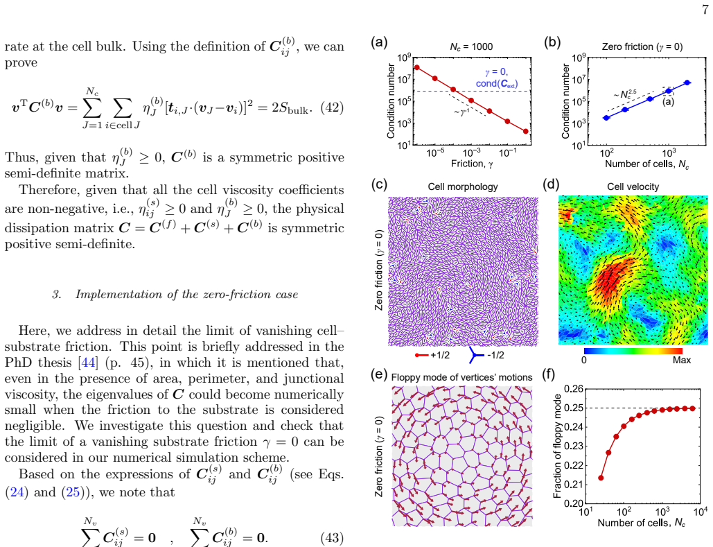
We show an example of simulating an active cell sheet without friction in Fig. 2(c, d). The numerical stability of the proposed framework is verified by analyzing the condition number of the friction-viscosity coefficient matrix (denoted cond(C)) and that of the augmented sadd...
2000
Reviewed August 2, 2026 · model on record in the stance chip above.
Discussion (0). Continue with ORCID to comment.