Residual Gaussian Splatting for Ultra Sparse-View CBCT Reconstruction
Pith reviewed 2026-05-07 08:10 UTC · model grok-4.3
The pith
Residual Gaussian Splatting recovers fine anatomical details from ultra-sparse CBCT views by separating geometric structure from residual textures.
A machine-rendered reading of the paper's core claim, the machinery that carries it, and where it could break.
Core claim
By introducing a spectrally-decoupled Gaussian representation that stratifies the volumetric field into a geometric base component and a residual detail component, Residual Gaussian Splatting converts explicit high-frequency fitting into a physically consistent implicit residual compensation task. A spectral-spatial collaborative optimization strategy then coordinates geometric anchoring with texture refinement to prevent spectral crosstalk, enabling the method to produce reconstructions that capture highly refined geometric textures while maintaining non-negative X-ray attenuation on clinical datasets.
What carries the argument
Spectrally-decoupled Gaussian representation that stratifies the volumetric field into a geometric base component and a residual detail component, paired with spectral-spatial collaborative optimization to handle high-frequency residuals.
If this is right
- Reconstructed images capture highly refined geometric textures in complex trabecular and vascular structures.
- The approach resolves the trade-off between artifact suppression and detail preservation.
- It yields superior visual fidelity compared to existing neural rendering baselines under ultra sparse-view conditions.
- The method maintains physical consistency with non-negative X-ray attenuation across clinical datasets.
Where Pith is reading between the lines
- The same base-plus-residual split could be tested on other explicit representations such as voxel grids or neural fields for similar inverse problems.
- If validated on larger cohorts, the technique could support clinical protocols that acquire CBCT with substantially fewer projections and lower patient dose.
- Applying the residual compensation stage to 4D or motion-affected data might address temporal inconsistencies in dynamic imaging.
- The wavelet-inspired multi-resolution handling suggests extensions to multi-modal fusion tasks where spectral separation is also needed.
Load-bearing premise
The decomposition into geometric base and residual detail components together with the joint optimization strategy can block spectral crosstalk and keep all attenuation values non-negative.
What would settle it
If the reconstructed volumes on clinical CBCT test sets still exhibit over-smoothing of trabecular patterns or produce negative attenuation values when compared to dense-view ground truth, the central claim would be falsified.
Figures
read the original abstract
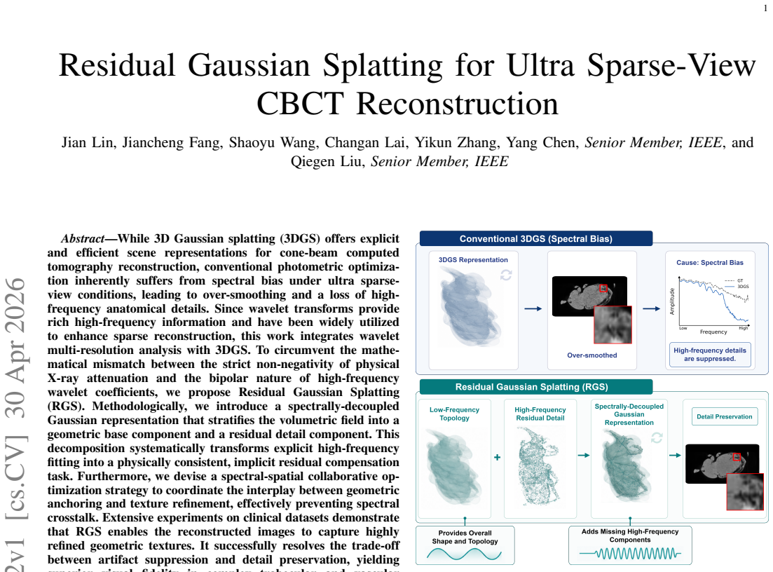
While 3D Gaussian splatting (3DGS) offers explicit and efficient scene representations for cone-beam computed tomography reconstruction, conventional photometric optimization inherently suffers from spectral bias under ultra sparse-view conditions, leading to over-smoothing and a loss of high-frequency anatomical details. Since wavelet transforms provide rich high-frequency information and have been widely utilized to enhance sparse reconstruction, this work integrates wavelet multi-resolution analysis with 3DGS. To circumvent the mathematical mismatch between the strict non-negativity of physical X-ray attenuation and the bipolar nature of high-frequency wavelet coefficients, we propose Residual Gaussian Splatting (RGS). Methodologically, we introduce a spectrally-decoupled Gaussian representation that stratifies the volumetric field into a geometric base component and a residual detail component. This decomposition systematically transforms explicit high-frequency fitting into a physically consistent, implicit residual compensation task. Furthermore, we devise a spectral-spatial collaborative optimization strategy to coordinate the interplay between geometric anchoring and texture refinement, effectively preventing spectral crosstalk. Extensive experiments on clinical datasets demonstrate that RGS enables the reconstructed images to capture highly refined geometric textures. It successfully resolves the trade-off between artifact suppression and detail preservation, yielding superior visual fidelity in complex trabecular and vascular structures compared to existing neural rendering baselines.
Editorial analysis
A structured set of objections, weighed in public.
Referee Report
Summary. The manuscript proposes Residual Gaussian Splatting (RGS) for ultra sparse-view CBCT reconstruction. It augments 3D Gaussian splatting with wavelet multi-resolution analysis via a spectrally-decoupled representation that decomposes the volumetric attenuation field into a geometric base component and a bipolar residual detail component. This converts explicit high-frequency fitting into an implicit residual compensation task. A spectral-spatial collaborative optimization strategy is introduced to coordinate the components and avoid crosstalk. The authors claim that experiments on clinical datasets demonstrate superior capture of refined geometric textures, resolution of the artifact-detail trade-off, and better visual fidelity in trabecular and vascular structures relative to existing neural rendering baselines.
Significance. If the central claims are substantiated, the work could meaningfully advance explicit scene representations for medical CT by reconciling the efficiency of 3DGS with wavelet-derived high-frequency content while respecting non-negativity. This addresses a practical limitation in sparse-view CBCT, where radiation dose reduction is clinically important, and offers a concrete decomposition strategy that may generalize beyond the current setting. The approach is technically novel relative to prior 3DGS adaptations in tomography.
major comments (2)
- [§3 (spectrally-decoupled Gaussian representation)] §3 (spectrally-decoupled Gaussian representation): the claim that the decomposition 'systematically transforms explicit high-frequency fitting into a physically consistent, implicit residual compensation task' and maintains 'physical consistency with non-negative X-ray attenuation' is load-bearing. The residual component is explicitly bipolar (high-frequency wavelet coefficients), yet the manuscript provides no explicit non-negativity mechanism (ReLU, softplus, projection, or loss penalty) on the summed field. In ultra-sparse regimes the photometric loss can still drive negative densities in low-signal voxels; without such a constraint or a proof that optimization preserves non-negativity, the physical-consistency guarantee does not follow from the construction.
- [§5 (experiments)] §5 (experiments): the abstract and results assert 'superior visual fidelity' and successful resolution of the artifact-detail trade-off on clinical datasets, but no quantitative metrics (PSNR, SSIM, MAE, or equivalent), error bars, or statistical tests are reported. Without these, together with ablations isolating the base/residual split and the collaborative optimization, it is impossible to verify that observed improvements exceed what could be obtained by post-hoc tuning of existing 3DGS baselines.
minor comments (2)
- [Abstract] The abstract would be strengthened by stating the precise number of views (e.g., '4–8 views') and naming the clinical datasets, allowing readers to immediately gauge the ultra-sparse regime.
- [§3] Notation for the base and residual Gaussians (e.g., how the wavelet coefficients are mapped to residual splat parameters) should be introduced with explicit equations rather than descriptive prose only.
Simulated Author's Rebuttal
We thank the referee for the insightful comments on our manuscript. We address each of the major comments below and will make the necessary revisions to strengthen the paper.
read point-by-point responses
-
Referee: §3 (spectrally-decoupled Gaussian representation): the claim that the decomposition 'systematically transforms explicit high-frequency fitting into a physically consistent, implicit residual compensation task' and maintains 'physical consistency with non-negative X-ray attenuation' is load-bearing. The residual component is explicitly bipolar (high-frequency wavelet coefficients), yet the manuscript provides no explicit non-negativity mechanism (ReLU, softplus, projection, or loss penalty) on the summed field. In ultra-sparse regimes the photometric loss can still drive negative densities in low-signal voxels; without such a constraint or a proof that optimization preserves non-negativity, the physical-consistency guarantee does not follow from the construction.
Authors: We thank the referee for highlighting this critical aspect of physical consistency. Upon re-examination, we recognize that while the base component is designed to be non-negative, an explicit safeguard on the summed field is indeed beneficial for ultra-sparse scenarios. In the revised manuscript, we will add a non-negativity enforcement mechanism, specifically applying a ReLU activation to the final attenuation values after summing the base and residual components, along with a regularization term in the loss to penalize any residual negative values. This will be detailed in the updated §3, including a short proof sketch that the optimization maintains non-negativity. The core decomposition remains valid as it converts high-frequency fitting to residual compensation within this constrained framework. revision: yes
-
Referee: §5 (experiments): the abstract and results assert 'superior visual fidelity' and successful resolution of the artifact-detail trade-off on clinical datasets, but no quantitative metrics (PSNR, SSIM, MAE, or equivalent), error bars, or statistical tests are reported. Without these, together with ablations isolating the base/residual split and the collaborative optimization, it is impossible to verify that observed improvements exceed what could be obtained by post-hoc tuning of existing 3DGS baselines.
Authors: We agree that quantitative metrics and ablations are necessary to fully substantiate the claims. The original submission prioritized visual and qualitative analysis to demonstrate the resolution of the artifact-detail trade-off in complex anatomical structures. For the revision, we will incorporate PSNR, SSIM, and MAE metrics with error bars computed over the clinical dataset cases. We will also add ablation experiments that isolate the effects of the base/residual decomposition and the spectral-spatial collaborative optimization. These will be compared to appropriately tuned 3DGS baselines to show the specific contributions of our method. The updated results section will include tables summarizing these quantitative findings and statistical significance where applicable. revision: yes
Circularity Check
Derivation chain is self-contained; new decomposition and optimization add independent content
full rationale
The paper begins from the established spectral bias of photometric 3DGS optimization under ultra-sparse views and the external literature on wavelets for sparse reconstruction. It then identifies the non-negativity mismatch with bipolar wavelet coefficients and introduces an explicit methodological choice: a spectrally-decoupled Gaussian representation that splits the field into a geometric base component plus a residual detail component. This split is presented as transforming the fitting task into implicit residual compensation, accompanied by a newly devised spectral-spatial collaborative optimization. No equation or claim reduces a 'prediction' or result to a fitted parameter or prior definition by construction. No self-citation is invoked as a load-bearing uniqueness theorem, no ansatz is smuggled via prior work, and no renaming of a known empirical pattern occurs. The central construction (base + residual + collaborative optimization) is therefore an independent modeling decision rather than a tautological re-expression of inputs. The derivation remains self-contained against external benchmarks.
Axiom & Free-Parameter Ledger
axioms (3)
- domain assumption Wavelet transforms provide rich high-frequency information useful for enhancing sparse reconstruction
- domain assumption 3D Gaussian splatting provides explicit and efficient scene representations for CBCT
- domain assumption X-ray attenuation coefficients are strictly non-negative
invented entities (1)
-
Residual Gaussian Splatting (RGS) with spectrally-decoupled representation
no independent evidence
Reference graph
Works this paper leans on
-
[1]
D. Donoho, “Compressed sensing,”IEEE Transactions on Information Theory, vol. 52, no. 4, pp. 1289–1306, 2006
work page 2006
-
[2]
Sparse MRI: The application of compressed sensing for rapid MR imaging,
M. Lustig, D. Donoho, and J. M. Pauly, “Sparse MRI: The application of compressed sensing for rapid MR imaging,”Magnetic Resonance in Medicine: An Official Journal of the International Society for Magnetic Resonance in Medicine, vol. 58, no. 6, pp. 1182–1195, 2007
work page 2007
-
[3]
Low-dose X-ray CT reconstruction via dictionary learning,
Q. Xu, H. Yu, X. Mou, L. Zhang, J. Hsieh, and G. Wang, “Low-dose X-ray CT reconstruction via dictionary learning,”IEEE Transactions on Medical Imaging, vol. 31, no. 9, pp. 1682–1697, Sep. 2012
work page 2012
-
[4]
K-SVD: An algorithm for designing overcomplete dictionaries for sparse representation,
M. Aharon, M. Elad, and A. Bruckstein, “K-SVD: An algorithm for designing overcomplete dictionaries for sparse representation,”IEEE Transactions on Signal Processing, vol. 54, no. 11, pp. 4311–4322, Nov. 2006
work page 2006
-
[5]
A theory for multiresolution signal decomposition: The wavelet representation,
S. Mallat, “A theory for multiresolution signal decomposition: The wavelet representation,”IEEE Transactions on Pattern Analysis and Machine Intelligence, vol. 11, no. 7, pp. 674–693, Jul. 1989
work page 1989
-
[6]
Deep learning for tomographic image reconstruction,
G. Wang, J. C. Ye, and B. De Man, “Deep learning for tomographic image reconstruction,”Nature Machine Intelligence, vol. 2, no. 12, pp. 737–748, Dec. 2020
work page 2020
-
[7]
Deep learning-based algorithms for low-dose CT imaging: A review,
H. Chen, Q. Li, L. Zhou, and F. Li, “Deep learning-based algorithms for low-dose CT imaging: A review,”European Journal of Radiology, vol. 172, Mar. 2024
work page 2024
-
[8]
Deep learning computed tomography,
T. W ¨urfl, F. C. Ghesu, V . Christlein, and A. Maier, “Deep learning computed tomography,” inMedical Image Computing and Computer- Assisted Intervention - MICCAI 2016, S. Ourselin, L. Joskowicz, M. R. Sabuncu, G. Unal, and W. Wells, Eds. Cham: Springer International Publishing, 2016, pp. 432–440
work page 2016
-
[9]
Convolutional neural network based metal artifact reduction in X-ray computed tomography,
Y . Zhang and H. Yu, “Convolutional neural network based metal artifact reduction in X-ray computed tomography,”IEEE Transactions on Medical Imaging, vol. 37, no. 6, pp. 1370–1381, Jun. 2018
work page 2018
-
[10]
Deep-neural-network- based sinogram synthesis for sparse-view CT image reconstruction,
H. Lee, J. Lee, H. Kim, B. Cho, and S. Cho, “Deep-neural-network- based sinogram synthesis for sparse-view CT image reconstruction,” IEEE Transactions on Radiation and Plasma Medical Sciences, vol. 3, no. 2, pp. 109–119, Mar. 2019
work page 2019
-
[11]
Stage-by-stage wavelet optimization refinement diffusion model for sparse-view CT reconstruc- tion,
K. Xu, S. Lu, B. Huang, W. Wu, and Q. Liu, “Stage-by-stage wavelet optimization refinement diffusion model for sparse-view CT reconstruc- tion,”IEEE Transactions on Medical Imaging, vol. 43, no. 10, pp. 3412– 3424, Oct. 2024
work page 2024
-
[12]
Generative modeling in sinogram domain for sparse-view CT reconstruction,
B. Guan, C. Yang, L. Zhang, S. Niu, M. Zhang, Y . Wang, W. Wu, and Q. Liu, “Generative modeling in sinogram domain for sparse-view CT reconstruction,”IEEE Transactions on Radiation and Plasma Medical Sciences, vol. 8, no. 2, pp. 195–207, Feb. 2024
work page 2024
-
[13]
Z. Li, D. Chang, Z. Zhang, F. Luo, Q. Liu, J. Zhang, G. Yang, and W. Wu, “Dual-domain collaborative diffusion sampling for multi-source stationary computed tomography reconstruction,”IEEE Transactions on Medical Imaging, vol. 43, no. 10, pp. 3398–3411, Oct. 2024
work page 2024
-
[14]
Hybrid-domain neural network processing for sparse-view CT reconstruction,
D. Hu, J. Liu, T. Lv, Q. Zhao, Y . Zhang, G. Quan, J. Feng, Y . Chen, and L. Luo, “Hybrid-domain neural network processing for sparse-view CT reconstruction,”IEEE Transactions on Radiation and Plasma Medical Sciences, vol. 5, no. 1, pp. 88–98, Jan. 2021
work page 2021
-
[15]
Genera- tive adversarial networks for noise reduction in low-dose CT,
J. M. Wolterink, T. Leiner, M. A. Viergever, and I. I ˇsgum, “Genera- tive adversarial networks for noise reduction in low-dose CT,”IEEE Transactions on Medical Imaging, vol. 36, no. 12, pp. 2536–2545, Dec. 2017
work page 2017
-
[16]
DuDoRNet: Learning a dual-domain recurrent network for fast MRI reconstruction with deep T1 prior,
B. Zhou and S. K. Zhou, “DuDoRNet: Learning a dual-domain recurrent network for fast MRI reconstruction with deep T1 prior,” inProceedings of the IEEE/CVF Conference on Computer Vision and Pattern Recogni- tion, 2020, pp. 4273–4282
work page 2020
-
[17]
DuDoNet: Dual domain network for CT metal artifact reduction,
W.-A. Lin, H. Liao, C. Peng, X. Sun, J. Zhang, J. Luo, R. Chellappa, and S. K. Zhou, “DuDoNet: Dual domain network for CT metal artifact reduction,” inProceedings of the IEEE/CVF Conference on Computer Vision and Pattern Recognition, 2019, pp. 10 512–10 521
work page 2019
-
[18]
Momentum-Net: Fast and convergent iterative neural network for inverse problems,
I. Y . Chun, Z. Huang, H. Lim, and J. A. Fessler, “Momentum-Net: Fast and convergent iterative neural network for inverse problems,”IEEE Transactions on Pattern Analysis and Machine Intelligence, vol. 45, no. 4, pp. 4915–4931, Apr. 2023
work page 2023
-
[19]
NeRF: Representing scenes as neural radiance fields for view synthesis,
B. Mildenhall, P. P. Srinivasan, M. Tancik, J. T. Barron, R. Ramamoorthi, and R. Ng, “NeRF: Representing scenes as neural radiance fields for view synthesis,”Communications of The Acm, vol. 65, no. 1, pp. 99– 106, Dec. 2021
work page 2021
-
[20]
3D Gaussian splatting for real-time radiance field rendering,
B. Kerbl, G. Kopanas, T. Leimk ¨uhler, and G. Drettakis, “3D Gaussian splatting for real-time radiance field rendering,” Aug. 2023
work page 2023
-
[21]
NAF: Neural attenuation fields for sparse- view CBCT reconstruction,
R. Zha, Y . Zhang, and H. Li, “NAF: Neural attenuation fields for sparse- view CBCT reconstruction,” inMedical Image Computing and Computer Assisted Intervention – MICCAI 2022, L. Wang, Q. Dou, P. T. Fletcher, S. Speidel, and S. Li, Eds. Cham: Springer Nature Switzerland, 2022, pp. 442–452
work page 2022
-
[22]
Radiative gaussian splatting for efficient x-ray novel view synthesis,
Y . Cai, Y . Liang, J. Wang, A. Wang, Y . Zhang, X. Yang, Z. Zhou, and A. Yuille, “Radiative gaussian splatting for efficient x-ray novel view synthesis,” inComputer Vision – ECCV 2024, A. Leonardis, E. Ricci, S. Roth, O. Russakovsky, T. Sattler, and G. Varol, Eds. Cham: Springer Nature Switzerland, 2025, pp. 283–299
work page 2024
-
[23]
R$ˆ2$-Gaussian: Rectifying radiative Gaussian splatting for tomographic reconstruction,
R. Zha, T. J. Lin, Y . Cai, J. Cao, Y . Zhang, and H. Li, “R$ˆ2$-Gaussian: Rectifying radiative Gaussian splatting for tomographic reconstruction,” Oct. 2024
work page 2024
-
[24]
3DGR-CT: Sparse- view CT reconstruction with a 3D Gaussian representation,
Y . Li, X. Fu, H. Li, S. Zhao, R. Jin, and S. K. Zhou, “3DGR-CT: Sparse- view CT reconstruction with a 3D Gaussian representation,”Medical Image Analysis, vol. 103, p. 103585, Jul. 2025
work page 2025
-
[25]
On the spectral bias of neural networks,
N. Rahaman, A. Baratin, D. Arpit, F. Draxler, M. Lin, F. Hamprecht, Y . Bengio, and A. Courville, “On the spectral bias of neural networks,” in Proceedings of the 36th International Conference on Machine Learning. PMLR, May 2019, pp. 5301–5310
work page 2019
-
[26]
What is cone-beam CT and how does it work?
W. C. Scarfe and A. G. Farman, “What is cone-beam CT and how does it work?”Dental Clinics of North America, vol. 52, no. 4, pp. 707–730, Oct. 2008
work page 2008
-
[27]
Clinical applications of cone-beam computed tomography in dental practice,
W. C. Scarfe, A. G. Farman, P. Sukovicet al., “Clinical applications of cone-beam computed tomography in dental practice,”Journal-Canadian Dental Association, vol. 72, no. 1, p. 75, 2006
work page 2006
-
[28]
Practical cone-beam algorithm,
L. A. Feldkamp, L. C. Davis, and J. W. Kress, “Practical cone-beam algorithm,”JOSA A, vol. 1, no. 6, pp. 612–619, Jun. 1984
work page 1984
-
[29]
M. Zwicker, H. Pfister, J. van Baar, and M. Gross, “EW A volume splatting,” inProceedings Visualization, 2001. VIS ’01., Oct. 2001, pp. 29–538
work page 2001
-
[30]
Mallat,A wavelet tour of signal processing
S. Mallat,A wavelet tour of signal processing. Elsevier, Sep. 1999
work page 1999
-
[31]
A fast iterative shrinkage-thresholding algo- rithm for linear inverse problems,
A. Beck and M. Teboulle, “A fast iterative shrinkage-thresholding algo- rithm for linear inverse problems,”SIAM Journal on Imaging Sciences, vol. 2, no. 1, pp. 183–202, 2009
work page 2009
-
[32]
B. Stephen, P. Neal, C. Eric, P. Borja, and E. Jonathan, “Distributed optimization and statistical learning via the alternating direction method of multipliers,”Foundations and Trends in Information Retrieval, vol. 3, no. 1, pp. 1–122, Jul. 2011
work page 2011
-
[33]
Improved compressed sensing-based algorithm for sparse-view CT image reconstruction,
Z. Zhu, K. Wahid, P. Babyn, D. Cooper, I. Pratt, and Y . Carter, “Improved compressed sensing-based algorithm for sparse-view CT image reconstruction,”Computational and Mathematical Methods in Medicine, vol. 2013, no. 1, p. 185750, 2013
work page 2013
-
[34]
G.-H. Chen, J. Tang, and S. Leng, “Prior image constrained compressed sensing (PICCS): A method to accurately reconstruct dynamic CT im- ages from highly undersampled projection data sets,”Medical Physics, vol. 35, no. 2, pp. 660–663, 2008
work page 2008
-
[35]
Sparse-view x-ray CT reconstruction via total generalized variation regularization,
S. Niu, Y . Gao, Z. Bian, J. Huang, W. Chen, G. Yu, Z. Liang, and J. Ma, “Sparse-view x-ray CT reconstruction via total generalized variation regularization,”Physics in Medicine & Biology, vol. 59, no. 12, p. 2997, May 2014
work page 2014
-
[36]
C. M. Sandino, J. Y . Cheng, F. Chen, M. Mardani, J. M. Pauly, and S. S. Vasanawala, “Compressed sensing: From research to clinical practice with deep neural networks: Shortening scan times for magnetic resonance imaging,”IEEE Signal Processing Magazine, vol. 37, no. 1, pp. 117–127, Jan. 2020
work page 2020
-
[37]
ISTA-Net: Interpretable optimization-inspired deep network for image compressive sensing,
J. Zhang and B. Ghanem, “ISTA-Net: Interpretable optimization-inspired deep network for image compressive sensing,” inProceedings of the IEEE Conference on Computer Vision and Pattern Recognition, 2018, pp. 1828–1837
work page 2018
-
[38]
State-of-the-art deep learning CT reconstruction algorithms in abdominal imaging,
A. Mileto, L. Yu, J. W. Revels, S. Kamel, M. A. Shehata, J. J. Ibarra- Rovira, V . K. Wong, A. M. Roman-Colon, J. M. Lee, K. M. Elsayes, and C. T. Jensen, “State-of-the-art deep learning CT reconstruction algorithms in abdominal imaging,”RadioGraphics, vol. 44, no. 12, p. e240095, Dec. 2024
work page 2024
discussion (0)
Sign in with ORCID, Apple, or X to comment. Anyone can read and Pith papers without signing in.