Reducing Annotation Burden for Femoral Cartilage Segmentation in Knee MRI via Cross-Sequence Transfer Learning
Pith reviewed 2026-05-12 03:05 UTC · model grok-4.3
The pith
Transfer learning across MRI sequences can reduce annotated images needed for femoral cartilage segmentation to as few as nine subjects.
A machine-rendered reading of the paper's core claim, the machinery that carries it, and where it could break.
Core claim
Optimizing a modified 2D U-Net on 507 DESS images from the Osteoarthritis Initiative allows subsequent fine-tuning on target sequences to reach performance plateaus with reduced training data: Cube-to-DESS transfer attains a Dice similarity coefficient of 0.903 with 9 subjects, matching same-sequence DESS training on 44 subjects, whereas DESS-to-Cube transfer plateaus at 0.802 with 24 subjects compared to a same-sequence baseline of 0.830.
What carries the argument
Bidirectional cross-sequence transfer learning by fine-tuning a pretrained modified 2D U-Net on increasing sizes of target-sequence training data while holding validation and test sets fixed.
If this is right
- Cube-to-DESS transfer matches the DESS same-sequence baseline DSC of 0.900 after fine-tuning on only 9 subjects.
- DESS-to-Cube transfer reaches a lower DSC plateau of 0.802 after fine-tuning on 24 subjects, below the same-sequence Cube baseline of 0.830.
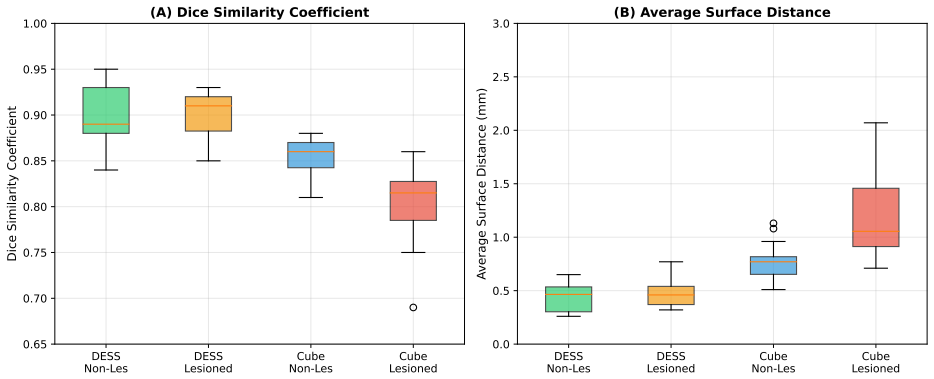
- Lesions have no significant effect on DESS segmentation accuracy but reduce Cube accuracy from 0.856 to 0.805.
- Same-sequence training yields higher accuracy on DESS than on Cube.
- The amount of target data needed to reach a performance plateau is strongly direction-dependent.
Where Pith is reading between the lines
- Clinics could favor acquiring the more transferable sequence when possible to minimize future annotation work for new models.
- The sequence-dependent effect of lesions implies that transfer models may require targeted validation or augmentation when pathology is present.
- The approach could be tested on additional MRI contrasts or other joint structures to see if similar annotation savings appear.
- Using fixed validation and test sets across experiments means the reported savings are tied to the specific held-out subjects from the two sites.
Load-bearing premise
The 44-subject subsets balanced for lesions from each sequence are representative enough of broader populations to reliably measure how much annotation can be reduced through transfer.
What would settle it
Repeating the cross-sequence experiments on a larger independent set of DESS and Cube knee MRI images and checking if the performance plateaus still occur at nine or 24 training subjects would confirm or refute the claimed reduction in annotation burden.
Figures


read the original abstract
Purpose: To develop and evaluate cross-sequence transfer learning for automatic femoral cartilage segmentation, testing bidirectional transfer between dual-echo steady-state (DESS) and sagittal proton density-weighted 3D fast spin-echo (Cube) sequences. Materials and Methods: We optimized a modified 2D U-Net on 507 DESS images from the Osteoarthritis Initiative (OAI). We then established same-sequence baselines using subject-level cross-validation on a subset of 44 OAI DESS images and 44 Cube images acquired at the Istituto Ortopedico Rizzoli, Bologna, Italy. Each subset included 22 non-lesioned and 22 lesioned subjects. Finally, we performed transfer learning across sequences by fine-tuning the pretrained models on the target sequence with increasing training set sizes to study convergence, while keeping validation and test sets fixed. Segmentations were evaluated using Dice similarity coefficient (DSC) and average surface distance (ASD). Lesion effects were assessed with two-sided Mann-Whitney U tests with Bonferroni correction. Results: Same-sequence training yielded higher accuracy on DESS than Cube (DSC, $0.900$ vs $0.830$; $P < .001$). Cube-to-DESS transfer matched DESS performance (DSC, $0.903 \pm 0.032$ vs $0.900 \pm 0.027$), reaching a performance plateau at 9 training subjects. DESS-to-Cube yielded a lower combined DSC ($0.802 \pm 0.049$ vs $0.830 \pm 0.042$), reaching a plateau at 24 training subjects. Lesions did not affect DESS ($P \ge .39$) but reduced Cube accuracy (DSC, $0.805$ vs $0.856$; $P < .001$). Conclusion: Transfer learning across sequences can substantially reduce target-sequence annotation requirements for femoral cartilage segmentation, but performance is direction- and sequence-dependent, and the effects of lesions on segmentation may vary across MRI sequences.
Editorial analysis
A structured set of objections, weighed in public.
Referee Report
Summary. The paper develops and evaluates bidirectional cross-sequence transfer learning with a modified 2D U-Net for femoral cartilage segmentation in knee MRI. It pre-trains on 507 OAI DESS volumes, establishes same-sequence baselines via subject-level cross-validation on 44-subject subsets (22 lesioned + 22 non-lesioned) from OAI DESS and Rizzoli Cube data, then fine-tunes the source model on increasing numbers of target-sequence subjects while holding validation and test sets fixed. Reported results include DSC 0.900 (DESS baseline) vs 0.830 (Cube baseline), Cube-to-DESS transfer reaching DSC 0.903 plateauing at 9 subjects, DESS-to-Cube reaching 0.802 plateauing at 24 subjects, and sequence-dependent lesion effects (no effect on DESS, reduced accuracy on Cube).
Significance. If the quantitative claims on annotation reduction hold under broader validation, the work provides concrete evidence that transfer learning can lower the target-sequence labeling effort for cartilage segmentation, with practical implications for scaling automated analysis in osteoarthritis cohorts. The direction- and sequence-specific findings, plus the differential lesion impact, are useful for guiding sequence choice in future studies.
major comments (1)
- [Results (44-subject subset experiments and transfer convergence curves)] The central claim that transfer learning substantially reduces target-sequence annotation requirements rests on performance plateaus observed at 9 subjects (Cube-to-DESS) and 24 subjects (DESS-to-Cube) together with the assertion that transfer matches or approaches same-sequence baselines. These quantities are derived from subject-level cross-validation on only 44 DESS and 44 Cube cases (22 lesioned/22 non-lesioned each) drawn from two sites, with fixed val/test splits and no external validation or larger-cohort replication. With this sample size the reported DSC values, Mann-Whitney lesion tests, and convergence curves are sensitive to subject selection, lesion distribution, and cross-site differences, undermining the reliability of the quantitative annotation-reduction estimates.
minor comments (3)
- [Materials and Methods] The abstract and methods description omit key implementation details required for reproducibility: exact preprocessing pipeline (intensity normalization, resampling, augmentation), U-Net architectural modifications, hyperparameter search procedure, and training schedule for both pre-training and fine-tuning stages.
- [Results] No error analysis, per-subject DSC distributions, or failure-case visualizations are provided to explain why Cube-to-DESS transfer succeeds with fewer subjects than the reverse direction.
- [Results (lesion-effect analysis)] The statement that lesions do not affect DESS (P ≥ .39) but reduce Cube accuracy would benefit from reporting the exact p-values after Bonferroni correction and the corresponding ASD results for completeness.
Simulated Author's Rebuttal
We thank the referee for the positive assessment of our work's significance and for the detailed feedback on the experimental design. We address the major comment point by point below.
read point-by-point responses
-
Referee: The central claim that transfer learning substantially reduces target-sequence annotation requirements rests on performance plateaus observed at 9 subjects (Cube-to-DESS) and 24 subjects (DESS-to-Cube) together with the assertion that transfer matches or approaches same-sequence baselines. These quantities are derived from subject-level cross-validation on only 44 DESS and 44 Cube cases (22 lesioned/22 non-lesioned each) drawn from two sites, with fixed val/test splits and no external validation or larger-cohort replication. With this sample size the reported DSC values, Mann-Whitney lesion tests, and convergence curves are sensitive to subject selection, lesion distribution, and cross-site differences, undermining the reliability of the quantitative annotation-reduction estimates.
Authors: We agree that the 44-subject subsets (balanced for lesion status) from two sites constitute a genuine limitation that affects the precision and generalizability of the reported plateau points and the exact annotation-reduction numbers. Our design deliberately used subject-level cross-validation with fixed validation and test sets across all training sizes to enable direct comparison of convergence behavior while controlling for test-set variability; the plateaus are visually evident in the provided curves and are accompanied by standard deviations. Nevertheless, we recognize that subject selection, lesion distribution, and cross-site factors could shift the specific thresholds of 9 and 24 subjects. In the revised manuscript we will expand the Discussion and Limitations sections to explicitly state that these quantitative estimates are preliminary, to report fold-wise variability where not already shown, and to recommend validation on larger multi-center cohorts before clinical translation. This revision does not alter the core directional findings or the same-sequence baseline comparisons but appropriately tempers the strength of the annotation-reduction claims. revision: partial
Circularity Check
No circularity: empirical ML study with independent held-out evaluations
full rationale
The paper is a purely empirical machine-learning study. It trains a modified 2D U-Net on OAI DESS data, establishes same-sequence baselines via subject-level cross-validation on 44-subject subsets, and measures transfer-learning convergence by fine-tuning on increasing target-sequence training sizes while holding validation/test sets fixed. All reported metrics (DSC, ASD) are computed directly on these independent splits; no equations, fitted parameters renamed as predictions, self-referential definitions, uniqueness theorems, or ansatzes appear. Claims rest on observable performance differences rather than any derivation that reduces to its own inputs by construction.
Axiom & Free-Parameter Ledger
free parameters (2)
- U-Net training hyperparameters
- Fine-tuning set sizes
axioms (2)
- domain assumption The modified 2D U-Net is an appropriate architecture for both DESS and Cube sequences without need for 3D or sequence-specific redesign.
- domain assumption The 44-subject subsets provide stable estimates of baseline and transfer performance.
Reference graph
Works this paper leans on
-
[1]
Shinnosuke Hada, Haruka Kaneko, Ryo Sadatsuki, Lizu Liu, Ippei Futami, Mayuko Kinoshita, An- warjan Yusup, Yoshitomo Saita, Yuji Takazawa, Hiroshi Ikeda, Kazuo Kaneko, and Muneaki Ishijima. The degeneration and destruction of femoral articular cartilage shows a greater degree of deterioration than that of the tibial and patellar articular cartilage in ear...
work page 2014
-
[2]
Wluka, Graeme Jones, Changhai Ding, and Flavia M
Yuanyuan Wang, Anita E. Wluka, Graeme Jones, Changhai Ding, and Flavia M. Cicuttini. Use mag- netic resonance imaging to assess articular cartilage.Therapeutic Advances in Musculoskeletal Dis- ease, 4(2):77–97, April 2012
work page 2012
-
[3]
Wolfgang Wirth, Christoph Ladel, Susanne Maschek, Anna Wisser, Felix Eckstein, and Frank Roemer. Quantitative measurement of cartilage morphology in osteoarthritis: Current knowledge and future directions.Skeletal Radiology, 52(11):2107–2122, November 2023
work page 2023
-
[4]
Sheheryar Khan, Muhammad Ammar Khawer, Junru Zhong, Rizwan Qureshi, Muhammad Asim, and Weitian Chen. Advancing deep learning based knee cartilage segmentation in MRI: Innovations, chal- lenges and applications.Osteoarthritis and Cartilage Open, 8(1):100702, March 2026
work page 2026
-
[5]
Nieminen, Simo Saarakkala, and Victor Casula
Egor Panfilov, Aleksei Tiulpin, Miika T. Nieminen, Simo Saarakkala, and Victor Casula. Deep learning-based segmentation of knee MRI for fully automatic subregional morphological assessment of cartilage tissues: Data from the Osteoarthritis Initiative.Journal of Orthopaedic Research, 40(5):1113– 1124, 2022
work page 2022
-
[6]
Berk Norman, Valentina Pedoia, and Sharmila Majumdar. Use of 2D U-Net Convolutional Neural Net- works for Automated Cartilage and Meniscus Segmentation of Knee MR Imaging Data to Determine Relaxometry and Morphometry.Radiology, 288(1):177–185, July 2018
work page 2018
-
[7]
Peterfy, Erika Schneider, and Michael C
Charles G. Peterfy, Erika Schneider, and Michael C. Nevitt. The osteoarthritis initiative: Report on the design rationale for the magnetic resonance imaging protocol for the knee.Osteoarthritis and Cartilage, 16(12):1433–1441, December 2008
work page 2008
-
[8]
Klaus M. Friedrich, Gert Reiter, Bernd Kaiser, Marius Mayerhöfer, Michael Deimling, Vladimir Jellus, Wilhelm Horger, Siegfried Trattnig, Mark Schweitzer, and Erich Salomonowitz. High-resolution car- tilage imaging of the knee at 3 T: Basic evaluation of modern isotropic 3D MR-sequences.European Journal of Radiology, 78(3):398–405, June 2011
work page 2011
-
[9]
Osamu Tokuda, Yuko Harada, Gen Shiraishi, Tetsuhisa Motomura, Kouji Fukuda, Motoichi Kimura, and Naofumi Matsunaga. MRI of the anatomical structures of the knee: The proton density-weighted fast spin-echo sequence vs the proton density-weighted fast-recovery fast spin-echo sequence.British Journal of Radiology, 85(1017):e686–e693, September 2012
work page 2012
-
[10]
Ahmad Waleed Salehi, Shakir Khan, Gaurav Gupta, Bayan Ibrahimm Alabduallah, Abrar Almjally, Hadeel Alsolai, Tamanna Siddiqui, and Adel Mellit. A Study of CNN and Transfer Learning in Medical Imaging: Advantages, Challenges, Future Scope.Sustainability, 15(7):5930, January 2023
work page 2023
-
[11]
Nima Tajbakhsh, Jae Y . Shin, Suryakanth R. Gurudu, R. Todd Hurst, Christopher B. Kendall, Michael B. Gotway, and Jianming Liang. Convolutional Neural Networks for Medical Image Analysis: Full Training or Fine Tuning?IEEE Transactions on Medical Imaging, 35(5):1299–1312, May 2016. 13
work page 2016
-
[12]
Chundru, Sibaji Gaj, Andreas Nanavati, Morgan H
Mingrui Yang, Ceylan Colak, Kishore K. Chundru, Sibaji Gaj, Andreas Nanavati, Morgan H. Jones, Carl S. Winalski, Naveen Subhas, and Xiaojuan Li. Automated knee cartilage segmentation for het- erogeneous clinical MRI using generative adversarial networks with transfer learning.Quantitative Imaging in Medicine and Surgery, 12(5):2620–2633, May 2022
work page 2022
-
[13]
Mohsen Ghafoorian, Alireza Mehrtash, Tina Kapur, Nico Karssemeijer, Elena Marchiori, Mehran Pesteie, Charles R. G. Guttmann, Frank-Erik de Leeuw, Clare M. Tempany, Bram van Ginneken, Andriy Fedorov, Purang Abolmaesumi, Bram Platel, and William M. Wells. Transfer Learning for Domain Adaptation in MRI: Application in Brain Lesion Segmentation. InMedical Ima...
work page 2017
-
[14]
Giulia Grenno, Giordano Valente, Giacomo Dal Fabbro, Luca Macchiarola, Alberto Grassi, Stefano Zaffagnini, and Fulvia Taddei. High tibial osteotomy leads to medial-lateral redistribution of knee internal forces during walking and stair ambulation and improves patient-reported outcomes.Knee Surgery, Sports Traumatology, Arthroscopy, 34(5):1787–1801, May 2026
work page 2026
-
[15]
Felix Ambellan, Alexander Tack, Moritz Ehlke, and Stefan Zachow. Automated segmentation of knee bone and cartilage combining statistical shape knowledge and convolutional neural networks: Data from the Osteoarthritis Initiative.Medical Image Analysis, 52:109–118, February 2019
work page 2019
-
[16]
Serena Bonaretti, Garry E. Gold, and Gary S. Beaupre. pyKNEEr: An image analysis workflow for open and reproducible research on femoral knee cartilage.PLOS ONE, 15(1):e0226501, January 2020
work page 2020
-
[17]
U-Net: Convolutional Networks for Biomedical Image Segmentation
Olaf Ronneberger, Philipp Fischer, and Thomas Brox. U-Net: Convolutional Networks for Biomedical Image Segmentation. In Nassir Navab, Joachim Hornegger, William M. Wells, and Alejandro F. Frangi, editors,Medical Image Computing and Computer-Assisted Intervention – MICCAI 2015, pages 234–
work page 2015
-
[18]
Springer International Publishing, 2015
work page 2015
-
[19]
Algorithms for Hyper-Parameter Optimization
James Bergstra, Rémi Bardenet, Yoshua Bengio, and Balázs Kégl. Algorithms for Hyper-Parameter Optimization. InAdvances in Neural Information Processing Systems, volume 24. Curran Associates, Inc., 2011
work page 2011
-
[20]
Jason Yosinski, Jeff Clune, Yoshua Bengio, and Hod Lipson. How transferable are features in deep neural networks? InAdvances in Neural Information Processing Systems, volume 27. Curran Asso- ciates, Inc., 2014
work page 2014
-
[21]
Transfusion: Understanding Trans- fer Learning for Medical Imaging
Maithra Raghu, Chiyuan Zhang, Jon Kleinberg, and Samy Bengio. Transfusion: Understanding Trans- fer Learning for Medical Imaging. InAdvances in Neural Information Processing Systems, volume 32. Curran Associates, Inc., 2019
work page 2019
-
[22]
Abdel Aziz Taha and Allan Hanbury. Metrics for evaluating 3D medical image segmentation: Analysis, selection, and tool.BMC Medical Imaging, 15:29, August 2015
work page 2015
-
[23]
Optuna: A Next-generation Hyperparameter Optimization Framework
Takuya Akiba, Shotaro Sano, Toshihiko Yanase, Takeru Ohta, and Masanori Koyama. Optuna: A Next-generation Hyperparameter Optimization Framework. InProceedings of the 25th ACM SIGKDD International Conference on Knowledge Discovery & Data Mining, pages 2623–2631. Association for Computing Machinery, 2019
work page 2019
-
[24]
Serena Bonaretti, Mojtaba Barzegari, Melissa Bevers, Steven Boyd, Andrew J Burghardt, Donnie Cameron, Francesco Chiumento, Gianluigi Crimi, Gerald Degenhart, Pholpat Durongbhan, et al. Open 14 and reproducible research in musculoskeletal imaging: why it matters and how to implement it with the guidelines of the open and reproducible musculoskeletal imagin...
work page 2026
-
[25]
Anthony A. Gatti and Monica R. Maly. Automatic knee cartilage and bone segmentation using multi- stage convolutional neural networks: Data from the osteoarthritis initiative.Magnetic Resonance Ma- terials in Physics, Biology and Medicine, 34(6):859–875, December 2021
work page 2021
-
[26]
Ankur J. Shah and Drushi Patel. Imaging update on cartilage.Journal of Clinical Orthopaedics and Trauma, 22:101610, September 2021. 15
work page 2021
discussion (0)
Sign in with ORCID, Apple, or X to comment. Anyone can read and Pith papers without signing in.