nASR: An End-to-End Trainable Neural Layer for Channel-Level EEG Artifact Subspace Reconstruction in Real-Time BCI
Pith reviewed 2026-06-30 20:08 UTC · model grok-4.3
The pith
nASR makes ASR thresholds trainable inside an end-to-end network so artifact removal and BCI decoding optimize together and run six to eight times faster.
A machine-rendered reading of the paper's core claim, the machinery that carries it, and where it could break.
Core claim
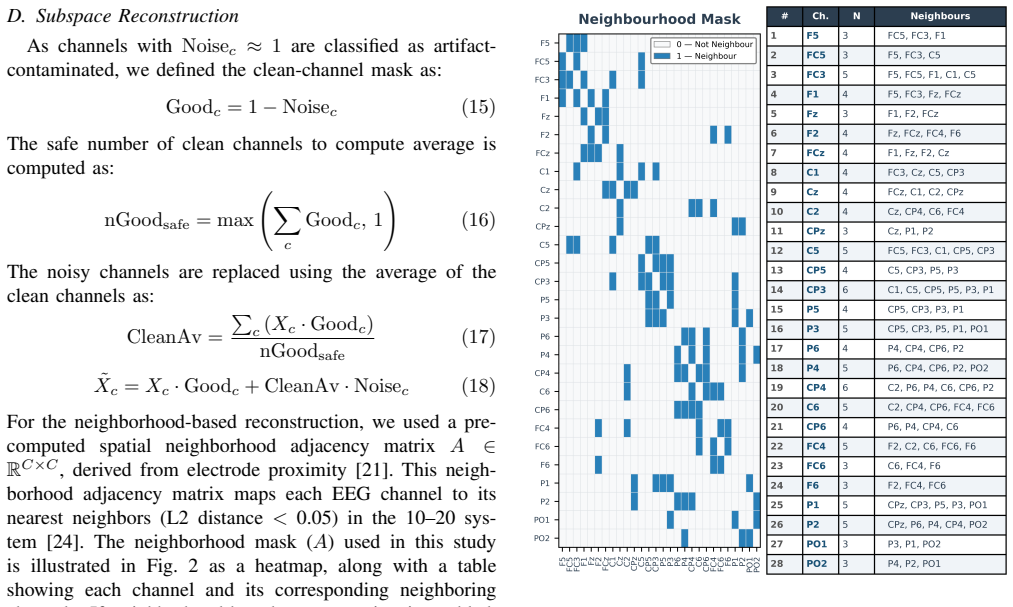
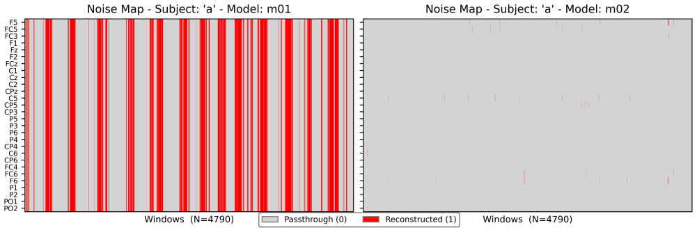
nASR is a Keras layer that introduces trainable thresholds K and L to perform artifact detection in PC variance space and eigen-spread-based identification of primary artifact channels, enabling selective channel-level subspace reconstruction that preserves clean channels and improves both accuracy and speed over fixed-parameter ASR.
What carries the argument
The nASR layer with trainable thresholds K (artifact detection in PC variance) and L (eigen-spread channel identification) that performs selective channel-level reconstruction inside the end-to-end network.
If this is right
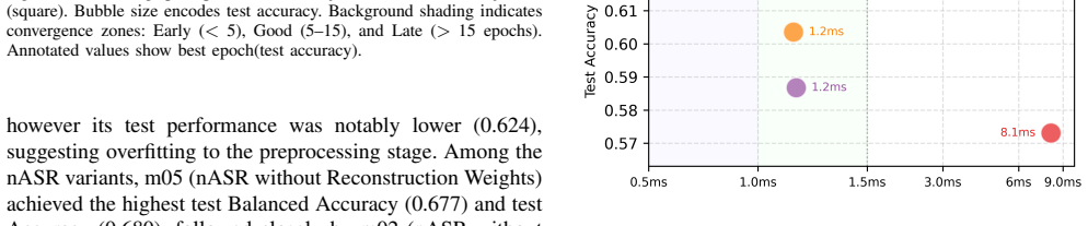
- nASR variants achieve higher test classification accuracy than standard ASR across the evaluated models.
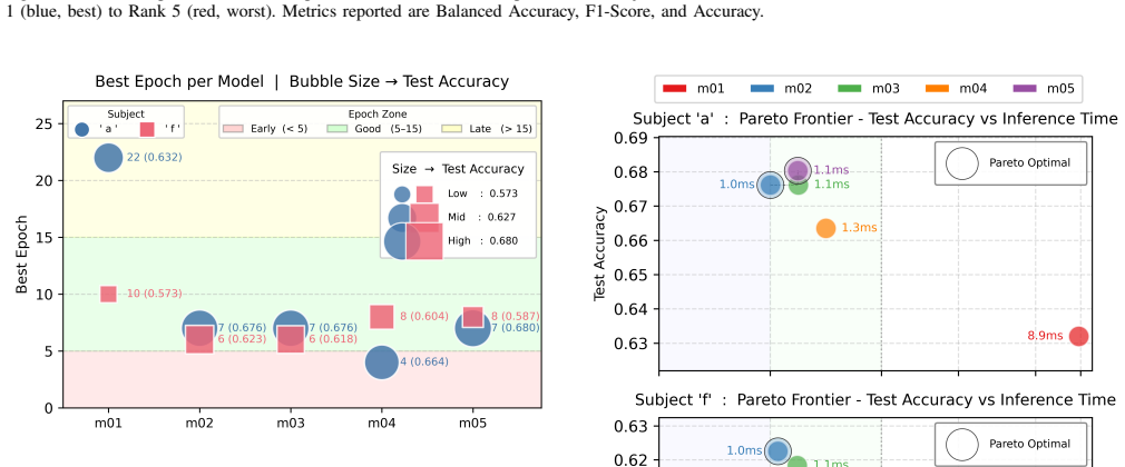
- Inference time drops by a factor of six to eight compared with traditional ASR.
- Selective channel-level reconstruction leaves unaffected channels unchanged.
- The layer supports real-time BCI by combining artifact cleaning with task optimization in one trainable module.
Where Pith is reading between the lines
- The design removes the need for manual threshold tuning when moving between users or sessions.
- Channel-level selectivity may reduce unintended distortion of spatial patterns used by other BCI decoders.
- Faster inference could allow the full pipeline to run on embedded hardware without external GPUs.
Load-bearing premise
Jointly optimizing thresholds K and L will preserve task-relevant neural features rather than overfit to the small training set or distort signals on new subjects.
What would settle it
Accuracy and feature-preservation metrics on held-out subjects or new recording sessions from the same BCI dataset where any nASR variant falls below traditional ASR performance.
Figures
read the original abstract
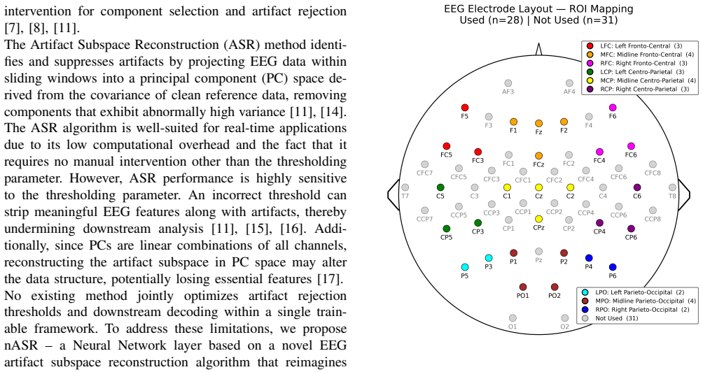
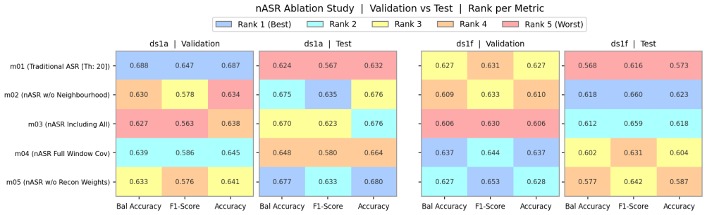
Electroencephalogram (EEG) signals are highly susceptible to artifacts, resulting in a low signal-to-noise ratio which makes extraction of meaningful neural information challenging. Artifact Subspace Reconstruction (ASR) is one of the most widely used artifact filtering techniques in EEG-based BCI applications, owing to its real-time applicability. ASR reconstructs artifact-free signals by operating in Principal Component (PC) space within sliding windows. However, ASR performance is critically sensitive to its threshold parameter - an incorrect threshold risks removing task-relevant neural features alongside artifacts. Furthermore, since PCs are linear combinations of all channels, subspace reconstruction in PC space may alter the underlying data structure, potentially discarding essential neural information. To address these limitations, we propose nASR, a novel end-to-end trainable Keras layer that jointly optimizes artifact rejection and downstream decoding. nASR introduces two trainable threshold parameters: K, which governs artifact detection in PC variance space, and L, which quantifies eigen-spread to pinpoint the primary artifact--contributing channels, enabling selective channel-level reconstruction that preserves clean channel information. An ablation study comprising five model variants (m01 - m05), evaluated across two subjects from the BCI Competition IV Dataset 1, confirms that nASR variants consistently outperform traditional ASR on test classification metrics, while achieving a 6-8x reduction in inference time, making nASR a strong candidate for real-time BCI applications demanding both low latency and high decoding performance.
Editorial analysis
A structured set of objections, weighed in public.
Referee Report
Summary. The paper proposes nASR, an end-to-end trainable Keras layer extending Artifact Subspace Reconstruction (ASR) for EEG artifact removal in BCI. It introduces two trainable thresholds K (for PC variance-based artifact detection) and L (for eigen-spread to identify artifact-contributing channels), enabling channel-level selective reconstruction. An ablation study with five model variants (m01-m05) on two subjects from BCI Competition IV Dataset 1 reports that nASR variants outperform traditional ASR on test classification metrics while achieving 6-8x faster inference, positioning it as suitable for real-time applications.
Significance. If the performance improvements and speed gains are confirmed under rigorous validation, nASR could meaningfully advance real-time BCI by jointly optimizing artifact rejection with downstream decoding, mitigating ASR's known threshold sensitivity and potential distortion of neural features in PC space. The end-to-end trainable design and inference speedup represent practical strengths for low-latency systems.
major comments (2)
- [Ablation study / Results] The ablation study (described in the abstract and presumably detailed in the Results or Experiments section) evaluates five variants on only two subjects and claims consistent outperformance without statistical tests, error bars, cross-subject validation, or details on training procedures and overfitting checks. EEG signals exhibit substantial inter-subject and session variability; N=2 provides no statistical power and does not test whether joint optimization of K and L preserves task features or merely fits the small sample, directly undermining the central claim of reliable superiority for real-time BCI.
- [Introduction / Method] The abstract notes ASR's sensitivity to threshold choice and potential PC-space distortion of data structure, yet the manuscript provides no concrete evidence (e.g., feature preservation metrics or comparisons on held-out sessions) that the trainable K/L parameters address these issues beyond the two-subject ablation. This leaves the core motivation for nASR unverified at the scale needed to support the performance claims.
minor comments (2)
- Clarify the exact definitions and initialization of the five model variants (m01-m05) and how they differ in use of trainable K/L versus fixed ASR parameters.
- The abstract's claim of 'channel-level reconstruction' would benefit from a brief equation or pseudocode snippet showing how L selects channels without altering the overall PC reconstruction pipeline.
Simulated Author's Rebuttal
We thank the referee for the constructive feedback. We address each major comment below and outline the revisions we will make to the manuscript.
read point-by-point responses
-
Referee: Ablation study evaluates five variants on only two subjects without statistical tests, error bars, cross-subject validation, or training details. N=2 lacks statistical power and does not verify if optimization preserves task features.
Authors: We concur that the limited sample size of two subjects restricts the robustness of our findings and does not allow for cross-subject generalization or statistical validation. This preliminary evaluation aimed to illustrate the potential of end-to-end training. We will revise the manuscript to include evaluations on additional subjects, statistical analyses, error bars, and explicit descriptions of the training process and measures against overfitting. revision: yes
-
Referee: The manuscript provides no concrete evidence (e.g., feature preservation metrics or comparisons on held-out sessions) that the trainable K/L parameters address ASR's threshold sensitivity and PC-space distortion beyond the two-subject ablation.
Authors: While the improved classification metrics in the ablation study suggest that the trainable thresholds help mitigate these issues through joint optimization, we agree that direct evidence is not provided. We will add feature preservation metrics and evaluations on held-out sessions in the revised version to better substantiate the motivation for nASR. revision: yes
Circularity Check
No circularity in derivation or performance claims
full rationale
The paper introduces nASR as an end-to-end trainable Keras layer with two new parameters K and L. Performance claims rest solely on an empirical ablation study (m01-m05 variants) evaluated on test metrics from two subjects in BCI Competition IV Dataset 1, plus reported inference-time speedup. No equations, derivations, or first-principles results are presented that reduce claimed outperformance to quantities defined by the fitted parameters themselves. The method is described as a new trainable component without self-definitional loops, fitted-input predictions, or load-bearing self-citations that collapse the central result to its inputs by construction. The derivation chain is therefore self-contained against external benchmarks.
Axiom & Free-Parameter Ledger
Reference graph
Works this paper leans on
-
[1]
Where does EEG come from and what does it mean?,
M. X. Cohen, “Where does EEG come from and what does it mean?,” Trends in Neurosciences, vol. 40, no. 4, pp. 208–218, Apr. 2017, doi: 10.1016/j.tins.2017.02.004
-
[2]
Electroencephalogram (EEG) and its background,
S. Siuly, Y . Li, and Y . Zhang, “Electroencephalogram (EEG) and its background,” inEEG Signal Analysis and Classification: Techniques and Applications, Cham, Switzerland: Springer International Publish- ing, 2016, pp. 3–21, doi: 10.1007/978-3-319-47653-7 1
-
[3]
Remote collection of electrophysiological data with brain wearables: Opportunities and challenges,
R. J. Sugden, V .-L. L. Pham-Kim-Nghiem-Phu, I. Campbell, A. Leon, and P. Diamandis, “Remote collection of electrophysiological data with brain wearables: Opportunities and challenges,”Bioelectronic Medicine, vol. 9, no. 1, p. 12, 2023, doi: 10.1186/s42234-023-00114-5
-
[4]
Deep learning for electroencephalogram (EEG) classification tasks: A review,
A. Craik, Y . He, and J. L. Contreras-Vidal, “Deep learning for electroencephalogram (EEG) classification tasks: A review,”Jour- nal of Neural Engineering, vol. 16, no. 3, p. 031001, 2019, doi: 10.1088/1741-2552/ab0ab5
-
[5]
Design and validation of a low-cost mobile eeg-based brain–computer interface,
A. Craiket al., “Design and validation of a low-cost mobile EEG- based brain–computer interface,”Sensors, vol. 23, no. 13, 2023, doi: 10.3390/s23135930
-
[6]
Eeg artifact removal: State-of-the-art and guidelines,
J. A. Uriguen and B. Garcia-Zapirain, “EEG artifact removal—state- of-the-art and guidelines,”Journal of Neural Engineering, vol. 12, no. 3, p. 031001, 2015, doi: 10.1088/1741-2560/12/3/031001
-
[7]
Removal of artifacts from EEG signals: A review,
X. Jiang, G.-B. Bian, and Z. Tian, “Removal of artifacts from EEG signals: A review,”Sensors, vol. 19, no. 5, p. 987, 2019, doi: 10.3390/s19050987
-
[8]
C. R. Rashmi and C. P. Shantala, “EEG artifacts detection and removal techniques for brain–computer interface applications: A systematic review,” 2022, doi: 10.19101/IJATEE.2021.874883
-
[9]
A. Kilicarslan and J. L. Contreras-Vidal, “Neuro-robotics: Rehabili- tation and restoration of walking using exoskeletons via non-invasive brain–machine interfaces,” inNeuroprosthetics and Brain-Computer Interfaces in Spinal Cord Injury, G. M ¨uller-Putz and R. Rupp, Eds. Cham, Switzerland: Springer International Publishing, 2021, pp. 143– 166, doi: 10.10...
-
[10]
EEG de-noising using SURE thresholding based on wavelet transforms,
G. Geetha and S. N. Geethalakshmi, “EEG de-noising using SURE thresholding based on wavelet transforms,”International Journal of Computer Applications, vol. 24, no. 6, pp. 29–33, Jun. 2011, doi: 10.5120/2948-3935
-
[11]
C.-Y . Chang, S.-H. Hsu, L. Pion-Tonachini, and T.-P. Jung, “Evaluation of artifact subspace reconstruction for automatic artifact components removal in multi-channel EEG recordings,”IEEE Transactions on Biomedical Engineering, vol. 67, no. 4, pp. 1114–1121, 2020, doi: 10.1109/TBME.2019.2930186
-
[12]
A robust adaptive denoising framework for real-time artifact removal in scalp EEG measurements,
A. Kilicarslan, R. G. Grossman, and J. L. Contreras-Vidal, “A robust adaptive denoising framework for real-time artifact removal in scalp EEG measurements,”Journal of Neural Engineering, vol. 13, no. 2, p. 026013, Feb. 2016, doi: 10.1088/1741-2560/13/2/026013
-
[14]
Real-time neuroimaging and cognitive mon- itoring using wearable dry EEG,
T. R. Mullenet al., “Real-time neuroimaging and cognitive mon- itoring using wearable dry EEG,”IEEE Transactions on Biomed- ical Engineering, vol. 62, no. 11, pp. 2553–2567, 2015, doi: 10.1109/TBME.2015.2481482
-
[15]
P. Anders, H. M ¨uller, N. Skjæret-Maroni, B. Vereijken, and J. Baumeister, “The influence of motor tasks and cut-off parameter selection on artifact subspace reconstruction in EEG recordings,” Medical & Biological Engineering & Computing, vol. 58, no. 11, pp. 2673–2683, Nov. 2020, doi: 10.1007/s11517-020-02252-3
-
[16]
The influence assessment of artifact subspace reconstruction on the EEG signal characteristics,
M. Plechawska-W ´ojcik, P. Augustynowicz, M. Kaczorowska, E. Zabielska-Mendyk, and D. Zapała, “The influence assessment of artifact subspace reconstruction on the EEG signal characteristics,” Applied Sciences, vol. 13, no. 3, 2023, doi: 10.3390/app13031605
-
[17]
A. Oladipupo Ibraheem, “Correlation of data reconstruction error and shrinkages in pair-wise distances under principal component analysis (PCA),”arXiv preprint arXiv:1412.6752, Dec. 2014, doi: 10.48550/arXiv.1412.6752
work page internal anchor Pith review Pith/arXiv arXiv doi:10.48550/arxiv.1412.6752 2014
-
[18]
A. Dapena, H. J. P ´erez-Iglesias, and V . Zarzoso, “Blind channel estimation based on maximizing the eigenvalue spread of cumulant matrices in (2 × 1) Alamouti’s coding schemes,”Wireless Commu- nications and Mobile Computing, vol. 12, no. 6, pp. 516–528, Apr. 2012, doi: 10.1002/wcm.992
-
[19]
T. D. Lagerlund, D. I. Rubin, and J. R. Daube, “V olume conduction,” inClinical Neurophysiology, 4th ed., New York, NY , USA: Oxford University Press, 2016, pp. 929–946, doi: 10.1093/med/9780190259631.003.0054
-
[20]
B. Blankertz, G. Dornhege, M. Krauledat, K. R. M ¨uller, and G. Curio, “The non-invasive Berlin brain–computer interface: Fast acquisition of effective performance in untrained subjects,”NeuroImage, vol. 37, no. 2, pp. 539–550, Aug. 2007, doi: 10.1016/j.neuroimage.2007.01.051
-
[21]
EEGReXferNet: A lightweight Gen-AI framework for EEG sub- space reconstruction via cross-subject transfer learning and channel- aware embedding,
S. Sarkar, P. Nabrzyski, S. Prasad, and J. L. Contreras-Vidal, “EEGReXferNet: A lightweight Gen-AI framework for EEG sub- space reconstruction via cross-subject transfer learning and channel- aware embedding,” inNeurIPS 2025 Workshop on Foundation Models for the Brain and Body, 2025. [Online]. Available: https://openreview.net/forum?id=TOW1by49Ec
2025
-
[22]
V . J. Lawhern, A. J. Solon, N. R. Waytowich, S. M. Gordon, C. P. Hung, and B. J. Lance, “EEGNet: A compact convolutional neural network for EEG-based brain–computer interfaces,”Journal of Neural Engineering, vol. 15, no. 5, p. 056013, Jul. 2018, doi: 10.1088/1741- 2552/aace8c
-
[23]
K. J. Popeet al., “Managing electromyogram contamination in scalp recordings: An approach identifying reliable beta and gamma EEG features of psychoses or other disorders,”Brain and Behavior, vol. 12, no. 9, p. e2721, 2022, doi: 10.1002/brb3.2721
-
[24]
The international 10–20 system revisited: Cartesian and spherical coordinates,
K. B. E. B ¨ocker, J. A. G. van Avermaete, and M. M. C. van den Berg-Lenssen, “The international 10–20 system revisited: Cartesian and spherical coordinates,”Brain Topography, vol. 6, no. 3, pp. 231– 235, 1994, doi: 10.1007/BF01187714
-
[25]
Which reference should we use for EEG and ERP practice?,
D. Yao, Y . Qin, S. Hu, L. Dong, M. L. Bringas Vega, and P. A. Vald´es Sosa, “Which reference should we use for EEG and ERP practice?,”Brain Topography, vol. 32, no. 4, pp. 530–549, 2019, doi: 10.1007/s10548-019-00707-x
discussion (0)
Sign in with ORCID, Apple, or X to comment. Anyone can read and Pith papers without signing in.