Visual Search Patterns in 3D Pancreatic Imaging: An Eye Tracking Study
Pith reviewed 2026-05-20 21:21 UTC · model grok-4.3
The pith
Eye tracking aligned with slice navigation in CT volumes allows analysis of radiologists' gaze behavior in 3D pancreatic imaging.
A machine-rendered reading of the paper's core claim, the machinery that carries it, and where it could break.
Core claim
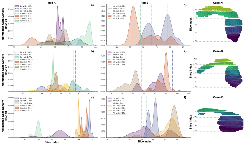
By collecting eye tracking data from two radiologists searching abdominal CTs of the pancreas and aligning eye gaze movements with slice navigation, the representation of the pancreas through the volume can be visualized and clinicians' gaze behavior can be analyzed in both space and time, providing an example for a taxonomy of eye movement data in 3D medical imaging.
What carries the argument
Alignment of eye gaze movements with slice navigation to visualize gaze in space and time across the CT volume.
If this is right
- Eye tracking data synchronized with slice navigation can reveal patterns in how radiologists search for pancreatic abnormalities in CT volumes.
- This method enables analysis of gaze behavior both spatially within each slice and temporally as slices are navigated.
- A taxonomy of such eye movements could help understand diagnostic approaches in radiology.
- Applications of eye tracking to 3D imaging extend beyond 2D settings to routine clinical volumetric scans.
Where Pith is reading between the lines
- Extending this to more radiologists might identify common search strategies or differences based on experience.
- Similar tracking could be applied to other 3D imaging tasks like MRI or ultrasound volumes.
- Findings might suggest improvements to image viewing software to better support natural search patterns.
Load-bearing premise
Eye-gaze data from the scrolling interface with only two participants accurately represents clinicians' typical diagnostic visual search behavior.
What would settle it
A study with additional radiologists using the same interface that produces inconsistent gaze patterns or a comparison showing major differences when using non-scrolling 3D viewing tools.
Figures
read the original abstract
Eye tracking has emerged as a powerful tool for examining visual perception and search strategies in various domains, including medicine. While it is relatively straightforward to apply in 2D settings, its use in 3D medical imaging remains challenging and not yet well explored. This gap is particularly relevant for radiology, where volumetric images such as computed tomography (CT) scans are routinely read by medical experts. Radiologists typically interpret these images by navigating through hundreds of 2D slices, most often viewed in the axial projection. A taxonomy of eye movement data during navigation through a CT volume could be valuable to understand how radiologists approach diagnostic tasks. As an example of the derived taxonomy, we asked two radiologists to search abdominal CTs of the pancreas. We collect eye tracking data and align eye gaze movements with slice navigation to visualize the representation of the pancreas through volume and analyze clinicians' gaze behavior in both space and time.
Editorial analysis
A structured set of objections, weighed in public.
Referee Report
Summary. The manuscript describes an eye-tracking study in which two radiologists navigated abdominal CT volumes of the pancreas. Gaze data are aligned with slice navigation to derive a taxonomy of visual search patterns in 3D imaging and to analyze gaze behavior in both space and time.
Significance. A validated taxonomy of radiologist visual search in volumetric CT could inform interface design and training, but the present demonstration rests on an n=2 sample with no reported quantitative metrics, clustering methods, or reliability measures, limiting its immediate impact.
major comments (2)
- [Abstract / Methods] Abstract and implied Methods: the taxonomy is derived from eye-tracking data of only two radiologists; no details are supplied on how gaze trajectories were segmented, clustered, or categorized into distinct patterns, nor is inter-rater reliability or stability across sessions reported. With n=2 any taxonomy necessarily conflates individual differences with general categories.
- [Abstract] Abstract: the central claim that the aligned gaze data faithfully capture diagnostic visual search behavior lacks supporting quantitative results, error analysis, or validation steps against the scrolling interface distortion noted in the skeptic's concern.
minor comments (2)
- [Methods] Clarify the exact CT acquisition parameters and display settings used during the eye-tracking sessions to allow replication.
- [Results] Add a figure showing example gaze trajectories overlaid on selected slices to illustrate the proposed taxonomy categories.
Simulated Author's Rebuttal
We thank the referee for the constructive comments on our manuscript. We have revised the paper to better frame the work as exploratory, provide additional methodological details, and include quantitative support for the gaze alignment approach.
read point-by-point responses
-
Referee: [Abstract / Methods] Abstract and implied Methods: the taxonomy is derived from eye-tracking data of only two radiologists; no details are supplied on how gaze trajectories were segmented, clustered, or categorized into distinct patterns, nor is inter-rater reliability or stability across sessions reported. With n=2 any taxonomy necessarily conflates individual differences with general categories.
Authors: We agree that n=2 limits generalizability and have revised the manuscript to explicitly describe the work as an initial exploration to begin deriving a taxonomy rather than a validated classification. The revised Methods section now details the segmentation of gaze trajectories via timestamp alignment between eye-tracker output and slice navigation logs, followed by qualitative categorization into observed patterns (e.g., sequential scrolling with targeted fixations versus broad overview sweeps) without automated clustering. Because only two participants were involved, formal inter-rater reliability statistics are not applicable; we instead report intra-participant consistency across repeated readings and have added this as an explicit limitation in the Discussion, noting that future studies with larger cohorts will be needed to separate individual differences from general patterns. revision: partial
-
Referee: [Abstract] Abstract: the central claim that the aligned gaze data faithfully capture diagnostic visual search behavior lacks supporting quantitative results, error analysis, or validation steps against the scrolling interface distortion noted in the skeptic's concern.
Authors: We have revised the abstract to temper the language, describing the alignment as a method for visualizing and analyzing gaze relative to the 3D volume rather than claiming faithful capture of all diagnostic behavior. The revised Results section now reports quantitative metrics including percentage of gaze time on pancreatic anatomy across slices and temporal dwell distributions. We have added an error analysis in Methods that quantifies alignment accuracy by comparing gaze coordinates to manually verified anatomical landmarks and accounts for potential scrolling latency in the interface; this provides an initial validation step against the noted distortion concerns. revision: yes
Circularity Check
No significant circularity in empirical observational study
full rationale
This paper is an observational eye-tracking study that collects gaze data from two radiologists navigating CT volumes of the pancreas and aligns it with slice navigation to describe visual search patterns. No mathematical derivations, equations, fitted parameters, or predictions are presented that could reduce to inputs by construction. The taxonomy emerges directly from the collected data rather than from any self-referential chain, self-citation load-bearing premise, or ansatz smuggled via prior work. The study is self-contained as an empirical demonstration with no load-bearing steps that collapse into their own definitions or fitted values.
Axiom & Free-Parameter Ledger
axioms (1)
- domain assumption Eye-tracking hardware and software accurately record gaze location on displayed 2D slices during manual navigation
Lean theorems connected to this paper
-
IndisputableMonolith/Foundation/RealityFromDistinction.leanreality_from_one_distinction unclear?
unclearRelation between the paper passage and the cited Recognition theorem.
A taxonomy of eye movement data during navigation through a CT volume could be valuable... we asked two radiologists to search abdominal CTs of the pancreas. We collect eye tracking data and align eye gaze movements with slice navigation
What do these tags mean?
- matches
- The paper's claim is directly supported by a theorem in the formal canon.
- supports
- The theorem supports part of the paper's argument, but the paper may add assumptions or extra steps.
- extends
- The paper goes beyond the formal theorem; the theorem is a base layer rather than the whole result.
- uses
- The paper appears to rely on the theorem as machinery.
- contradicts
- The paper's claim conflicts with a theorem or certificate in the canon.
- unclear
- Pith found a possible connection, but the passage is too broad, indirect, or ambiguous to say the theorem truly supports the claim.
Reference graph
Works this paper leans on
-
[1]
Cognitive Research: Principles and Implications , volume=
What do we know about volumetric medical image interpretation?: a review of the basic science and medical image perception literatures , author=. Cognitive Research: Principles and Implications , volume=. 2019 , publisher=
work page 2019
-
[2]
Characterizing search, recognition, and decision in the detection of lung nodules on CT scans: elucidation with eye tracking , author=. Radiology , volume=. 2015 , publisher=
work page 2015
-
[3]
IEEE Transactions on Visualization and Computer Graphics , volume=
Visual analytics of a computer-aided diagnosis system for pancreatic lesions , author=. IEEE Transactions on Visualization and Computer Graphics , volume=. 2019 , publisher=
work page 2019
-
[4]
Journal of Medical Imaging , volume=
Review of prospects and challenges of eye tracking in volumetric imaging , author=. Journal of Medical Imaging , volume=. 2016 , publisher=
work page 2016
-
[5]
Scanners and drillers: characterizing expert visual search through volumetric images , author=. Journal of vision , volume=. 2013 , publisher=
work page 2013
-
[6]
Cancer Detection and Diagnosis: A Handbook of Emerging Technologies , pages=
Reading the Reader: Utilizing Eye Movements and Machine Learning to Enhance Accuracy during Diagnostic Visual Search , author=. Cancer Detection and Diagnosis: A Handbook of Emerging Technologies , pages=. 2025 , publisher=
work page 2025
-
[7]
Mercan, E. and Shapiro, L.G. and others , title =. J Digit Imaging , volume =. 2018 , month =. doi:10.1007/s10278-017-9990-5 , pmid =
-
[8]
Journal of Medical Imaging , volume=
Comparing search patterns in digital breast tomosynthesis and full-field digital mammography: an eye tracking study , author=. Journal of Medical Imaging , volume=. 2017 , publisher=
work page 2017
- [9]
-
[10]
Wu, CC and Wolfe, JM , title =. Vision (Basel) , volume =. 2019 , month =. doi:10.3390/vision3020032 , pmid =
-
[11]
An interactive eye-tracking system for measuring radiologists' visual fixations in volumetric CT images: Implementation and initial eye-tracking accuracy validation , author=. Medical physics , volume=. 2021 , publisher=
work page 2021
-
[12]
American Journal of Roentgenology , volume=
Prospective evaluation of reader performance on MDCT in characterization of cystic pancreatic lesions and prediction of cyst biologic aggressiveness , author=. American Journal of Roentgenology , volume=. 2011 , publisher=
work page 2011
-
[13]
Artificial intelligence in pancreatic cancer , author=. Theranostics , volume=
- [14]
- [15]
- [16]
-
[17]
Kassner, M and Patera, W and Bulling, A , title =. Adjunct Proceedings of the 2014 ACM International Joint Conference on Pervasive and Ubiquitous Computing , series =
work page 2014
-
[18]
Radiology: Artificial Intelligence , volume =
Wasserthal, J and Breit, H-C and others , title =. Radiology: Artificial Intelligence , volume =. 2023 , doi =
work page 2023
-
[19]
ORB: An efficient alternative to SIFT or SURF , year=
Rublee, E and Rabaud, V and others , booktitle=. ORB: An efficient alternative to SIFT or SURF , year=
discussion (0)
Sign in with ORCID, Apple, or X to comment. Anyone can read and Pith papers without signing in.