Local Structural Signatures of Shear Bands in Metallic Glasses via Electron Nanodiffraction
Pith reviewed 2026-06-29 21:50 UTC · model grok-4.3
The pith
Plastic deformation in metallic glasses arises from coordinated nanoscale structural transformations.
A machine-rendered reading of the paper's core claim, the machinery that carries it, and where it could break.
Core claim
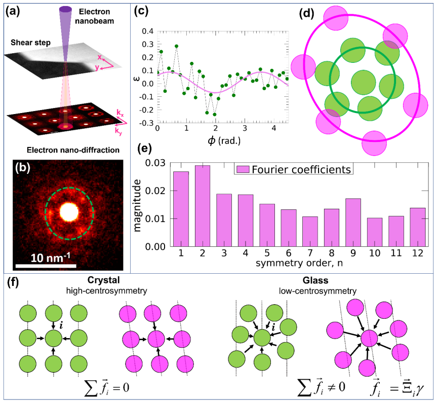
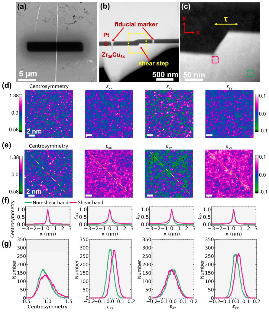
By preparing inverted cross-sectional transmission electron microscopy lamellae of shear bands formed during bending and employing cryogenic ion polishing to minimize preparation artefacts, the intrinsic atomic structure is preserved. Mapping of local centrosymmetry and strain from electron nano-diffraction reveals nanoscale, stripe-like regions oriented at 45 degrees to the applied strain where strain has localized. These regions exhibit a high density of local atomic structures that have transformed to configurations with reduced centrosymmetry and increased magnitudes of shear and normal strain.
What carries the argument
Parameters derived from electron nano-diffraction that quantify local centrosymmetry and strain on cryogenically polished inverted cross-sectional lamellae.
If this is right
- Plastic deformation localizes into nanoscale stripe-like regions with transformed atomic structures.
- These regions show reduced centrosymmetry together with higher shear and normal strain.
- The transformations are coordinated across the plastic zone beneath surface shear steps.
- Direct experimental mapping of these local changes explains shear-band formation in metallic glasses.
Where Pith is reading between the lines
- The same diffraction parameters could be tested on shear bands formed under tension or compression rather than bending.
- Atomistic simulations could be checked against the measured density of low-centrosymmetry sites inside the stripes.
- If the preparation method works, it offers a template for studying other disordered solids where ion damage has previously obscured structure.
- The 45-degree orientation suggests a link to maximum shear planes that could be used to predict band locations in components.
Load-bearing premise
Cryogenic ion polishing of the inverted cross-sectional lamellae sufficiently minimizes preparation artefacts so that the measured local centrosymmetry and strain reflect the intrinsic undeformed atomic structure.
What would settle it
Finding identical centrosymmetry and strain distributions inside and outside the shear-band zones, or observing the same stripe patterns in samples prepared by standard room-temperature ion milling.
Figures
read the original abstract
Structural changes in a glass due to deformation are subtle and difficult to quantify using conventional imaging and diffraction techniques. Additionally, transmission electron microscopy (TEM) sample preparation using energetic ions often causes structural modifications that are challenging to detect in disordered materials. By preparing inverted cross-sectional transmission electron microscopy lamellae of shear bands formed during bending, and employing cryogenic ion polishing to minimize preparation artefacts, we preserve the intrinsic atomic structure. Using sensitive, new parameters derived from electron nano-diffraction, we directly probe the local nano-scale structure in the plastic zone beneath surface shear steps in metallic glasses. Mapping of local centrosymmetry and strain reveals nanoscale, stripe-like regions oriented at 45 degree to the applied strain where strain has localized. These regions exhibit a high density of local atomic structures that have transformed to configurations with reduced centrosymmetry and increased magnitudes of shear and normal strain. Our results demonstrate that plastic deformation in metallic glasses arises from coordinated nanoscale structural transformations, providing direct experimental insight into a long-standing problem.
Editorial analysis
A structured set of objections, weighed in public.
Referee Report
Summary. The paper claims that by preparing inverted cross-sectional TEM lamellae of shear bands in metallic glasses via cryogenic ion polishing and applying new parameters from electron nanodiffraction, local mappings of centrosymmetry and strain reveal stripe-like regions at 45° to the applied strain with reduced centrosymmetry and elevated shear/normal strain; these are interpreted as evidence that plastic deformation arises from coordinated nanoscale structural transformations.
Significance. If the preparation artefacts are demonstrably absent and the mappings are reproducible, the work would supply direct experimental access to the local atomic configurations inside shear bands, a long-standing gap in metallic-glass plasticity research. The approach of combining cross-sectional geometry with nanodiffraction parameters is potentially enabling for disordered materials.
major comments (2)
- [Abstract / Methods] Abstract and Methods: No quantitative validation is presented (e.g., centrosymmetry histograms or strain distributions comparing deformed vs. undeformed regions of the same lamella, or cryogenic vs. room-temperature polishing controls). Because the central claim requires that the observed reductions in centrosymmetry and increases in strain are intrinsic rather than ion-induced, this omission is load-bearing.
- [Abstract] Abstract: The support for the claim of 'coordinated nanoscale structural transformations' cannot be assessed; the text supplies neither error bars, statistical tests, nor comparison to undeformed controls, leaving the strength of the stripe-like features and their 45° orientation unquantified.
minor comments (1)
- [Abstract] The term 'inverted cross-sectional lamellae' is introduced without a schematic or explicit description of the geometry relative to the bending axis; a figure clarifying the orientation would aid reproducibility.
Simulated Author's Rebuttal
We thank the referee for the careful reading and constructive comments. We address each major point below and will revise the manuscript accordingly to strengthen the quantitative support for our claims.
read point-by-point responses
-
Referee: [Abstract / Methods] Abstract and Methods: No quantitative validation is presented (e.g., centrosymmetry histograms or strain distributions comparing deformed vs. undeformed regions of the same lamella, or cryogenic vs. room-temperature polishing controls). Because the central claim requires that the observed reductions in centrosymmetry and increases in strain are intrinsic rather than ion-induced, this omission is load-bearing.
Authors: We agree that direct quantitative comparisons are required to rule out ion-induced artifacts. In the revised manuscript we will add (i) centrosymmetry and strain histograms with error bars for deformed versus undeformed regions extracted from the same lamella and (ii) equivalent data from control lamellae prepared by room-temperature ion polishing. These additions will be placed in a new subsection of the Methods and in the Results to demonstrate that the observed reductions in centrosymmetry and elevations in strain are intrinsic to the shear-band material. revision: yes
-
Referee: [Abstract] Abstract: The support for the claim of 'coordinated nanoscale structural transformations' cannot be assessed; the text supplies neither error bars, statistical tests, nor comparison to undeformed controls, leaving the strength of the stripe-like features and their 45° orientation unquantified.
Authors: We will revise the abstract to include a concise statement of the statistical significance of the stripe-like features. In the main text and supplementary figures we will add (i) error bars on all reported strain and centrosymmetry values, (ii) direct side-by-side comparisons to undeformed control regions, and (iii) quantitative measures (angular histograms and Kolmogorov-Smirnov tests) confirming the 45° orientation preference. These changes will allow readers to assess the strength of the evidence for coordinated structural transformations. revision: yes
Circularity Check
No circularity; purely observational experimental study with no derivations or self-referential predictions.
full rationale
The manuscript presents experimental TEM/nanodiffraction mapping of centrosymmetry and strain in shear bands of metallic glasses. No equations, fitted parameters, predictions, or theoretical derivations are described that could reduce to inputs by construction. Claims rest on direct measurements after sample preparation, with no self-citation chains or ansatzes invoked as load-bearing steps. The work is self-contained against external benchmarks as an observational report.
Axiom & Free-Parameter Ledger
axioms (1)
- domain assumption Cryogenic ion polishing of inverted cross-sectional lamellae preserves the intrinsic atomic structure of metallic glasses without detectable artefacts.
Reference graph
Works this paper leans on
-
[1]
T. G. Ingram, Proc. R. Soc. Lond. A 1934, 145, 362
1934
-
[2]
P. B. Hirsch, M. J. Whelan, Philos. Trans. R. Soc. A 1960, 252, 499
1960
-
[3]
M. J. Hÿtch, E. Snoeck, R. Kilaas, Ultramicroscopy 1998, 74, 131
1998
-
[4]
W. L. Johnson, J. Lu, M. D. Demetriou, Intermetallics (Barking). 2002, 10, 1039
2002
-
[5]
P. M. Anderson, J. P. Hirth, J. Lothe, Theory of Dislocations , Cambridge University Press, 2017
2017
-
[6]
C. P. Royall, S. R. Williams, Phys. Rep. 2015, 560, 1
2015
-
[7]
R. M. O. Mota, E. T. Lund, S. Sohn, D. J. Browne, D. C. Hofmann, S. Curtarolo, A. van de Walle, J. Schroers, Commun. Mater. 2021, 2, 23
2021
-
[8]
W. H. Wang, Y. Yang, T. G. Nieh, C. T. Liu, Intermetallics (Barking). 2015, 67, 81
2015
-
[9]
M. L. Falk, J. S. Langer, Phys. Rev. E 1998, 57, 7192
1998
-
[10]
Nicolas, E
A. Nicolas, E. E. Ferrero, K. Martens, J. -L. Barrat, Rev. Mod. Phys. 2018, 90, 45006
2018
-
[11]
E. T. Lund, S. Sohn, A. van de Walle, S. Curtarolo, D. Hofmann, J. Schroers, Materialia (Oxf). 2025, 40, 102408
2025
-
[12]
L. H. Dai, in Adiabatic Shear Localization (Second Edition) , Elsevier, Oxford, 2012, 311–361
2012
-
[13]
V Sergueeva, N
A. V Sergueeva, N. A. Mara, J. D. Kuntz, D. J. Branagan, A. K. Mukherjee, Mater. Sci. Eng. A 2004, 383, 219
2004
-
[14]
G. N. Yang, B. A. Sun, S. Q. Chen, Y. Shao, K. F. Yao, J. Alloys Compd. 2017, 695, 3457
2017
-
[15]
M. J. Kramer, D. J. Sordelet, A. F. Bastarows, X. Tan, S. B. Biner, J. Non-Cryst. Solids 2005, 351, 2159
2005
-
[16]
A. S. Argon, Acta Metall. 1979, 27, 47. 30
1979
-
[17]
T. C. Pekin, J. Ding, C. Gammer, B. Ozdol, C. Ophus, M. Asta, R. O. Ritchie, A. M. Minor, Nat. Commun. 2019, 10, 2445
2019
-
[18]
Glushko, R
O. Glushko, R. Pippan, D. Şopu, C. Mitterer, J. Eckert, Nat. Commun. 2024, 15, 5601
2024
-
[19]
R. Maaß, J. F. Löffler, Adv. Funct. Mater. 2015, 25, 2353
2015
-
[20]
Y. Leng, T. H. Courtney, J. Mater. Sci. 1991, 26, 588
1991
-
[21]
Hieronymus-Schmidt, H
V. Hieronymus-Schmidt, H. Rösner, G. Wilde, A. Zaccone, Phys. Rev. B 2017, 95, 134111
2017
-
[22]
Y. Cao, J. Li, B. Kou, C. Xia, Z. Li, R. Chen, H. Xie, T. Xiao, W. Kob, L. Hong, others, Nat. Commun. 2018, 9, 2911
2018
-
[23]
Niiyama, M
T. Niiyama, M. Wakeda, T. Shimokawa, S. Ogata, Phys. Rev. E 2019, 100, 43002
2019
-
[24]
E. D. Bøjesen, T. C. Petersen, A. V. Martin, M. Weyland, A. C. Y. Liu, JPhys Mater.: Materials 2020, 3
2020
-
[25]
Rösner, A
H. Rösner, A. Bera, A. Zaccone, Phys. Rev. B 2024, 110, 14107
2024
-
[26]
M. H. Lee, E. S. Park, R. T. Ott, B. S. Kim, J. Eckert, Appl. Phys. Lett. 2014, 105, 61906
2014
-
[27]
Dasgupta, H
R. Dasgupta, H. G. E. Hentschel, I. Procaccia, Phys. Rev. Lett. 2012, 109, 255502
2012
-
[28]
D. Şopu, A. Stukowski, M. Stoica, S. Scudino, Phys. Rev. Lett. 2017, 119
2017
-
[29]
Baggioli, I
M. Baggioli, I. Kriuchevskyi, T. W. Sirk, A. Zaccone, Phys. Rev. Lett. 2021, 127 015501
2021
-
[30]
Zhang, H
L. Zhang, H. Zhang, Prog. Mater. Sci. 2025, 152, 101472
2025
-
[31]
Q. Qiao, L. Wang, C. W. Tam, X. Gong, X. Dong, Y. Lin, W. I. Lam, H. Qian, D. Guo, D. Zhang, C. T. Kwok, L. M. Tam, Mater. Sci. Eng. A 2024, 902
2024
-
[32]
X. Meng, W. Wang, Y. Xie, N. Wang, X. Ma, J. Dong, J. Gao, T. Yang, Y. Huang, Mater. Charact. 2026, 231
2026
-
[33]
L. Shao, C. Liu, Y. Chen, X. Zhang, N. Xue, W. Li, Y. Wu, Y. Liu, K. Sajjad, S. Liu, Y. Wang, X. Tong, Z. Liu, B. Jiang, J. Huang, L. Zhu, Mater. Des. 2026, 264
2026
-
[34]
A. C. Y. Liu, D. M. Paganin, L. Bourgeois, P. N. H. Nakashima, R. T. Ott, M. J. Kramer, Phys. Rev. B Condens. Matter Mater. Phys. 2011, 84
2011
-
[35]
C. Liu, V. Roddatis, P. Kenesei, R. Maaß, Acta Mater. 2017, 140, 206
2017
-
[36]
X. Mu, M. R. Chellali, E. Boltynjuk, D. Gunderov, R. Z. Valiev, H. Hahn, C. Kübel, Y. Ivanisenko, L. Velasco, Adv. Mater. 2021, 33, 2007267
2021
-
[37]
Gammer, C
C. Gammer, C. Ophus, T. C. Pekin, J. Eckert, A. M. Minor, Appl. Phys. Lett. 2018, 112. 31
2018
-
[38]
S. Kang, D. Wang, A. Caron, C. Minnert, K. Durst, C. Kübel, X. Mu, Adv. Mater. 2023, 35
2023
-
[39]
Ophus, Microsc
C. Ophus, Microsc. Microanal. 2019, 25, 563
2019
-
[40]
A. C. Y. Liu, M. J. Neish, G. Stokol, G. A. Buckley, L. A. Smillie, M. D. De Jonge, R. T. Ott, M. J. Kramer, L. Bourgeois, Phys. Rev. Lett. 2013, 110
2013
-
[41]
J. M. Gibson, M. M. J. Treacy, T. Sun, N. J. Zaluzec, Phys. Rev. Lett. 2010, 105, 125504
2010
-
[42]
A. C. Y. Liu, G. R. Lumpkin, T. C. Petersen, J. Etheridge, L. Bourgeois, Acta Crystallogr. A Found. Adv. 2015, 71, 473
2015
-
[43]
G. A. C. Ortiz, M. Islam, G. H. Yoo, J. Y. Kim, S. Im, Y. Wang, Y. Wang, Y. Fan, Y. Wang, E. S. Park, J. Hwang, Acta Mater. 2025, 298, 121402
2025
-
[44]
V Martin, E
A. V Martin, E. D. Bøjesen, T. C. Petersen, C. Hu, M. J. Biggs, M. Weyland, A. C. Y. Liu, Small 2020, 16, 2000828
2020
-
[45]
A. C. Y. Liu, R. F. Tabor, M. D. de Jonge, S. T. Mudie, T. C. Petersen, Proc. Natl. Acad. Sci. 2017, 114, 10344
2017
-
[46]
A. C. Y. Liu, E. D. Bøjesen, R. F. Tabor, S. T. Mudie, A. Zaccone, P. Harrowell, T. C. Petersen, Sci. Adv. 2022, 8
2022
-
[47]
A. C. Y. Liu, H. Pham, A. Bera, T. C. Petersen, T. W. Sirk, S. T. Mudie, R. F. Tabor, J. Nunez-Iglesias, A. Zaccone, M. Baggioli, Acta Crystallogr. A 2026, 82
2026
-
[48]
Milkus, A
R. Milkus, A. Zaccone, Phys. Rev. B 2016, 93, 94204
2016
-
[49]
Schlegel, J
M. Schlegel, J. Brujic, E. M. Terentjev, A. Zaccone, Sci. Rep. 2016, 6
2016
-
[50]
C. E. Maloney, A. Lemaıtre, Phys. Rev. E 2006, 74, 16118
2006
-
[51]
Zhang, A
Y. Zhang, A. L. Greer, Appl. Phys. Lett. 2006, 89, 71907
2006
-
[52]
J. Luo, L. Huang, Y. Shi, B. Deng, Acta Mater. 2023, 248, 118787
2023
-
[53]
M. W. Tate, P. Purohit, D. Chamberlain, K. X. Nguyen, R. Hovden, C. S. Chang, P. Deb, E. Turgut, J. T. Heron, D. G. Schlom, D. C. Ralph, G. D. Fuchs, K. S. Shanks, H. T. Philipp, D. A. Muller, S. M. Gruner, Microsc. Microanal. 2016, 22, 237
2016
-
[54]
A. C. Y. Liu, R. F. Tabor, L. Bourgeois, M. D. De Jonge, S. T. Mudie, T. C. Petersen, Phys. Rev. Lett. 2016, 116
2016
-
[55]
Neukirch, B
S. Neukirch, B. Audoly, Proc. R. Soc. A : Mathematical, Physical and Engineering Sciences 2021, 477, 20210548
2021
-
[56]
C. Tang, H. Peng, Y. Chen, M. Ferry, J. Appl. Phys. 2016, 120
2016
-
[57]
L. Li, E. R. Homer, C. A. Schuh, Acta Mater. 2013, 61, 3347. 32
2013
-
[58]
R. Maaß, K. Samwer, W. Arnold, C. A. Volkert, Appl. Phys. Lett. 2014, 105, 171902
2014
-
[59]
R. Maaß, P. Birckigt, C. Borchers, K. Samwer, C. A. Volkert, Acta Mater. 2015, 98, 94
2015
-
[60]
Y. Z. Lu, M. Q. Jiang, X. Lu, Z. X. Qin, Y. J. Huang, J. Shen, Phys. Rev. Appl. 2018, 9, 14023
2018
-
[61]
Ozawa, L
M. Ozawa, L. Berthier, G. Biroli, A. Rosso, G. Tarjus, Proc. Natl. Acad. Sci. 2018, 115, 6656
2018
-
[62]
Y. F. Gao, L. Wang, H. Bei, T. G. Nieh, Acta Mater. 2011, 59, 4159
2011
-
[63]
R. D. Conner, W. L. Johnson, N. E. Paton, W. D. Nix, J. Appl. Phys. 2003, 94, 904
2003
-
[64]
Tian, Y.-J
Z.-L. Tian, Y.-J. Wang, Y. Chen, L.-H. Dai, Phys. Rev. B 2017, 96, 94103
2017
-
[65]
E. J. Kirkland, Advanced Computing in Electron Microscopy , Springer, Cham, Switzerland, 2021
2021
-
[66]
J. Dong, H. Peng, H. Wang, Y. Tong, Y. Wang, W. Dmowski, T. Egami, B. Sun, W. Wang, H. Bai, Nat. Phys. 2023, 19, 1896
2023
-
[67]
Parisi, I
G. Parisi, I. Procaccia, C. Rainone, M. Singh, Proc. Natl. Acad. Sci. 2017, 114, 5577
2017
-
[68]
A. D. S. Parmar, S. Kumar, S. Sastry, Phys. Rev. X 2019, 9, 21018
2019
-
[69]
Zaccone, Theory of Disordered Solids, Springer, Cham, Switzerland, 2023
A. Zaccone, Theory of Disordered Solids, Springer, Cham, Switzerland, 2023
2023
-
[70]
Zaccone, E
A. Zaccone, E. Scossa-Romano, Phys. Rev. B 2011, 83, 184205
2011
-
[71]
S. H. Chen, K. C. Chan, L. Xia, Intermetallics (Barking). 2013, 43, 38
2013
-
[72]
J. S. Langer, Scr. Mater. 2006, 54, 375
2006
-
[73]
Schall, D
P. Schall, D. A. Weitz, F. Spaepen, Science (1979). 2007, 318, 1895
1979
-
[74]
K. E. Jensen, D. A. Weitz, F. Spaepen, Phys. Rev. E 2014, 90, 42305
2014
-
[75]
Wochner, C
P. Wochner, C. Gutt, T. Autenrieth, T. Demmer, V. Bugaev, A. D. Ortiz, A. Duri, F. Zontone, G. Grübel, H. Dosch, Proc. Natl. Acad. Sci. 2009, 106, 11511
2009
-
[76]
Savitzky, M
Abraham. Savitzky, M. J. E. Golay, Anal. Chem. 1964, 36, 1627
1964
-
[77]
Bohlender, D
D. Bohlender, D. Durand, P. Dowler, C. B. Markwardt, Non-Linear Least-Squares Fitting in IDL with MPFIT, 2009
2009
-
[78]
T. Aste, M. Saadatfar, A. Sakellariou, T. J. Senden, Phys. A: Stat. Mech. Appl. 2004, 339, 16. 33
2004
-
[79]
Y. Yang, J. Zhou, F. Zhu, Y. Yuan, D. J. Chang, D. S. Kim, M. Pham, A. Rana, X. Tian, Y. Yao, S. J. Osher, A. K. Schmid, L. Hu, P. Ercius, J. Miao, Nature 2021, 592
2021
discussion (0)
Sign in with ORCID, Apple, or X to comment. Anyone can read and Pith papers without signing in.