ARMA-C3: A Contrastive ARMA Convolutional Framework for Unsupervised and Semi-supervised Classification
Pith reviewed 2026-06-29 22:16 UTC · model grok-4.3
The pith
ARMA-C3 learns node representations on image graphs via contrastive learning and graph-cut regularization for unsupervised and semi-supervised biomedical classification.
A machine-rendered reading of the paper's core claim, the machinery that carries it, and where it could break.
Core claim
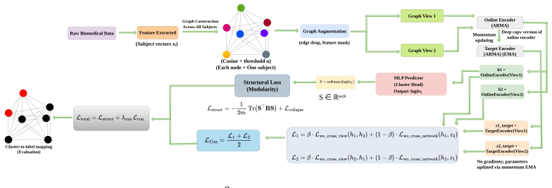
ARMA-C3 is a unified unsupervised and semi-supervised graph learning framework for node classification based on contrastive learning and graph-cut regularization to learn structurally meaningful and discriminative representations by modeling samples or images as graph nodes and exploiting inter-sample relationships.
What carries the argument
ARMA-C3: a contrastive ARMA convolutional graph model that combines autoregressive moving-average layers, contrastive objectives, and graph-cut regularization on a sample graph for node classification.
If this is right
- The framework produces competitive or superior binary classification accuracy on ADNI, NIFD, BreastMNIST, PneumoniaMNIST, and a liver ultrasound dataset.
- Performance remains strong under limited supervision and severe class imbalance.
- Representations generalize across different biomedical imaging modalities.
- The method outperforms classical clustering, state-of-the-art machine-learning models, and existing graph-based deep-learning approaches in multiple evaluation settings.
Where Pith is reading between the lines
- The same graph-construction step could be reused to surface patient subgroups that standard pipelines ignore.
- Because the method works with very few labels, it might reduce the annotation burden for new imaging modalities.
- Cross-modal robustness suggests the learned embeddings could transfer to non-imaging clinical variables such as genetics or lab values.
Load-bearing premise
Modeling biomedical samples as nodes in a graph and using their pairwise relationships will capture subject-level dependencies that ordinary feature-based classifiers miss.
What would settle it
On the same five datasets, if ARMA-C3 underperforms standard supervised baselines or simple clustering methods when only 10 percent of labels are available, the performance advantage claim would be refuted.
Figures
read the original abstract
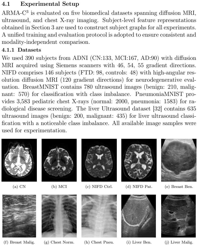
In biomedical and neurodegenerative disorders, accurate and early disease identification remains challenging due to the scarcity of labeled data and the complexity of imaging patterns. To address these challenges, we introduce ARMA-C3, a unified unsupervised and semi-supervised graph learning framework for node classification based on contrastive learning and graph-cut regularization to learn structurally meaningful and discriminative representations. By modeling samples or images as graph nodes and exploiting inter-sample relationships, the proposed framework captures subject-level dependencies that conventional machine learning methods typically overlook. We conduct extensive binary classification experiments across five clinically relevant datasets: the Alzheimer's Disease Neuroimaging Initiative (ADNI), the Neuroimaging in Frontotemporal Dementia (NIFD) dataset, and three medical imaging benchmarks (BreastMNIST, PneumoniaMNIST, and a liver ultrasound dataset). Experimental results demonstrate that ARMA-C3 achieves competitive and frequently superior performance compared to classical clustering techniques, state-of-the-art machine learning models, and existing graph-based deep learning approaches across multiple evaluation settings, particularly under limited supervision and severe class imbalance. The proposed framework further demonstrates robust representation learning and strong cross-modal generalization across diverse biomedical imaging modalities.
Editorial analysis
A structured set of objections, weighed in public.
Referee Report
Summary. The manuscript proposes ARMA-C3, a unified unsupervised and semi-supervised graph learning framework for node classification that combines contrastive learning with graph-cut regularization. Samples or images are modeled as graph nodes to exploit inter-sample relationships and capture subject-level dependencies overlooked by conventional ML methods. Extensive binary classification experiments are reported on five biomedical datasets (ADNI, NIFD, BreastMNIST, PneumoniaMNIST, and a liver ultrasound dataset), with claims of competitive or superior performance versus classical clustering, state-of-the-art ML models, and existing graph-based deep learning approaches, particularly under limited supervision and severe class imbalance, plus robust cross-modal generalization.
Significance. If the performance gains are attributable to the graph-based inter-sample modeling rather than the contrastive objective alone, the framework could offer a useful advance for representation learning in label-scarce and imbalanced biomedical imaging settings. The unified handling of unsupervised and semi-supervised regimes and reported cross-modal results would strengthen its potential impact if isolated and verified.
major comments (1)
- [Abstract and §4 (Experiments)] Abstract and §4 (Experiments): the central claim that 'modeling samples or images as graph nodes and exploiting inter-sample relationships' drives the reported gains (especially under limited supervision and imbalance) is load-bearing but unsupported by ablation. No experiments isolate the contribution of the graph construction or ARMA convolution by removing graph edges or replacing ARMA with a non-graph backbone while holding the contrastive objective fixed; without such controls the attribution to subject-level dependencies remains unverified.
Simulated Author's Rebuttal
We thank the referee for the constructive feedback. We address the major comment point by point below.
read point-by-point responses
-
Referee: [Abstract and §4 (Experiments)] Abstract and §4 (Experiments): the central claim that 'modeling samples or images as graph nodes and exploiting inter-sample relationships' drives the reported gains (especially under limited supervision and imbalance) is load-bearing but unsupported by ablation. No experiments isolate the contribution of the graph construction or ARMA convolution by removing graph edges or replacing ARMA with a non-graph backbone while holding the contrastive objective fixed; without such controls the attribution to subject-level dependencies remains unverified.
Authors: We agree that the manuscript lacks the precise ablations requested to isolate the graph construction and ARMA convolution while holding the contrastive objective fixed. The reported experiments include comparisons to non-graph baselines and other graph-based methods, but these do not control for the contrastive component in the exact manner described. In the revised manuscript we will add the suggested controls: a contrastive baseline using a non-graph backbone (standard CNN or MLP) with identical contrastive loss, and a graph version with edges removed (disconnected nodes). These results will be included in §4 to better attribute gains to inter-sample modeling under limited supervision and imbalance. revision: yes
Circularity Check
No significant circularity; empirical claims rest on external benchmarks
full rationale
The manuscript presents ARMA-C3 as an empirical graph-based contrastive framework evaluated on five external biomedical datasets (ADNI, NIFD, BreastMNIST, etc.). No equations, derivations, or parameter-fitting steps are described that reduce by construction to the inputs (no self-definitional relations, no fitted quantities renamed as predictions, no load-bearing self-citations). The modeling choice of samples as graph nodes is an explicit architectural decision whose contribution is asserted via performance comparisons against independent baselines; these comparisons are externally falsifiable and do not rely on internal re-labeling of the same quantities. The work is therefore self-contained against external benchmarks.
Axiom & Free-Parameter Ledger
Reference graph
Works this paper leans on
-
[1]
J. Yang, R. Shi, B. Ni, Medmnist: A large-scale lightweight benchmark for 2d and 3d biomedical image classification, Scientific Data 8 (2021) 1–10
2021
-
[2]
F. Caso, F. Agosta, M. Filippi, Insights into white matter damage in alzheimer’s disease: from postmortem to in vivo diffusion tensor mri studies, Neurodegenerative Diseases 16 (2016) 26–33. 37
2016
-
[3]
Acosta-Cabronero, P
J. Acosta-Cabronero, P. J. Nestor, Diffusion tensor imaging in alzheimer’s disease: insights into the limbic-diencephalic network and methodological considerations, Frontiers in aging neuroscience 6 (2014) 266
2014
-
[4]
Kantarci, M
K. Kantarci, M. Senjem, R. Avula, B. Zhang, A. Samikoglu, S. Weigand, S. Przybelski, H. Edmonson, P. Vemuri, D. Knopman, et al., Diffusion tensor imaging and cognitive function in older adults with no dementia, Neurology 77 (2011) 26–34
2011
-
[5]
Scarselli, M
F. Scarselli, M. Gori, A. C. Tsoi, M. Hagenbuchner, G. Monfardini, The graph neural network model, IEEE transactions on neural networks 20 (2008) 61–80
2008
-
[6]
C. R. Jack, D. S. Knopman, W. J. Jagust, R. C. Petersen, M. W. Weiner, P. S. Aisen, L. M. Shaw, P. Vemuri, H. J. Wiste, S. D. Weigand, et al., Tracking pathophysiological processes in alzheimer’s disease: an updated hypothetical model of dynamic biomarkers, The lancet neurol- ogy 12 (2013) 207–216
2013
-
[7]
Varol, A
E. Varol, A. Sotiras, C. Davatzikos, A. D. N. Initiative, et al., Hydra: Revealing heterogeneity of imaging and genetic patterns through a mul- tiple max-margin discriminative analysis framework, Neuroimage 145 (2017) 346–364
2017
-
[8]
D. I. Shuman, S. K. Narang, P. Frossard, A. Ortega, P. Vandergheynst, The emerging field of signal processing on graphs: Extending high- dimensional data analysis to networks and other irregular domains, IEEE Signal Processing Magazine 30 (2013) 83–98
2013
-
[9]
Ortega, P
A. Ortega, P. Frossard, J. Kovačević, J. M. F. Moura, P. Vandergheynst, Graph signal processing: Overview, challenges, and applications, Pro- ceedings of the IEEE 106 (2018) 808–828
2018
-
[10]
F. M. Bianchi, D. Grattarola, C. Alippi, Graph neural networks with convolutional arma filters, in: IEEE Transactions on Pattern Analysis and Machine Intelligence, IEEE, 2021
2021
- [11]
-
[12]
Y. Chen, L. Wu, M. Zaki, Iterative deep graph learning for graph neural networks: Better and robust node embeddings, Advances in neural information processing systems 33 (2020) 19314–19326
2020
-
[13]
J. Wen, E. Thibeau-Sutre, M. Diaz-Melo, J. Samper-González, A. Routier, S. Bottani, D. Dormont, S. Durrleman, N. Burgos, O. Col- liot, et al., Convolutional neural networks for classification of alzheimer’s disease: overview and reproducible evaluation, Medical image analysis 63 (2020) 101694
2020
-
[14]
Parisot, S
S. Parisot, S. I. Ktena, E. Ferrante, M. Lee, R. Guerrero, B. Glocker, D. Rueckert, Disease prediction using graph convolutional networks: ap- plication to autism spectrum disorder and alzheimer’s disease, Medical image analysis 48 (2018) 117–130
2018
-
[15]
J. MacQueen, Some methods for classification and analysis of multivari- ate observations, in: Proceedings of the Fifth Berkeley Symposium on Mathematical Statistics and Probability, volume 1, University of Cali- fornia Press, 1967, pp. 281–297
1967
- [16]
-
[17]
Y. Wang, X. Shen, Y. Yuan, Y. Du, M. Li, S. X. Hu, J. L. Crowley, D. Vaufreydaz, Tokencut: Segmenting objects in images and videos with self-supervised transformer and normalized cut, IEEE transactions on pattern analysis and machine intelligence 45 (2023) 15790–15801
2023
- [18]
-
[19]
Liu, J.Li, Y.Chen, R
Y. Liu, J.Li, Y.Chen, R. Wu, E. Wang, J. Zhou, S.Tian, S.Shen, X. Fu, C. Meng, et al., Revisiting modularity maximization for graph cluster- ing: A contrastive learning perspective, in: Proceedings of the 30th ACM SIGKDD Conference on Knowledge Discovery and Data Mining, 2024, pp. 1968–1979. 39
2024
-
[20]
T. N. Kipf, M. Welling, Semi-supervised classification with graph con- volutional networks, in: International Conference on Learning Repre- sentations, 2017
2017
-
[21]
Veličković, G
P. Veličković, G. Cucurull, A. Casanova, A. Romero, P. Lio, Y. Bengio, Graph attention networks, in: International Conference on Learning Representations, 2018
2018
-
[22]
X. Luo, G. Dong, J. Wu, A. Beheshti, J. Yang, S. Xue, An interpretable brain graph contrastive learning framework for brain disorder analy- sis, in: Proceedings of the 17th ACM International Conference on Web Search and Data Mining, 2024, pp. 1074–1077
2024
-
[23]
D. Chen, L. Yao, M. Liu, Z. Shen, Y. Hu, Z. Song, Q. Wang, L. Zhang, Self-supervised learning with adaptive graph structure and function rep- resentation for cross-dataset brain disorder diagnosis, in: International Conference on Medical Image Computing and Computer-Assisted Inter- vention, Springer, 2024, pp. 612–622
2024
-
[24]
W. Meng, R. Inampudi, X. Zhang, J. Xu, Y. Huang, M. Xie, J. Bian, R. Yin, An interpretable population graph network to identify rapid progression of alzheimer’s disease using uk biobank, medRxiv (2024)
2024
-
[25]
Gasteiger, A
J. Gasteiger, A. Bojchevski, S. Günnemann, Predict then propagate: Graph neural networks meet personalized pagerank, in: International Conference on Learning Representations, 2018
2018
- [26]
-
[27]
P. Veličković, W. Fedus, W. L. Hamilton, P. Liò, Y. Bengio, R. D. Hjelm, Deep graph infomax, International Conference on Learning Representations (2019). URL:https://arxiv.org/abs/1809.10341
work page internal anchor Pith review Pith/arXiv arXiv 2019
-
[28]
Distilling the Knowledge in a Neural Network
G. Hinton, O. Vinyals, J. Dean, Distilling the knowledge in a neural network, arXiv preprint arXiv:1503.02531 (2015)
work page internal anchor Pith review Pith/arXiv arXiv 2015
-
[29]
Tsitsulin, J
A. Tsitsulin, J. Palowitch, B. Perozzi, E. Müller, Graph clustering with graph neural networks, Journal of Machine Learning Research 24 (2023) 1–21. 40
2023
-
[30]
Caron, H
M. Caron, H. Touvron, I. Misra, H. Jégou, J. Mairal, P. Bojanowski, A. Joulin, Emerging properties in self-supervised vision transformers, in: Proceedings of the IEEE/CVF international conference on computer vision, 2021, pp. 9650–9660
2021
-
[31]
K. He, X. Zhang, S. Ren, J. Sun, Deep residual learning for image recognition, CVPR (2016)
2016
-
[32]
Y. Xu, B. Zheng, X. Liu, T. Wu, J. Ju, S. Wang, Y. Lian, H. Zhang, T. Liang, Y. Sang, R. Jiang, G. Wang, J. Ren, T. Chen, Annotated ul- trasound liver images, 2022. URL:https://doi.org/10.5281/zenodo. 7272660. doi:10.5281/zenodo.7272660
-
[33]
Di Paola, G
M. Di Paola, G. Spalletta, C. Caltagirone, In vivo structural neu- roanatomy of corpus callosum in alzheimer’s disease and mild cogni- tive impairment using different mri techniques: a review, Journal of Alzheimer’s disease 20 (2010) 67–95
2010
-
[34]
Walterfang, E
M. Walterfang, E. Luders, J. C. Looi, P. Rajagopalan, D. Velakoulis, P. M. Thompson, O. Lindberg, P. Östberg, L. E. Nordin, L. Svensson, et al., Shape analysis of the corpus callosum in alzheimer’s disease and frontotemporal lobar degeneration subtypes, Journal of Alzheimer’s Disease 40 (2014) 897–906
2014
-
[35]
K. Hua, J. Zhang, S. Wakana, H. Jiang, X. Li, D. S. Reich, P. A. Cal- abresi, J. J. Pekar, P. C. van Zijl, S. Mori, Tract probability maps in stereotaxic spaces: analyses of white matter anatomy and tract-specific quantification, Neuroimage 39 (2008) 336–347
2008
-
[36]
Oishi, K
K. Oishi, K. Akhter, M. Mielke, C. Ceritoglu, J. Zhang, H. Jiang, X. Li, L. Younes, M. I. Miller, P. C. van Zijl, et al., Multi-modal mri analysis with disease-specific spatial filtering: initial testing to predict mild cog- nitive impairment patients who convert to alzheimer’s disease, Frontiers in neurology 2 (2011) 54
2011
-
[37]
M. A. Nowrangi, P. B. Rosenberg, The fornix in mild cognitive impair- ment and alzheimer’s disease, Frontiers in aging neuroscience 7 (2015) 1
2015
-
[38]
Kantarci, C
K. Kantarci, C. G. Schwarz, R. I. Reid, S. A. Przybelski, T. G. Lesnick, S. M. Zuk, M. L. Senjem, J. L. Gunter, V. Lowe, M. M. Machulda, 41 et al., White matter integrity determined with diffusion tensor imag- ing in older adults without dementia: influence of amyloid load and neurodegeneration, JAMA neurology 71 (2014) 1547–1554
2014
-
[39]
S. J. Teipel, M. J. Grothe, M. Filippi, A. Fellgiebel, M. Dyrba, G. B. Frisoni, T.Meindl, A.L.Bokde, H.Hampel, S.Klöppel, etal., Fractional anisotropy changes in alzheimer’s disease depend on the underlying fiber tract architecture: a multiparametric dti study using joint independent component analysis, Journal of Alzheimer’s Disease 41 (2014) 69–83
2014
-
[40]
Douaud, S
G. Douaud, S. Jbabdi, T. E. Behrens, R. A. Menke, A. Gass, A. U. Monsch, A. Rao, B. Whitcher, G. Kindlmann, P. M. Matthews, et al., Dti measures in crossing-fibre areas: increased diffusion anisotropy re- veals early white matter alteration in mci and mild alzheimer’s disease, Neuroimage 55 (2011) 880–890
2011
-
[41]
D. J. Madden, J. Spaniol, M. C. Costello, B. Bucur, L. E. White, R. Cabeza, S. W. Davis, N. A. Dennis, J. M. Provenzale, S. A. Huettel, Cerebral white matter integrity mediates adult age differences in cogni- tive performance, Journal of cognitive neuroscience 21 (2008) 289–302
2008
-
[42]
M. M. Mielke, N. A. Kozauer, K. C. G. Chan, M. George, J. Toroney, M. Zerrate, K. Bandeen-Roche, M.-C. Wang, J. Pekar, S. Mori, et al., Regionally-specificdiffusiontensorimaginginmildcognitiveimpairment and alzheimer’s disease, Neuroimage 46 (2009) 47–55
2009
-
[43]
Villain, M
N. Villain, M. Fouquet, J.-C. Baron, F. Mézenge, B. Landeau, V. de La Sayette, F. Viader, F. Eustache, B. Desgranges, G. Chételat, Sequential relationships between grey matter and white matter atrophy and brain metabolic abnormalities in early alzheimer’s disease, Brain 133 (2010) 3301–3314
2010
-
[44]
Zhuang, W
L. Zhuang, W. Wen, W. Zhu, J. Trollor, N. Kochan, J. Crawford, S. Rep- permund, H. Brodaty, P. Sachdev, White matter integrity in mild cog- nitive impairment: a tract-based spatial statistics study, Neuroimage 53 (2010) 16–25
2010
-
[45]
Zhang, N
Y. Zhang, N. Schuff, G.-H. Jahng, W. Bayne, S. Mori, L. Schad, S. Mueller, A.-T. Du, J. Kramer, K. Yaffe, et al., Diffusion tensor 42 imaging of cingulum fibers in mild cognitive impairment and alzheimer disease, Neurology 68 (2007) 13–19
2007
-
[46]
Yasmin, S
H. Yasmin, S. Aoki, O. Abe, Y. Nakata, N. Hayashi, Y. Masutani, M. Goto, K. Ohtomo, Tract-specific analysis of white matter pathways in healthy subjects: a pilot study using diffusion tensor mri, Neurora- diology 51 (2009) 831–840
2009
-
[47]
Garyfallidis, M
E. Garyfallidis, M. Brett, B. Amirbekian, A. Rokem, S. Van Der Walt, M. Descoteaux, I. Nimmo-Smith, D. Contributors, Dipy, a library for the analysis of diffusion mri data, Frontiers in neuroinformatics 8 (2014) 8
2014
-
[48]
F. M. Bianchi, D. Grattarola, C. Alippi, Spectral clustering with graph neural networks for graph pooling, in: International conference on ma- chine learning, PMLR, 2020, pp. 874–883
2020
-
[49]
Cortes, V
C. Cortes, V. Vapnik, Support-vector networks, Machine learning 20 (1995) 273–297
1995
-
[50]
Chen, Xgboost: A scalable tree boosting system, Cornell University (2016)
T. Chen, Xgboost: A scalable tree boosting system, Cornell University (2016)
2016
-
[51]
D. E. Rumelhart, G. E. Hinton, R. J. Williams, Learning representations by back-propagating errors, nature 323 (1986) 533–536
1986
-
[52]
Breiman, Random forests, Machine learning 45 (2001) 5–32
L. Breiman, Random forests, Machine learning 45 (2001) 5–32. 43
2001
discussion (0)
Sign in with ORCID, Apple, or X to comment. Anyone can read and Pith papers without signing in.