High-temperature instability of artificial cuprorivaite: a study using thermal analysis, X-ray powder diffractometry and polarized light microscopy
Pith reviewed 2026-06-29 21:27 UTC · model grok-4.3
The pith
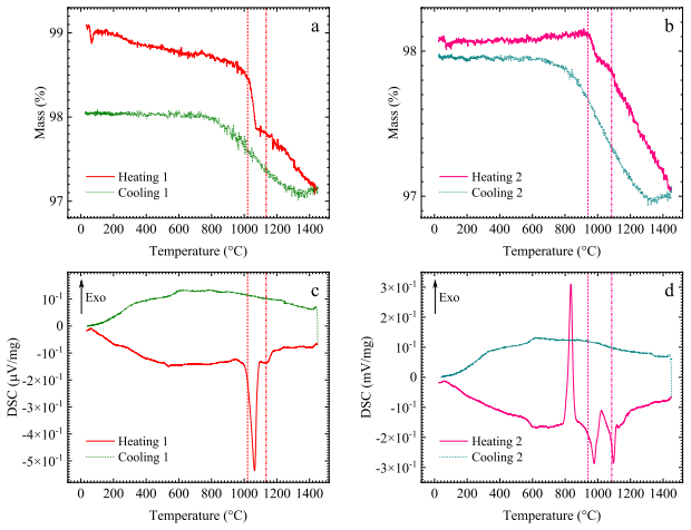
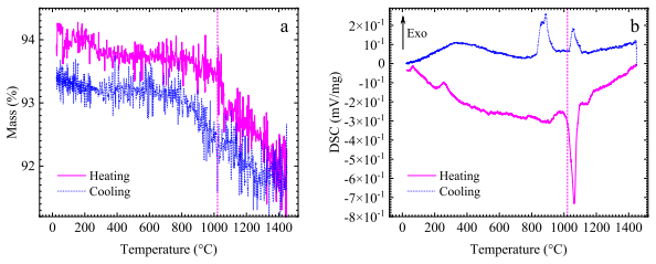
CaCuSi4O10 decomposes irreversibly by incongruent melting starting at about 1020°C.
A machine-rendered reading of the paper's core claim, the machinery that carries it, and where it could break.
Core claim
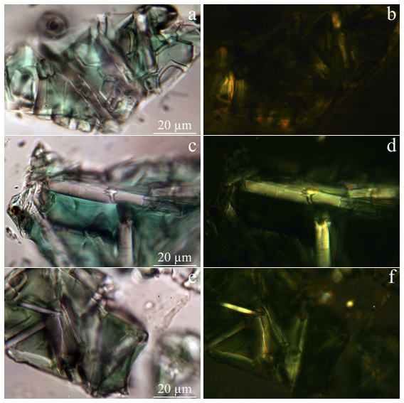
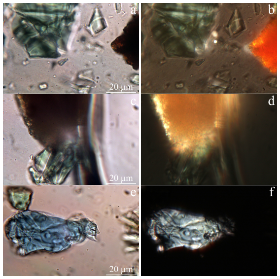
CaCuSi4O10 powder decomposes by incongruent melting starting at about 1020°C, shown by an endothermic DSC peak with minimum at 1064.4°C. The decomposition is irreversible; cyclic annealings to 1450°C do not re-synthesize it. After two annealings to 1450°C, it transforms into acicular monoclinic tridymite crystals fused with green glass of composition CuO-Cu2O-CaO-SiO2, with tridymite to glass weight ratio about 12:13.
What carries the argument
Incongruent melting process of CaCuSi4O10 tracked via differential scanning calorimetry combined with post-annealing X-ray powder diffractometry and polarized light microscopy for phase identification.
If this is right
- Decomposition is irreversible and subsequent cyclic annealings up to 1450°C do not cause re-synthesis of CaCuSi4O10.
- The material transforms into acicular crystals of monoclinic tridymite fused with green glass of CuO-Cu2O-CaO-SiO2 composition.
- The weight ratio of tridymite to glass is about 12:13 after two successive annealings to 1450°C.
- The process begins at a temperature of about 1020°C with the DSC peak minimum at 1064.4°C.
Where Pith is reading between the lines
- This establishes a firm upper temperature limit beyond which cuprorivaite cannot be maintained or recovered by thermal means alone.
- The specific glass composition produced may serve as a reference point for studying related copper silicate systems.
- Any application involving heating above 1020°C must account for permanent conversion to the two-phase mixture.
Load-bearing premise
The observed DSC peak and post-heating XRD and microscopy patterns unambiguously identify incongruent melting and the exact product phases without contributions from sample impurities, atmosphere interactions, or instrumental artifacts.
What would settle it
Re-detection of CaCuSi4O10 in XRD patterns or microscopy after cooling from 1450°C would contradict the claim of irreversible decomposition.
Figures
read the original abstract
CaCuSi$_4$O$_{10}$ powder was studied by differential scanning calorimetry and thermogravimetry methods in the range from room temperature to 1450$\,^{\circ}$C at heating and cooling rates of 20$\,^{\circ}$C/min. The process of decomposition of cuprorivaite, the composition and transformations of its decomposition products during successive heat treatments were also studied by powder X-ray diffraction and polarization optical microscopy techniques. It was found that CaCuSi$_4$O$_{10}$ starts to decompose by incongruent melting at a temperature of about 1020$\,^{\circ}$C, with the minimum of the endothermic DSC peak associated with this process being at 1064.4$\,^{\circ}$C. CaCuSi$_4$O$_{10}$ decomposes irreversibly and subsequent cyclic annealings up to a temperature of 1450$\,^{\circ}$C at heating and cooling rates of 20$\,^{\circ}$C/min do not cause its re-synthesis. CaCuSi$_4$O$_{10}$ transforms into a two-phase system consisting of acicular crystals of monoclinic tridymite fused with green glass with the composition CuO$\,-\,$Cu$_2$O$\,-\,$CaO$\,-\,$SiO$_2$, with the weight ratio of tridymite to glass being about $12:13$, as a result of two successive annealings up to the temperature of 1450$\,^{\circ}$C.
Editorial analysis
A structured set of objections, weighed in public.
Referee Report
Summary. The manuscript reports a multi-technique experimental study of synthetic CaCuSi4O10 (cuprorivaite) powder using DSC/TG from room temperature to 1450°C at 20°C/min heating/cooling rates, combined with post-anneal powder XRD and polarized light microscopy. The central claims are that the compound undergoes irreversible incongruent melting starting at ~1020°C (DSC endotherm minimum at 1064.4°C), transforms into monoclinic tridymite plus a CuO-Cu2O-CaO-SiO2 glass (weight ratio ~12:13 after two anneals), and shows no re-synthesis upon thermal cycling.
Significance. If the phase assignments and temperature values hold after addressing experimental controls, the work supplies new data on the high-temperature limits of cuprorivaite relevant to pigment synthesis and materials stability. The orthogonal use of thermal analysis, diffraction, and optical microscopy is a positive feature for phase identification, though the lack of quantitative uncertainties and controls limits the strength of the reported onset temperature and irreversibility conclusions.
major comments (3)
- [Thermal analysis] Abstract and thermal analysis description: the DSC onset of ~1020°C and peak minimum of 1064.4°C are presented as the decomposition temperature without reported calibration against standards, replicate runs, error bars, or raw curve data, which directly supports the precise numerical claim but leaves it only moderately substantiated.
- [Experimental methods] Experimental methods (implied by the DSC/TG description): no atmosphere (air, inert, or controlled pO2) is specified for the thermal runs. The reported glass composition involves mixed Cu valence states, so uncontrolled redox or atmosphere effects could contribute to the endotherm and undermine the assignment to incongruent melting of pure CaCuSi4O10.
- [Phase identification] Phase identification and results: the decomposition products and 12:13 tridymite:glass ratio are identified from post-anneal XRD and microscopy, but no pre- or post-heating chemical analysis (e.g., EDS or ICP) is described to confirm starting-material purity or exclude impurity contributions (excess SiO2 or CuO) that could produce overlapping thermal events.
minor comments (2)
- The abstract states the weight ratio of 12:13 but does not indicate how it was quantified (integrated XRD intensities, image analysis, or mass balance), nor are uncertainties provided.
- Sample synthesis and initial purity characterization of the CaCuSi4O10 powder are not detailed, which is needed to support the claim that observed signals arise solely from the target phase.
Simulated Author's Rebuttal
We thank the referee for the constructive review and the opportunity to clarify and strengthen the manuscript. We respond point-by-point to the major comments below and indicate where revisions will be made.
read point-by-point responses
-
Referee: [Thermal analysis] Abstract and thermal analysis description: the DSC onset of ~1020°C and peak minimum of 1064.4°C are presented as the decomposition temperature without reported calibration against standards, replicate runs, error bars, or raw curve data, which directly supports the precise numerical claim but leaves it only moderately substantiated.
Authors: We agree that the manuscript would benefit from explicit reporting of calibration and uncertainties. The DSC/TG instrument was calibrated with standard reference materials before the experiments; we will add this detail to the methods section along with the onset determination procedure (baseline-tangent intersection). An estimated uncertainty of ±5 °C will be stated based on instrument specifications and heating rate. Replicate runs were not performed due to limited synthetic sample quantity, which we will note as a limitation. Raw DSC/TG curves will be supplied as supplementary material. revision: yes
-
Referee: [Experimental methods] Experimental methods (implied by the DSC/TG description): no atmosphere (air, inert, or controlled pO2) is specified for the thermal runs. The reported glass composition involves mixed Cu valence states, so uncontrolled redox or atmosphere effects could contribute to the endotherm and undermine the assignment to incongruent melting of pure CaCuSi4O10.
Authors: The measurements were performed in static air, the default condition for the instrument. We will revise the experimental section to state this explicitly. The mixed Cu valence in the resulting glass is consistent with the observed decomposition products and does not alter the phase assignment supported by post-anneal XRD and microscopy; however, we acknowledge that specifying the atmosphere removes ambiguity and will do so. revision: yes
-
Referee: [Phase identification] Phase identification and results: the decomposition products and 12:13 tridymite:glass ratio are identified from post-anneal XRD and microscopy, but no pre- or post-heating chemical analysis (e.g., EDS or ICP) is described to confirm starting-material purity or exclude impurity contributions (excess SiO2 or CuO) that could produce overlapping thermal events.
Authors: The starting powder was confirmed as phase-pure cuprorivaite by initial XRD, and decomposition products were identified by post-anneal XRD peak matching plus polarized-light microscopy morphology. No EDS or ICP data were collected. We will add a sentence noting reliance on diffraction and optical methods without bulk chemical verification and that the 12:13 ratio is an estimate from integrated XRD intensities. This is a fair observation; the multi-technique approach still supports the reported phase assemblage. revision: partial
Circularity Check
No circularity: pure experimental measurement report with no derivations or self-referential fits
full rationale
The paper is a straightforward experimental study reporting DSC/TG curves, post-anneal XRD patterns, and optical microscopy observations on CaCuSi4O10 powder. No equations, fitted parameters, predictions derived from models, or self-citations appear in the provided text. The central claims (onset of decomposition ~1020°C, irreversibility, phase products) are presented as direct results of the measurements and standard phase identification, without any reduction to inputs by construction or load-bearing self-referential steps. This is the expected outcome for an empirical thermal analysis paper.
Axiom & Free-Parameter Ledger
axioms (2)
- domain assumption DSC endothermic peaks reliably indicate incongruent melting onset
- domain assumption XRD and polarized-light microscopy unambiguously identify the final crystalline and glassy phases
Reference graph
Works this paper leans on
-
[1]
CaCuSi 4O10, in: J
Cuprorivaite. CaCuSi 4O10, in: J. W. Anthony, R. A. Bideaux, K. W. Bladh, M. C. Nichols (Eds.), Handbook of Mineralogy, V ol. II, Mineralogical Society of Amer- ica, Chantilly, V A, USA, 2003. URL http://www.handbookofmineralogy.org/pdfs/cuprorivaite.pdf
2003
-
[2]
URL https://www.mindat.org/min-1189.html
Cuprorivaite, Hudson Institute of Mineralogy, Keswick, V A, USA, dba Min- dat.org (2019). URL https://www.mindat.org/min-1189.html
2019
-
[3]
L. Fabbrizzi, Painting conditioned by chemistry: the case of Egyptian and ultramarine blue pigments, ChemTexts 11 (9) (2025) 1–20.doi:10.1007/ BF00376668. URL https://doi.org/10.1007/s40828-025-00204-8
-
[4]
Riederer, Egyptian blue, in: E
J. Riederer, Egyptian blue, in: E. W. FitzHugh (Ed.), Artists' Pigments: A Handbook of their History and Characteristics, V ol. 3, National Gallery of Art, Washington, USA, 2012, pp. 23–45. URL https://www.nga.gov/content/dam/ngaweb/research/publications/pdfs/ artists-pigments-vol3.pdf
2012
-
[5]
A. P. Laurie, The Materials of the Painter’s Craft in Europe and Egypt: from Earliest Times to the End of the XVIIth Century, with Some Account of Their Preparation and Use, T. N. Foulis, London & Edinburgh, UK, 1910. URL https://archive.org/details/cu31924016809927/page/n12/mode/2up
1910
-
[6]
H. Jaksch, W. Seipel, K. L. Weiner, A. E. Goresy, Egyptian blue – Cuprorivaite a window to ancient Egyptian technology, Naturwiss. 70 (11) (1983) 525–535. doi:10.1007/BF00376668. URL https://doi.org/10.1007/BF00376668
-
[7]
G. D. Hatton, A. J. Shortland, M. S. Tite, The production technology of Egyptian blue and green frits from second millennium BC Egypt and Mesopotamia, J. Ar- 28 chaeol. Sci. 35 (6) (2008) 1591–1604.doi:10.1016/j.jas.2007.11.008. URL https://doi.org/10.1016/j.jas.2007.11.008
-
[8]
E. Aloiz, J. G. Douglas, A. Nagel, Painted plaster and glazed brick fragments from Achaemenid Pasargadae and Persepolis, Iran, Heritage Science 4 (1) (2016) 3.doi:10.1186/s40494-016-0072-7. URL https://doi.org/10.1186/s40494-016-0072-7
-
[9]
N. Easthaugh, V . Walsh, T. Chaplin, R. Siddall, Pigment Compendium: A Dic- tionary of Historical Pigments, Routledge, London, UK, 2004.doi:10.4324/ 9780080473765. URL https://doi.org/10.4324/9780080473765
-
[10]
Couleurs Fines et Matériel pour la Peinture a l’Huile, Lefranc, Paris, France, 1930, Ch
Fabrique de Couleurs & Vernis. Couleurs Fines et Matériel pour la Peinture a l’Huile, Lefranc, Paris, France, 1930, Ch. Couleurs extra-fines en tubes, p. 10
1930
-
[11]
S. A. Pisareva, I. N. Shibanova, I. F. Kadikova, E. A. Morozova, T. V . Yuryeva, I. B. Afanasyev, V . A. Yuryev, Identification of CaCuSi4O10 (Egyptian blue) in the “Birch. Spring” painting by Robert Falk (1907) using photoluminescence, J. Cult. Herit. (2021).doi:10.1016/j.culher.2021.05.005. URL https://doi.org/10.1016/j.culher.2021.05.005
-
[12]
Berke, The invention of blue and purple pigments in ancient times, Chem
H. Berke, The invention of blue and purple pigments in ancient times, Chem. Soc. Rev. 36 (2007) 15–30.doi:10.1039/B606268G. URL http://doi.org/10.1039/B606268G
-
[13]
Panagiotaki, M
M. Panagiotaki, M. S. Tite, Y . Maniatis, Egyptian Blue in Egypt and beyond: the Aegean and the Near East, in: P. Kousoulis, N. Lazaridis (Eds.), Proceedings of the Tenth International Congress of Egyptologists, University of the Aegean, Rhodes 22–29 May 2008, V ol. II of Orientalia Lovaniensia Analecta 241, Peeters, Leuven – Paris – Bristol, CT, 2015, pp...
2008
-
[14]
A. K. Marketou, The pigment production site of the ancient agora of Kos (Greece): Revisiting the material evidence, Thiasos 8.1 (2019) 61–80. 29 URL http://www.thiasos.eu/wp-content/uploads/2019/06/02-2019-Marketou_ 20190610.pdf
2019
-
[15]
Y . I. Grenberg, S. A. Pisareva, A. B. Levstein, The study of the paint layer compo- sition in the samples of mural paintings of Erebuni, Preliminary report on research project (topic 7.3), The All-Union Scientific-Research Institute for Restoration, Moscow, USSR, in Russian (1982).doi:10.13140/RG.2.2.25685.17129. URL https://doi.org/10.13140/RG.2.2.25685.17129
-
[16]
S. A. Pisareva, V . N. Kireeva, The study of the paint layer composition in the samples of mural paintings of Erebuni, in: Y . I. Grenberg (Ed.), Investigations of monumental painting of Armenia and Novgorod, no. 6 in Culture and Arts in the USSR. Series: Restoration of Historical and Cultural Monuments. Express Information, The Lenin State Library of the...
-
[17]
G. Dardeniz, E. Akalßn, Coloring Urartu: A local variety of Egyptian blue in Anatolia, Paléorient 51 (2025) 89–98.doi:10.4000/15apg. URL https://doi.org/10.4000/15apg
-
[18]
G. M. Ingo, A. Çilingiro ˘glu, G. D. Carlo, A. Batmaz, T. D. Caro, C. Riccucci, E. I. Parisi, F. Faraldi, Egyptian Blue cakes from the Ayanis fortress (Eastern Anatolia, Turkey): micro-chemical and -structural investigations for the identification of manufacturing process and provenance, J. Archaeol. Sci. 40 (12) (2013) 4283– 4290.doi:10.1016/j.jas.2013.0...
-
[19]
Ö. Ormanci, Non-destructive characterization of Egyptian Blue cakes and wall painting fragments from the east of Lake Van, Turkey, Spectrochim. Acta A 229 30 (2020) 117889.doi:10.1016/j.saa.2019.117889. URL https://doi.org/10.1016/j.saa.2019.117889
-
[20]
R. Linn, E. H. Cline, A. Yasur-Landau, The advantages of visible induced lumi- nescence technique for the investigation of Aegean-style wall painting. A case study from Tel Kabri, Israel, in: J. Becker, J. Jungfleisch, C. von Rüden (Eds.), Tracing Technoscapes. The Production of Bronze Age Wall Paintings in the East- ern Mediterranean, Sidestone Press, Le...
2018
-
[21]
M. Ganio, J. Salvant, J. Williams, L. Lee, O. Cossairt, M. Walton, Investi- gating the use of Egyptian blue in Roman Egyptian portraits and panels from Tebtunis, Egypt, Appl. Phys. A 121 (3) (2015) 813–821.doi:10.1007/ s00339-015-9424-5. URL https://doi.org/10.1007/s00339-015-9424-5
-
[22]
J. Salvant, J. Williams, M. Ganio, F. Casadio, C. Daher, K. Sutherland, L. Mon- ico, F. Vanmeert, S. De Meyer, K. Janssens, C. Cartwright, M. Walton, A Roman Egyptian painting workshop: Technical investigation of the portraits from Tebtu- nis, Egypt, Archaeometry 60 (4) (2018) 815–833.doi:10.1111/arcm.12351. URL https://doi.org/10.1111/arcm.12351
-
[23]
G. E. Veresotskaya, Restoration of a fragment of the “Horsemen” archae- ological painting from Old Nisa, in: Artistic Heritage. Conservation, Re- search, Restoration, no. 23(53), The State Research Institute for Restoration, Moscow, Russia, 2006, pp. 166–174, 190, 191, Also in: ARTconservation, https://web.archive.org/web/20121111061508/http://art-con.ru/...
-
[24]
A. M. Rosenqvist, Analyser an skerd og skjold fra Bø-funnet (Analysis of sword and shield from the Bø-find), Viking 23 (1959) 29–34. URL https://www.duo.uio.no/bitstream/handle/10852/37585/1959-vol-23.pdf
1959
-
[25]
M. G. Canti, J. L. Heathcote, Microscopic Egyptian blue (synthetic cuprorivaite) from sediments at two archaeological sites in West Central England, J. Archaeol. 31 Sci. 29 (8) (2002) 831–836.doi:10.1006/jasc.2001.0717. URL http://doi.org/10.1006/jasc.2001.0717
-
[26]
M. Nicola, L. M. Seymour, M. Aceto, E. Priola, R. Gobetto, A. Masic, Late production of Egyptian blue: synthesis from brass and its character- istics, Archaeol. Anthrop. Sci. 11 (10) (2019) 5377–5392.doi:10.1007/ s12520-019-00873-w. URL https://doi.org/10.1007/s12520-019-00873-w
-
[27]
Review of energy-efficient train control and timetabling
M. Nicola, M. Aceto, V . Gheroldi, R. Gobetto, G. Chiari, Egyptian blue in the Castelseprio mural painting cycle. Imaging and evidence of a non-traditional manufacture, J. Archaeol. Sci. Rep. 19 (2018) 465–475.doi:10.1016/j. jasrep.2018.03.031. URL https://doi.org/10.1016/j.jasrep.2018.03.031
work page doi:10.1016/j 2018
-
[28]
A. Lluveras, A. Torrents, P. Girález, M. Vendrell-Saz, Evidence for the use of Egyptian blue in an 11th century mural altarpiece by SEM–EDS, FTIR and SR XRD (church of Sant Pere, Terrassa, Spain), Archaeometry 52 (2) (2010) 308–319.doi:10.1111/j.1475-4754.2009.00481.x. URL https://doi.org//10.1111/j.1475-4754.2009.00481.x
-
[29]
J. Bredal-Jørgensen, J. Sanyova, V . Rask, M. L. Sargent, R. H. Therkildsen, Striking presence of Egyptian blue identified in a painting by Giovanni Bat- tista Benvenuto from 1524, Anal. Bioanal. Chem. 401 (4) (2011) 1433–1439. doi:10.1007/s00216-011-5140-y. URL https://doi.org/10.1007/s00216-011-5140-y
-
[30]
A. Sgamellotti, C. Anselmi, An evergreen blue. Spectroscopic properties of Egyp- tian blue from pyramids to Raphael, and beyond, Inorganica Chim. Acta 530 (2022) 120699.doi:10.1016/j.ica.2021.120699. URL https://doi.org/10.1016/j.ica.2021.120699
-
[31]
S. M. Borisov, C. Würth, U. Resch-Genger, I. Klimant, New life of ancient pig- ments: Application in high-performance optical sensing materials, Anal. Chem. 32 85 (19) (2013) 9371–9377.doi:10.1021/ac402275g. URL https://doi.org/10.1021/ac402275g
-
[32]
G. Selvaggio, S. Kruss, Preparation, properties and applications of near-infrared fluorescent silicate nanosheets, Nanoscale 14 (2022) 9553–9575.doi:10.1039/ D2NR02967G. URL https://doi.org/10.1039/D2NR02967G
-
[33]
P. Sobik, O. Jeremiasz, P. Nowak, A. Sala, B. Pawłowski, G. Kulesza-Matlak, A. Sypie´n, K. Drabczyk, Towards efficient luminescent solar energy concentrator using cuprorivaite infrared phosphor (CaCuSi4O10)—effect of dispersing method on photoluminescence intensity, Materials 14 (14) (2021) 3952.doi:10.3390/ ma14143952. URL https://doi.org/10.3390/ma14143952
-
[34]
J. L. Tyler, R. L. Sacci, J. Ning, D. R. Mullins, K. Liang, J. Nanda, J. Sun, M. Naguib, Egyptian blue: from pigment to battery electrodes, RSC Adv. 11 (32) (2021) 19885–19889.doi:10.1039/D1RA00956G. URL http://dx.doi.org/10.1039/D1RA00956G
-
[35]
G. Selvaggio, A. Chizhik, R. Nißler, l. Kuhlemann, D. Meyer, L. Vuong, H. Preiß, N. Herrmann, F. A. Mann, Z. Lv, T. A. Oswald, A. Spreinat, L. Erpenbeck, J. Großhans, V . Karius, A. Janshoff, J. Pablo Giraldo, S. Kruss, Exfoliated near infrared fluorescent silicate nanosheets for (bio)photonics, Nat. Commun. 11 (1) (2020) 1495.doi:10.1038/s41467-020-15299...
-
[36]
G. Selvaggio, N. Herrmann, B. Hill, R. Dervi¸ so˘glu, S. Jung, M. Weitzel, M. Di- narvand, D. Stalke, L. Andreas, S. Kruss, Covalently functionalized Egyptian blue nanosheets for near-infrared bioimaging, ACS Appl. Bio Mater. (2022). doi:10.1021/acsabm.2c00872. URL https://doi.org/10.1021/acsabm.2c00872
-
[37]
C. He, C. Dong, L. Yu, Y . Chen, Y . Hao, Ultrathin 2D inorganic ancient pigment decorated 3D-printing scaffold enables photonic hyperthermia of osteosarcoma in 33 NIR-II biowindow and concurrently augments bone regeneration, Adv. Sci. 8 (19) (2021) 2101739.doi:10.1002/advs.202101739. URL https://doi.org/10.1002/advs.202101739
-
[38]
B. Errington, G. Lawson, S. W. Lewis, G. D. Smith, Micronised egyptian blue pigment: A novel near-infrared luminescent fingerprint dusting powder, Dyes Pigm. 132 (2016) 310–315.doi:10.1016/j.dyepig.2016.05.008. URL https://doi.org/10.1016/j.dyepig.2016.05.008
-
[39]
S. Shahbazi, J. V . Goodpaster, G. D. Smith, T. Becker, S. W. Lewis, Studies into exfoliation and coating of egyptian blue in methanol for application to the detection of latent fingermarks, Sci. Justice 62 (4) (2022) 455–460.doi:10. 1016/j.scijus.2022.05.004. URL https://doi.org/10.1016/j.scijus.2022.05.004
-
[40]
S. Shahbazi, J. V . Goodpaster, G. D. Smith, T. Becker, S. W. Lewis, Prepara- tion, characterization, and application of a lipophilic coated exfoliated Egyptian blue for near-infrared luminescent latent fingermark detection, Forensic Chem. 18 (2020) 100208.doi:10.1016/j.forc.2019.100208. URL https://doi.org/10.1016/j.forc.2019.100208
-
[41]
Y . Chen, M. Kan, Q. Sun, P. Jena, Structure and properties of egyptian blue mono- layer family: XCuSi4O10 (X=Ca, Sr, and Ba), J. Phys. Chem. Lett. 7 (3) (2016) 399–405.doi:10.1021/acs.jpclett.5b02770. URL https://doi.org/10.1021/acs.jpclett.5b02770
-
[42]
D. Johnson-McDaniel, C. A. Barrett, A. Sharafi, T. T. Salguero, Nanoscience of an ancient pigment, J. Am. Chem. Soc. 135 (2013) 1677–1679.doi:10.1021/ ja310587c. URL https://doi.org/10.1021/ja310587c
-
[43]
G. Pozza, D. Ajò, G. Chiari, F. D. Zuane, M. Favaro, Photoluminescence of the inorganic pigments Egyptian blue, Han blue and Han purple, J. Cult. Herit. 1 (4) (2000) 393–398.doi:10.1016/S1296-2074(00)01095-5. URL https://doi.org/10.1016/S1296-2074(00)01095-5 34
-
[44]
G. Accorsi, G. Verri, M. Bolognesi, N. Armaroli, C. Clementi, C. Miliani, A. Romani, The exceptional near-infrared luminescence properties of cuprori- vaite (Egyptian blue), Chem. Commun. 23 (2009) 3392–3394.doi:10.1039/ B902563D. URL https://doi.org/10.1039/B902563D
-
[45]
P. Berdahl, S. K. Boocock, G. C.-Y . Chan, S. S. Chen, R. M. Levinson, M. A. Zalich, High quantum yield of the Egyptian blue family of infrared phosphors (MCuSi4O10, M=Ca, Sr, Ba), J. Appl. Phys. 123 (19) (2018) 193103.doi: 10.1063/1.5019808. URL https://doi.org/10.1063/1.5019808
-
[46]
Y .-J. Li, S. Ye, C.-H. Wang, X.-M. Wang, Q.-Y . Zhang, Temperature-dependent near-infrared emission of highly concentrated Cu 2+ in CaCuSi4O10 phosphor, J. Mater. Chem. C 2 (48) (2014) 10395–10402.doi:10.1039/C4TC01966K. URL http://dx.doi.org/10.1039/C4TC01966K
-
[47]
L. Binet, J. Lizion, S. Bertaina, D. Gourier, Magnetic and new optical properties in the UV−visible range of the Egyptian blue pigment cuprorivaite CaCuSi 4O10, J. Phys. Chem. C 125 (45) (2021) 25189–25196.doi:10.1021/acs.jpcc. 1c06060. URL https://doi.org/10.1021/acs.jpcc.1c06060
-
[48]
V . A. Yuryev, T. V . Yuryeva, I. F. Kadikova, S. A. Malykhin, A. A. Klimenko, K. V . Chizh, Photoluminescence and cathodoluminescence of CaCu(Si 2O5)2, Opt. Mater. 140 (2023) 113892.doi:10.1016/j.optmat.2023.113892. URL https://doi.org/10.1016/j.optmat.2023.113892
-
[49]
S. A. Kozyukhin, S. I. Bezzubov, A. V . Gavrikov, A. V . Semeno, A. N. Samarin, V . V . Glushkov, High-frequency electron spin resonance study of a single crystal of Egyptian blue at 4.2 k, Mendeleev Commun. 36 (2026) 1–3.doi:10.71267/ mencom.7907. URL https://doi.org/10.71267/mencom.7907 35
-
[50]
R. Jenkins, R. L. Snyder, Introduction to X-ray Powder Diffractometry, Wiley, New York, 1996.doi:10.1002/9781118520994. URL https://doi.org/10.1002/9781118520994
-
[51]
S. Gražulis, D. Chateigner, R. T. Downs, A. T. Yokochi, M. Quirós, L. Lutterotti, E. Manakova, J. Butkus, P. Moeck, A. Le Bail, Crystallography Open Database – an open-access collection of crystal structures, J. Appl. Crystallogr. 42 (2009) 726–729.doi:10.1107/S0021889809016690. URL https://doi.org/10.1107/S0021889809016690
-
[52]
R. L. Feller, M. Bayard, Terminology and procedures used in the systematic examination of pigment particles with the polarizing microscope, in: R. L. Feller (Ed.), Artists' Pigments: A Handbook of their History and Characteristics, V ol. 1, National Gallery of Art, Washington, USA, 1986, pp. 285–298. URL https://www.nga.gov/content/dam/ngaweb/research/pub...
1986
-
[53]
Inoué, Polarization microscopy, Curr
S. Inoué, Polarization microscopy, Curr. Protoc. Cell Biol. 13 (1) (2002) 4.9.1– 4.9.27.doi:10.1002/0471143030.cb0409s13. URL https://doi.org/10.1002/0471143030.cb0409s13
-
[54]
N. Eastaugh, V . Walsh, Part 2 – Optical Microscopy of Historical Pigments, in: The Pigment Compendium: A Dictionary and Optical Microscopy of Historical Pigments, Routledge, NY , USA, 2008, p. 507.doi:10.4324/9780080454573. URL https://doi.org/10.4324/9780080454573
-
[55]
N. Eastaugh, V . Walsh, Optical microscopy, in: J. H. Stoner, R. Rushfield (Eds.), Conservation of Easel Paintings, Routledge, London, 2020, Ch. 18.doi:10. 4324/9780429399916. URL https://doi.org/10.4324/9780429399916
-
[56]
W. Bensch, M. Schur, Crystal structure of calcium copper phyllo- decaoxotetrasilicate, CaCuSi 4O10, Z. Kristallogr. Cryst. Mater. 210 (7) (1995) 530–530.doi:10.1524/zkri.1995.210.7.530. URL https://doi.org/10.1524/zkri.1995.210.7.530 36
-
[57]
H. Yang, C. T. Prewitt, Crystal structure and compressibility of a two-layer polytype of pseudowollastonite (CaSiO3), Am. Mineral. 84 (1999) 1902–1905. doi:10.2138/am-1999-11-1217. URL https://doi.org/10.2138/am-1999-11-1217
-
[58]
J. I. Langford, D. Louër, High-resolution powder diffraction studies of cop- per(II) oxide, J. Appl. Crystallogr. 24 (2) (1991) 149–155.doi:10.1107/ S0021889890012092. URL https://doi.org/10.1107/S0021889890012092
-
[59]
Sato, X-ray study of tridymite, Mineral
M. Sato, X-ray study of tridymite, Mineral. J. 4 (2) (1963) 115–130.doi:10. 2465/minerj1953.4.115. URL https://doi.org/10.2465/minerj1953.4.115
-
[60]
Kihara, Thermal change in unit-cell dimensions, and a hexagonal structure of tridymite, Z
K. Kihara, Thermal change in unit-cell dimensions, and a hexagonal structure of tridymite, Z. Kristallogr. Cryst. Mater. 148 (3–4) (1978) 237–254.doi:doi: 10.1524/zkri-1978-3-406. URL https://doi.org/10.1524/zkri-1978-3-406
-
[61]
P. Niggli, XII. Die Kristallstruktur einiger Oxyde I, Z. Kristallogr. Cryst. Mater. 57 (1–6) (1922) 253–299.doi:doi:10.1524/zkri.1922.57.1.253. URL https://doi.org/10.1524/zkri.1922.57.1.253
-
[62]
G. Fiquet, P. Richet, G. Montagnac, High-temperature thermal expansion of lime, periclase, corundum and spinel, Phys. Chem. Min. 27 (1999) 103–111.doi:doi: 10.1007/s002690050246. URL https://doi.org/10.1007/s002690050246
-
[63]
Dollase, W
W. Dollase, W. Baur, The superstructure of meteoritic low tridymite solved by computer simulation, Am. Mineral. 61 (9–10) (1976) 971–978. URL https://msaweb.org/AmMin/AM61/AM61_971.pdf
1976
-
[64]
M. S. Tite, M. Bimson, M. R. Cowell, The technology of Egyptian Blue, in: M. Bimson, I. C. Freestone (Eds.), Early Vitreous Materials, British Museum Occasional Paper, No. 56, British Museum, London, UK, 1987, pp. 39–46. 37
1987
-
[65]
K. A. Shahid, F. P. Glasser, Thermal properties of tridymite: 25°C–300°C, J. Therm. Anal. Calorim. 2 (2) (1970) 181–190.doi:10.1007/BF01911349. URL https://doi.org/10.1007/BF01911349
-
[66]
H. A. Graetsch, O. W. Flörke, X-ray powder diffraction patterns and phase rela- tionship of tridymite modifications, Z. Kristallogr. Cryst. Mater. 195 (1–4) (1991) 31–48.doi:10.1524/zkri.1991.195.14.31. URL https://doi.org/10.1524/zkri.1991.195.14.31
-
[67]
P. J. Heaney, Structure and chemistry of the low-pressure silica polymorphs, in: P. J. Heaney, C. T. Prewitt, G. V . Gibbs (Eds.), Silica: Physical Behav- ior, Geochemistry, and Materials Applications, V ol. 29 of Reviews in Miner- alogy, De Gruyter, Berlin, Boston, 1994, Ch. 1, pp. 1–40.doi:10.1515/ 9781501509698-006. URL https://doi.org/10.1515/97815015...
-
[68]
T. Hirose, K. Kihara, M. Okuno, S. Fujinami, K. Shinoda, X-ray, DTA and Ra- man studies of monoclinic tridymite and its higher temperature orthorhombic modification with varying temperature, J. Miner. Petrol. Sci. 100 (2005) 55–69. doi:10.2465/JMPS.100.55. URL https://doi.org/10.2465/JMPS.100.55
-
[69]
Huang, P
W.-L. Huang, P. J. Wyllie, Melting and subsolidus phase relationships for CaSiO3 to 35 kilobars pressure, Am. Mineral. 60 (3–4) (1975) 213–217. URL https://pubs.geoscienceworld.org/msa/ammin/article-abstract/60/3-4/ 213/542957/Melting-and-subsolidus-phase-relationships-for?redirectedFrom= fulltext
1975
-
[70]
Y . V . Seryotkin, E. V . Sokol, S. N. Kokh, Natural pseudowollastonite: Crystal structure, associated minerals, and geological context, Lithos 134–135 (2012) 75–90.doi:10.1016/j.lithos.2011.12.010. URL https://doi.org/10.1016/j.lithos.2011.12.010
-
[71]
S. H. Abd El Rahim, A. A. Melegy, E. M. A. Hamzawy, Wollastonite- pseudowollastonite from silica fume, limestone and glass cullet composite, In- 38 terceram. 66 (6) (2017) 232–236.doi:10.1007/BF03401217. URL https://doi.org/10.1007/BF03401217
-
[72]
D. Pushcharovsky, L. Bindi, Secrets from the Depths of Space and Earth: un- raveling newly discovered high-pressure polymorphs in meteorites and diamond inclusions, Minerals 15 (2) (2025) 144.doi:10.3390/min15020144. URL https://10.3390/min15020144
-
[73]
URL https://www.mindat.org/min-4015.html 39
Tridymite, Hudson Institute of Mineralogy, Keswick, V A, USA, dba Mindat.org. URL https://www.mindat.org/min-4015.html 39
discussion (0)
Sign in with ORCID, Apple, or X to comment. Anyone can read and Pith papers without signing in.