MORI-Seg: Learning Morphological Geometry for Instance Segmentation without Instance Annotations
Pith reviewed 2026-06-29 13:49 UTC · model grok-4.3
The pith
A neural network can learn to split connected semantic regions into separate instances by modeling distance fields and boundary bands from semantic masks alone.
A machine-rendered reading of the paper's core claim, the machinery that carries it, and where it could break.
Core claim
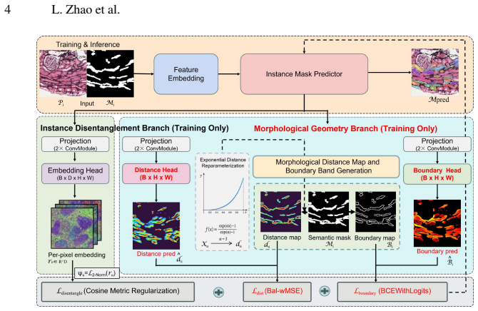
MORI-Seg learns morphology-aware geometric representations directly from semantic masks by jointly modeling object-centric distance fields and boundary-band representations to encode interior structure and contact interfaces. A class-conditioned feature disentanglement module further promotes intra-instance coherence and inter-instance separation. Under semantic-only supervision, the model decomposes connected semantic regions into distinct instance masks in an end-to-end manner.
What carries the argument
Joint modeling of object-centric distance fields and boundary-band representations inside a class-conditioned feature disentanglement module that extracts instance-level geometry from semantic inputs.
If this is right
- Instance-level masks become available for morphometric quantification without new instance annotations.
- Separation accuracy improves in adherent and crowded regions compared with classical post-processing pipelines.
- The end-to-end framework produces more reliable downstream quantitative studies on pathology datasets.
- The approach generalizes across different semantic-to-instance learning baselines that rely on similar geometric cues.
Where Pith is reading between the lines
- The same distance-field plus boundary-band approach could transfer to other domains where objects of one class touch frequently but instance labels are unavailable.
- If the geometric representations prove stable, the method could reduce annotation effort for large-scale medical image collections.
- Extensions might test whether adding weak instance cues during training further refines the disentanglement module without full supervision.
Load-bearing premise
Semantic masks alone contain enough geometric information for the learned distance fields and boundary representations to reliably separate instances.
What would settle it
Running the trained model on a test set of crowded kidney images with known manual instance annotations and checking whether separation accuracy exceeds that of standard heuristic post-processing methods such as watershed on the same semantic masks.
Figures

read the original abstract
Instance-level quantification of kidney functional units is essential for morphometric analysis, yet most publicly available pathology datasets provide only semantic segmentation annotations, where adjacent structures of the same class are merged into single regions. This prevents reliable instance-level analysis and limits downstream quantitative studies. Existing heuristic post-processing methods often yield suboptimal instance separation, particularly in crowded and adherent regions, while deep learning-based instance segmentation approaches typically require intensive instance-level annotations that are costly and labor-intensive to obtain. We propose MORI-Seg, a deep learning framework that enables instance segmentation without requiring instance-level annotations. Instead of heuristic splitting or instance supervision, MORI-Seg learns morphology-aware geometric representations directly from semantic masks by jointly modeling object-centric distance fields and boundary-band representations to encode interior structure and contact interfaces. A class-conditioned feature disentanglement module further promotes intra-instance coherence and inter-instance separation. Under semantic-only supervision, MORI-Seg decomposes connected semantic regions into distinct instance masks in an end-to-end manner. Experiments demonstrate improved instance separation accuracy and more reliable morphometric quantification compared with classical post-processing pipelines and representative semantic-to-instance learning approaches. The official implementation is publicly available at https://github.com/ddrrnn123/MORI-Seg.
Editorial analysis
A structured set of objections, weighed in public.
Referee Report
Summary. The paper proposes MORI-Seg, a deep learning framework for instance segmentation of kidney functional units without instance-level annotations. It learns object-centric distance fields and boundary-band representations directly from semantic masks, augmented by a class-conditioned feature disentanglement module, to decompose connected semantic regions into distinct instances in an end-to-end manner. The abstract claims this yields improved instance separation accuracy and more reliable morphometric quantification compared to classical post-processing and other semantic-to-instance methods.
Significance. If validated with quantitative evidence, the approach could reduce annotation costs for instance-level analysis in pathology by leveraging widely available semantic datasets. The joint modeling of distance fields and boundary bands to encode interior structure and contact interfaces offers a structured way to recover instance geometry from morphology alone.
major comments (2)
- [Abstract] Abstract: The central claim of 'improved instance separation accuracy' and 'more reliable morphometric quantification' is asserted without any quantitative metrics, error analysis, dataset details, ablation results, or evaluation protocol. This is load-bearing for assessing whether the decomposition succeeds under semantic-only supervision.
- [Method] Method description (as summarized in abstract): The end-to-end decomposition in adherent regions depends on the network implicitly recovering per-instance separation from semantic masks via learned distance fields and boundary bands. Since connected components supply no explicit per-instance gradient, this reduces to discovering morphological regularities (e.g., shape or spacing priors) that are not guaranteed to be stable or unique; the manuscript provides no concrete test or failure-case analysis for when these priors are violated.
minor comments (1)
- [Abstract] The public GitHub link is a positive for reproducibility; the abstract would be strengthened by naming the specific pathology datasets and instance counts used in experiments.
Simulated Author's Rebuttal
We thank the referee for the constructive feedback. We respond to each major comment below and note planned revisions where appropriate.
read point-by-point responses
-
Referee: [Abstract] Abstract: The central claim of 'improved instance separation accuracy' and 'more reliable morphometric quantification' is asserted without any quantitative metrics, error analysis, dataset details, ablation results, or evaluation protocol. This is load-bearing for assessing whether the decomposition succeeds under semantic-only supervision.
Authors: The abstract serves as a high-level summary. The full manuscript reports quantitative results on instance separation (including AP and object-level Dice) and morphometric errors, with dataset details, ablations, and protocols detailed in the Experiments section. We will revise the abstract to incorporate key quantitative gains over baselines for improved clarity. revision: yes
-
Referee: [Method] Method description (as summarized in abstract): The end-to-end decomposition in adherent regions depends on the network implicitly recovering per-instance separation from semantic masks via learned distance fields and boundary bands. Since connected components supply no explicit per-instance gradient, this reduces to discovering morphological regularities (e.g., shape or spacing priors) that are not guaranteed to be stable or unique; the manuscript provides no concrete test or failure-case analysis for when these priors are violated.
Authors: The framework supervises distance fields and boundary bands directly from semantic masks, with the class-conditioned disentanglement module encouraging separation based on learned morphological patterns. Experiments demonstrate gains in crowded regions versus post-processing and other semantic-to-instance baselines. We will add a dedicated limitations subsection with concrete failure-case analysis for scenarios where morphological regularities may not hold (e.g., extreme shape variability). revision: yes
Circularity Check
No circularity: method is a standard end-to-end learned representation from semantic supervision
full rationale
The abstract and description frame MORI-Seg as training a network to predict object-centric distance fields and boundary-band representations directly from semantic masks, followed by a class-conditioned disentanglement module. No equations, fitted parameters renamed as predictions, self-citations, or ansatzes are provided that would make any claimed output equivalent to the input by construction. The derivation chain is a conventional supervised learning pipeline whose outputs are not forced by redefinition of the inputs.
Axiom & Free-Parameter Ledger
Reference graph
Works this paper leans on
-
[1]
Biomedical Signal Processing and Control112, 108724 (2026)
Abera, D.E., Zaki, N., Qin, W.: Style transformation and distance map guided nucleus in- stance segmentation via multi task learning. Biomedical Signal Processing and Control112, 108724 (2026)
2026
-
[2]
Electronics9(3), 503 (2020)
Altini, N., Cascarano, G.D., Brunetti, A., Marino, F., Rocchetti, M.T., Matino, S., Venere, U., Rossini, M., Pesce, F., Gesualdo, L., et al.: Semantic segmentation framework for glomeruli detection and classification in kidney histological sections. Electronics9(3), 503 (2020)
2020
-
[3]
Barisoni, L., Nast, C.C., Jennette, J.C., Hodgin, J.B., Herzenberg, A.M., Lemley, K.V ., Con- way, C.M., Kopp, J.B., Kretzler, M., Lienczewski, C., Avila-Casado, C., Bagnasco, S., Sethi, S., Tomaszewski, J., Gasim, A.H., Hewitt, S.M.: Digital pathology evaluation in the multi- center nephrotic syndrome study network (NEPTUNE). Clinical Journal of the Amer...
work page doi:10.2215/cjn 2013
-
[4]
Journal of the American Society of Nephrology32(1), 52–68 (2021)
Bouteldja, N., Klinkhammer, B.M., Bülow, R.D., Droste, P., Otten, S.W., V on Stillfried, S.F., Moellmann, J., Sheehan, S.M., Korstanje, R., Menzel, S., et al.: Deep learning–based seg- mentation and quantification in experimental kidney histopathology. Journal of the American Society of Nephrology32(1), 52–68 (2021)
2021
-
[5]
Medical image analysis36, 135– 146 (2017)
Chen, H., Qi, X., Yu, L., Dou, Q., Qin, J., Heng, P.A.: Dcan: Deep contour-aware networks for object instance segmentation from histology images. Medical image analysis36, 135– 146 (2017)
2017
-
[6]
In: Proceedings of SPIE–the International Society for Optical Engineering
Chen, J., Wang, Y ., Deng, R., Liu, Q., Cui, C., Yao, T., Liu, Y ., Zhong, J., Fogo, A.B., Yang, H., et al.: Spatial pathomics toolkit for quantitative analysis of podocyte nuclei with histology and spatial transcriptomics data in renal pathology. In: Proceedings of SPIE–the International Society for Optical Engineering. vol. 12933, p. 1293310 (2024)
2024
-
[7]
In: International conference on med- ical image computing and computer-assisted intervention
Chen, L., Strauch, M., Merhof, D.: Instance segmentation of biomedical images with an object-aware embedding learned with local constraints. In: International conference on med- ical image computing and computer-assisted intervention. pp. 451–459. Springer (2019)
2019
-
[8]
In: IS&T International Symposium on Electronic Imaging
Deng, R., Cui, C., Liu, Q., Yao, T., Remedios, L.W., Bao, S., Landman, B.A., Wheless, L.E., Coburn, L.A., Wilson, K.T., et al.: Segment anything model (sam) for digital pathology: Assess zero-shot segmentation on whole slide imaging. In: IS&T International Symposium on Electronic Imaging. vol. 37, pp. COIMG–132 (2025)
2025
-
[9]
IEEE Transactions on Biomedical Engineering70(9), 2636–2644 (2023) 10 L
Deng, R., Liu, Q., Cui, C., Yao, T., Long, J., Asad, Z., Womick, R.M., Zhu, Z., Fogo, A.B., Zhao, S., et al.: Omni-seg: A scale-aware dynamic network for renal pathological image segmentation. IEEE Transactions on Biomedical Engineering70(9), 2636–2644 (2023) 10 L. Zhao et al
2023
-
[10]
In: International Conference on Medical Image Computing and Computer-Assisted Intervention
Deng, R., Liu, Q., Cui, C., Yao, T., Xiong, J., Bao, S., Li, H., Yin, M., Wang, Y ., Zhao, S., et al.: Hats: Hierarchical adaptive taxonomy segmentation for panoramic pathology image analysis. In: International Conference on Medical Image Computing and Computer-Assisted Intervention. pp. 155–166. Springer (2024)
2024
-
[11]
In: Proceedings of the IEEE/CVF conference on computer vision and pattern recognition
Deng, R., Liu, Q., Cui, C., Yao, T., Yue, J., Xiong, J., Yu, L., Wu, Y ., Yin, M., Wang, Y ., et al.: Prpseg: Universal proposition learning for panoramic renal pathology segmentation. In: Proceedings of the IEEE/CVF conference on computer vision and pattern recognition. pp. 11736–11746 (2024)
2024
-
[12]
In: Medical Imaging with Deep Learning (2025)
Deng, R., Yang, Y ., Pisapia, D.J., Liechty, B.L., Zhu, J., Xiong, J., Guo, J., Lu, Z., Wang, J., Yao, X., et al.: Casc-ai: Consensus-aware self-corrective learning for cell segmentation with noisy labels. In: Medical Imaging with Deep Learning (2025)
2025
-
[13]
arXiv preprint arXiv:2502.07288 (2025)
Deng, R., Yao, T., Tang, Y ., Guo, J., Lu, S., Xiong, J., Yu, L., Cap, Q.H., Cai, P., Lan, L., et al.: Kpis 2024 challenge: Advancing glomerular segmentation from patch-to slide-level. arXiv preprint arXiv:2502.07288 (2025)
-
[14]
Cur- rent Pathobiology Reports7(3), 73–84 (2019)
Gupta, R., Kurc, T., Sharma, A., Almeida, J.S., Saltz, J.: The emergence of pathomics. Cur- rent Pathobiology Reports7(3), 73–84 (2019)
2019
-
[15]
IEEE transactions on pattern analysis and machine intelligence (4), 532–550 (1987)
Haralick, R.M., Sternberg, S.R., Zhuang, X.: Image analysis using mathematical morphol- ogy. IEEE transactions on pattern analysis and machine intelligence (4), 532–550 (1987)
1987
-
[16]
In: Medical Imaging with Deep Learning (2026)
He, X., Deng, R., Prince, M.R., Sabuncu, M.R.: Instabound: Instance-boundary signed dis- tance regression. In: Medical Imaging with Deep Learning (2026)
2026
-
[17]
Kidney international99(1), 86–101 (2021)
Jayapandian, C.P., Chen, Y ., Janowczyk, A.R., Palmer, M.B., Cassol, C.A., Sekulic, M., Hodgin, J.B., Zee, J., Hewitt, S.M., O’Toole, J., et al.: Development and evaluation of deep learning–based segmentation of histologic structures in the kidney cortex with multiple his- tologic stains. Kidney international99(1), 86–101 (2021)
2021
-
[18]
The American Journal of Pathology191(8), 1431– 1441 (2021)
Jiang, L., Chen, W., Dong, B., Mei, K., Zhu, C., Liu, J., Cai, M., Yan, Y ., Wang, G., Zuo, L., et al.: A deep learning-based approach for glomeruli instance segmentation from multi- stained renal biopsy pathologic images. The American Journal of Pathology191(8), 1431– 1441 (2021)
2021
-
[19]
kpmp.org(2026), accessed: 02/09/2026
Kidney Precision Medicine Project: Kidney precision medicine project.https://www. kpmp.org(2026), accessed: 02/09/2026. The results here are in whole or part based upon data generated by the KPMP
2026
-
[20]
In: Proceedings of the IEEE/CVF con- ference on computer vision and pattern recognition
Li, R., He, C., Zhang, Y ., Li, S., Chen, L., Zhang, L.: Sim: Semantic-aware instance mask generation for box-supervised instance segmentation. In: Proceedings of the IEEE/CVF con- ference on computer vision and pattern recognition. pp. 7193–7203 (2023)
2023
-
[21]
IEEE Transactions on Image Pro- cessing31, 6893–6906 (2022)
Liang, T., Chu, X., Liu, Y ., Wang, Y ., Tang, Z., Chu, W., Chen, J., Ling, H.: Cbnet: A com- posite backbone network architecture for object detection. IEEE Transactions on Image Pro- cessing31, 6893–6906 (2022)
2022
-
[22]
arXiv preprint arXiv:2212.07784 (2022)
Lyu, C., Zhang, W., Huang, H., Zhou, Y ., Wang, Y ., Liu, Y ., Zhang, S., Chen, K.: Rtmdet: An empirical study of designing real-time object detectors. arXiv preprint arXiv:2212.07784 (2022)
-
[23]
Journal of Systems Architecture53(4), 210–226 (2007)
Rambabu, C., Chakrabarti, I.: An efficient immersion-based watershed transform method and its prototype architecture. Journal of Systems Architecture53(4), 210–226 (2007)
2007
-
[24]
In: Med- ical Imaging 2026: Digital and Computational Pathology
Shi, T., He, X., Fang, H., Ikemura, K., Sabuncu, M.R., Yang, Y ., Deng, R.: Mˆ 3-glodets: Multi-region and multi-scale analysis of fine-grained diseased glomerular detection. In: Med- ical Imaging 2026: Digital and Computational Pathology. vol. 13932, pp. 258–266. SPIE (2026)
2026
-
[25]
Siméoni, O., V o, H.V ., Seitzer, M., Baldassarre, F., Oquab, M., Jose, C., Khalidov, V ., Szafraniec, M., Yi, S., Ramamonjisoa, M., et al.: Dinov3. arXiv preprint arXiv:2508.10104 (2025)
work page internal anchor Pith review Pith/arXiv arXiv 2025
-
[26]
YOLOv12: Attention-Centric Real-Time Object Detectors
Tian, Y ., Ye, Q., Doermann, D.: Yolov12: Attention-centric real-time object detectors. arXiv preprint arXiv:2502.12524 (2025) MORI-Seg 11
work page internal anchor Pith review Pith/arXiv arXiv 2025
-
[27]
In: Euro- pean conference on computer vision
Tian, Z., Shen, C., Chen, H.: Conditional convolutions for instance segmentation. In: Euro- pean conference on computer vision. pp. 282–298. Springer (2020)
2020
-
[28]
IEEE Geoscience and Remote Sensing Letters23, 1–5 (2025)
Wang, R., Xu, Y ., Wu, Z., Wei, Z.: Distilling object detectors with scale-conscious knowledge in remote sensing images. IEEE Geoscience and Remote Sensing Letters23, 1–5 (2025)
2025
-
[29]
Computers in Biology and Medicine186, 109670 (2025)
Wang, X., Zhang, J., Xu, Y ., Huang, Y ., Ming, W., Jiao, Y ., Liu, B., Fan, X., Xu, J.: Glo-net: A dual task branch based neural network for multi-class glomeruli segmentation. Computers in Biology and Medicine186, 109670 (2025)
2025
-
[30]
Advances in Neural information processing systems33, 17721–17732 (2020)
Wang, X., Zhang, R., Kong, T., Li, L., Shen, C.: Solov2: Dynamic and fast instance segmen- tation. Advances in Neural information processing systems33, 17721–17732 (2020)
2020
-
[31]
Artificial Intelli- gence in Medicine164, 103111 (2025)
Xiong, Z., He, J., Valkema, P., Nguyen, T.Q., Naesens, M., Kers, J., Verbeek, F.J.: Advances in kidney biopsy lesion assessment through dense instance segmentation. Artificial Intelli- gence in Medicine164, 103111 (2025)
2025
-
[32]
International Journal of Computer Assisted Radiology and Surgery20(2), 225–236 (2025)
Yang, F., He, Q., Wang, Y ., Zeng, S., Xu, Y ., Ye, J., He, Y ., Guan, T., Wang, Z., Li, J.: Unsupervised stain augmentation enhanced glomerular instance segmentation on pathology images. International Journal of Computer Assisted Radiology and Surgery20(2), 225–236 (2025)
2025
-
[33]
In: IS&T International Symposium on Electronic Imaging
Yang, Y ., Wang, Y ., Yao, T., Deng, R., Yin, M., Zhao, S., Yang, H., Huo, Y .: Pyspatial: A high-speed whole slide image pathomics toolkit. In: IS&T International Symposium on Electronic Imaging. vol. 37, pp. HPCI–177 (2025)
2025
-
[34]
Communica- tions of the ACM27(3), 236–239 (1984)
Zhang, T.Y ., Suen, C.Y .: A fast parallel algorithm for thinning digital patterns. Communica- tions of the ACM27(3), 236–239 (1984)
1984
-
[35]
arXiv preprint arXiv:2508.15208 (2025)
Zhao, L., Yang, Y ., Zhu, Y ., Yang, H., Huo, Y ., Simonson, P.D., Ikemura, K., Sabuncu, M.R., Yang, Y ., Deng, R.: Dymorph-b2i: Dynamic and morphology-guided binary-to-instance seg- mentation for renal pathology. arXiv preprint arXiv:2508.15208 (2025)
-
[36]
Journal of Fungi10(3), 171 (2024)
Zhou, Y ., Reynolds, T.B.: Innovations in antifungal drug discovery among cell envelope synthesis enzymes through structural insights. Journal of Fungi10(3), 171 (2024)
2024
discussion (0)
Sign in with ORCID, Apple, or X to comment. Anyone can read and Pith papers without signing in.