Adaptive Temporal Gating of Longitudinal Magnetic Resonance Imaging for Alzheimer's Prediction
Pith reviewed 2026-06-29 13:15 UTC · model grok-4.3
The pith
TAF-Net with adaptive temporal gating on paired MRIs predicts MCI-to-AD conversion more accurately than single-timepoint methods.
A machine-rendered reading of the paper's core claim, the machinery that carries it, and where it could break.
Core claim
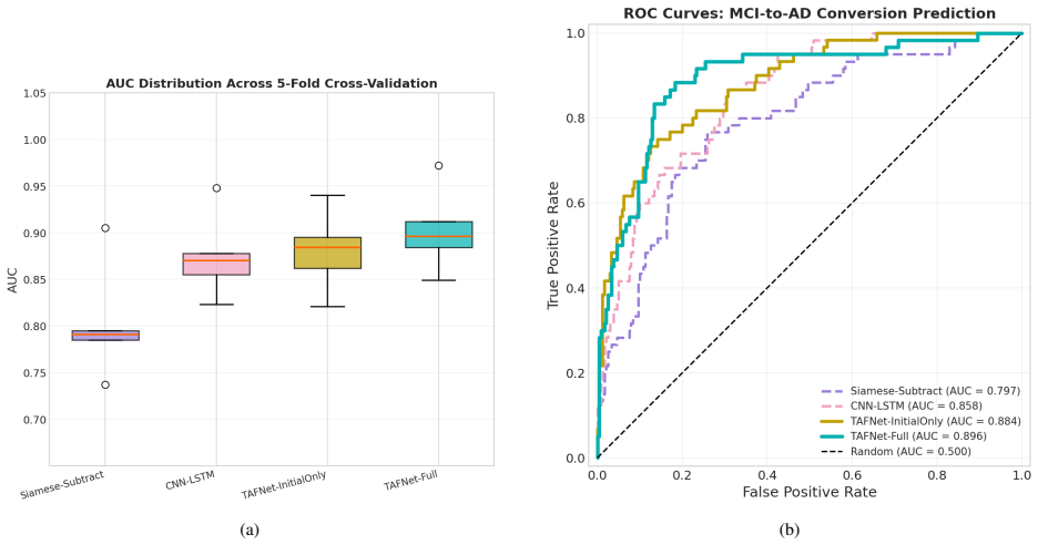
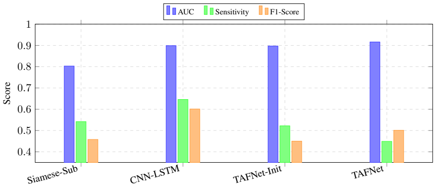
TAF-Net achieves the highest discriminative performance among all evaluated methods using only structural MRI for three-year MCI-to-AD conversion prediction, significantly outperforming the strongest baseline while approaching the performance of multimodal methods that require PET, CSF, or genetic data.
What carries the argument
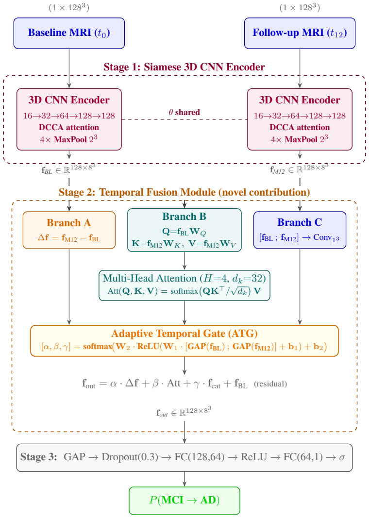
The Adaptive Temporal Gate within the Temporal Fusion Module, which learns patient-specific weightings to synthesize explicit structural change, region-to-region temporal cross-attention, and bilateral feature concatenation from paired longitudinal 3D MRI scans.
If this is right
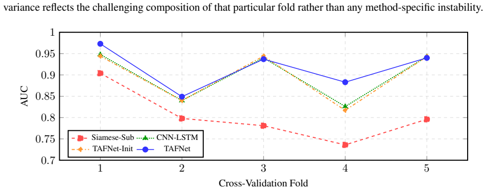
- Longitudinal fusion reduces predictive variance by 48% compared to single-timepoint evaluation.
- The architecture matches baseline performance using only a fraction of the training data.
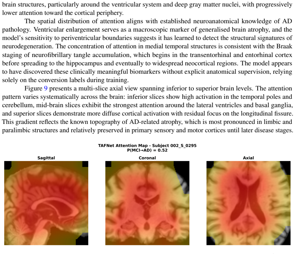
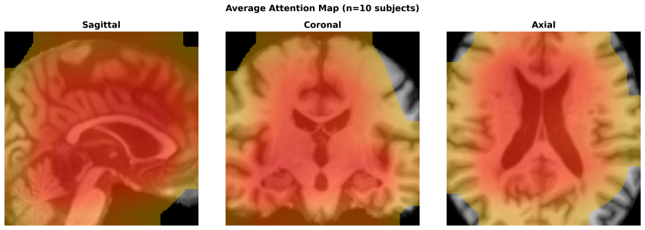
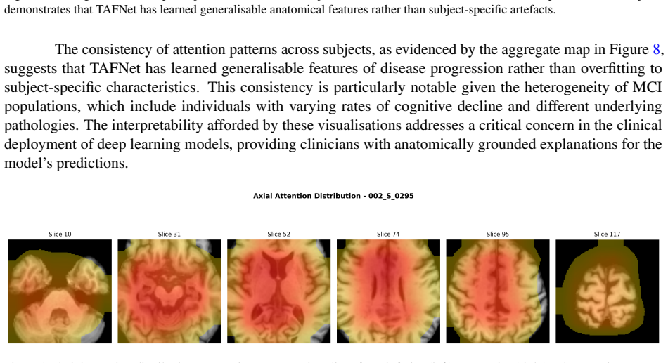
- Spatial attention maps align with established AD pathology in the medial temporal lobe and ventricles.
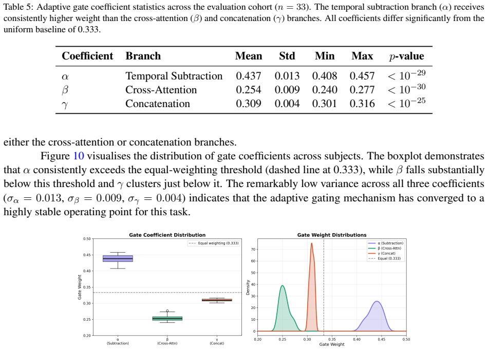
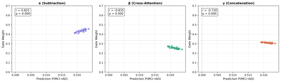
- The gating mechanism prioritizes explicit volumetric change, showing strong positive correlation to conversion risk.
Where Pith is reading between the lines
- If the adaptive weighting proves robust across datasets, it could allow for more efficient use of follow-up scans in clinical monitoring.
- The method's data efficiency suggests potential for training on smaller, more diverse cohorts where longitudinal data is limited.
- Extending the temporal fusion to more than two time points might further improve trajectory modeling for longer-term predictions.
Load-bearing premise
The paired longitudinal scans from the ADNI cohort represent typical patient trajectories that apply beyond this specific dataset.
What would settle it
Testing TAF-Net on a separate longitudinal MRI cohort where it does not significantly outperform the strongest single-timepoint baseline would challenge the central performance claim.
Figures
read the original abstract
Predicting conversion from Mild Cognitive Impairment (MCI) to Alzheimer's Disease (AD) is critical for early intervention. Current deep learning paradigms predominantly rely on cross-sectional structural MRI, neglecting prognostic value in patient-specific anatomical trajectories. We introduce the Temporal Adaptive Fusion Network (TAF-Net), a hybrid CNN-Transformer architecture that models paired longitudinal 3D MRI scans. Central to TAF-Net is a Temporal Fusion Module governed by an Adaptive Temporal Gate, which learns patient-specific weightings to synthesize three spatiotemporal representations: explicit structural change, region-to-region temporal cross-attention, and bilateral feature concatenation. Evaluated on the Alzheimer's Disease Neuroimaging Initiative cohort for three-year MCI-to-AD conversion prediction, TAF-Net achieved the highest discriminative performance among all evaluated methods using only structural MRI, significantly outperforming the strongest baseline and approaching multimodal methods requiring PET, CSF, or genetic data. The architecture exhibited exceptional data efficiency, matching baseline performance with a fraction of training data. Ablation studies demonstrate that longitudinal fusion improves discrimination while reducing predictive variance by 48% compared to single-timepoint evaluation. Interpretability analyses reveal spatial attention aligned with established AD pathology in the medial temporal lobe and ventricles, while the gating mechanism prioritizes explicit volumetric change with strong positive correlation to conversion risk.
Editorial analysis
A structured set of objections, weighed in public.
Referee Report
Summary. The paper introduces TAF-Net, a hybrid CNN-Transformer architecture with a Temporal Fusion Module controlled by an Adaptive Temporal Gate. The gate learns patient-specific weightings from paired longitudinal 3D structural MRI scans to synthesize explicit structural change, region-to-region temporal cross-attention, and bilateral feature concatenation representations. On the ADNI cohort, the model is evaluated for three-year MCI-to-AD conversion prediction and claims the highest performance among structural-MRI-only methods, significantly outperforming the strongest baseline while approaching multimodal methods that use PET, CSF, or genetic data. Additional claims include exceptional data efficiency (matching baselines with a fraction of training data), a 48% reduction in predictive variance from longitudinal fusion versus single-timepoint evaluation, and interpretability where spatial attention aligns with medial temporal lobe and ventricular pathology and the gate prioritizes volumetric change correlated with conversion risk.
Significance. If the central performance and ablation results hold under broader validation, the work would demonstrate concrete value in modeling patient-specific longitudinal trajectories from structural MRI alone for prognostic tasks in Alzheimer's disease. The reported data-efficiency gains and variance reduction would be particularly relevant for settings with limited labeled scans. The alignment of attention maps with established AD pathology provides a useful interpretability anchor. These elements, if substantiated with full methods and statistics, could support reduced reliance on multimodal or invasive biomarkers.
major comments (2)
- [Abstract and Evaluation] Abstract/Evaluation section: All reported performance numbers (highest AUC among structural-MRI methods, approaching multimodal baselines) and the 48% variance reduction are measured exclusively on the ADNI cohort for 3-year MCI-to-AD conversion. No external validation cohort, multi-site hold-out set, or domain-shift experiment is described, so the generalizability of the learned Adaptive Temporal Gate and patient-specific spatiotemporal representations remains untested. This is load-bearing for the claim that the architecture provides broadly applicable prognostic improvement.
- [Results] Results/Ablation section: The statement that longitudinal fusion 'significantly' outperforms the strongest baseline and reduces predictive variance by 48% is presented without accompanying statistical tests, confidence intervals, or cross-validation details. This makes it difficult to judge whether the reported gains are robust or could be explained by cohort-specific factors.
minor comments (2)
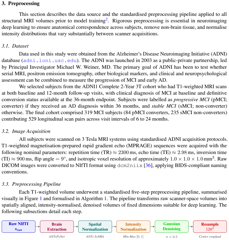
- [Abstract] Abstract: The architecture description refers to three spatiotemporal representations but does not name the precise fusion equations or the input dimensions of the paired scans; adding a short equation or diagram reference would aid immediate comprehension.
- [Interpretability analyses] The interpretability paragraph states attention is 'aligned with established AD pathology' but does not quantify the overlap (e.g., Dice with atlas regions) or report the correlation coefficient between gate weights and conversion risk; these details would strengthen the claim.
Simulated Author's Rebuttal
We thank the referee for the constructive feedback. We address each major comment below with proposed revisions to improve statistical rigor and transparency on generalizability.
read point-by-point responses
-
Referee: [Abstract and Evaluation] Abstract/Evaluation section: All reported performance numbers (highest AUC among structural-MRI methods, approaching multimodal baselines) and the 48% variance reduction are measured exclusively on the ADNI cohort for 3-year MCI-to-AD conversion. No external validation cohort, multi-site hold-out set, or domain-shift experiment is described, so the generalizability of the learned Adaptive Temporal Gate and patient-specific spatiotemporal representations remains untested. This is load-bearing for the claim that the architecture provides broadly applicable prognostic improvement.
Authors: We agree the evaluation is confined to ADNI and that this limits claims of broad applicability. ADNI remains the largest public longitudinal structural MRI resource for MCI-to-AD prediction. In revision we will: (1) add an explicit limitations paragraph in the discussion stating that results are ADNI-specific and that external validation is required for clinical translation, and (2) report an internal domain-shift experiment (train on one ADNI acquisition site, test on the others) to quantify robustness to scanner/protocol variation within the available data. We cannot introduce a new external cohort in this revision. revision: partial
-
Referee: [Results] Results/Ablation section: The statement that longitudinal fusion 'significantly' outperforms the strongest baseline and reduces predictive variance by 48% is presented without accompanying statistical tests, confidence intervals, or cross-validation details. This makes it difficult to judge whether the reported gains are robust or could be explained by cohort-specific factors.
Authors: The omission of statistical details was an oversight. The revised manuscript will include: (i) full description of the 5-fold stratified cross-validation protocol, (ii) 95% bootstrap confidence intervals for all reported AUC/accuracy values, (iii) p-values from DeLong’s test for AUC comparisons against the strongest baseline and from a paired t-test on per-fold prediction variances for the 48% reduction claim, and (iv) clarification that the variance reduction is computed across the same folds. These additions will be placed in the Results and supplementary material. revision: yes
Circularity Check
No circularity in derivation or performance claims
full rationale
The paper introduces an empirical CNN-Transformer architecture (TAF-Net) with an Adaptive Temporal Gate for longitudinal MRI fusion and reports its discriminative performance on the ADNI cohort for 3-year MCI-to-AD conversion. No equations, parameter-fitting steps, or self-citations are shown that reduce any claimed result to an input by construction. The architecture definitions, fusion module, and evaluation metrics are independent of the headline AUC numbers; the single-cohort limitation is a generalization concern, not a circularity issue under the specified patterns.
Axiom & Free-Parameter Ledger
axioms (1)
- domain assumption Paired longitudinal 3D MRI scans from the same patients are available and contain prognostic signal beyond single timepoint scans.
invented entities (1)
-
Adaptive Temporal Gate
no independent evidence
Reference graph
Works this paper leans on
-
[1]
Teunissen, Jeffrey Cummings, and Wiesje M
Philip Scheltens, Bart De Strooper, Miia Kivipelto, Henne Holstege, Gaël Chételat, Charlotte E. Teunissen, Jeffrey Cummings, and Wiesje M. van der Flier. Alzheimer’s disease.The Lancet, 397 (10284):1577–1590, 2021. doi: 10.1016/S0140-6736(20)32205-4
-
[2]
Jack, David S
Clifford R. Jack, David S. Knopman, William J. Jagust, Leslie M. Shaw, Paul S. Aisen, Michael W. Weiner, Ronald C. Petersen, and John Q. Trojanowski. Hypothetical model of dynamic biomarkers of the Alzheimer’s pathological cascade.The Lancet Neurology, 9(1):119–128, 2010. doi: 10.1016/ S1474-4422(09)70299-6
2010
-
[3]
Clifford R. Jack, David S. Knopman, William J. Jagust, Ronald C. Petersen, Michael W. Weiner, Paul S. Aisen, Leslie M. Shaw, Prashanthi Vemuri, Heather J. Wiste, Stephen D. Weigand, Timothy G. Lesnick, Vernon S. Pankratz, Michael C. Donohue, and John Q. Trojanowski. Tracking pathophysiological processes in Alzheimer’s disease: an updated hypothetical mode...
-
[4]
Sperling, Paul S
Reisa A. Sperling, Paul S. Aisen, Laurel A. Beckett, David A. Bennett, Suzanne Craft, Anne M. Fagan, Takeshi Iwatsubo, Clifford R. Jack, Jeffrey Kaye, Thomas J. Montine, Denise C. Park, Eric M. Reiman, Christopher C. Rowe, Eric Siemers, Yaakov Stern, Kristine Yaffe, Maria C. Carrillo, Bill Thies, Marcelle Morrison-Bogorad, Molly V . Wagster, and Creighton...
-
[5]
doi: 10.1016/j.jalz.2011.03.003
-
[6]
Alzheimers Dement.7(3), 270–279 (2011).https: //doi.org/10.1016/j.jalz.2011.03.008 10 G
Marilyn S. Albert, Steven T. DeKosky, Dennis Dickson, Bruno Dubois, Howard H. Feldman, Nick C. Fox, Anthony Gamst, David M. Holtzman, William J. Jagust, Ronald C. Petersen, Peter J. Snyder, Maria C. Carrillo, Bill Thies, and Creighton H. Phelps. The diagnosis of mild cognitive impairment due to Alzheimer’s disease: Recommendations from the National Instit...
-
[7]
Petersen, Barbara Caracciolo, Carol Brayne, Serge Gauthier, Vesna Jelic, and Laura Fratiglioni
Ronald C. Petersen, Barbara Caracciolo, Carol Brayne, Serge Gauthier, Vesna Jelic, and Laura Fratiglioni. Mild cognitive impairment: a concept in evolution.Journal of Internal Medicine, 275(3): 214–228, 2014. doi: 10.1111/joim.12190
-
[8]
Mitchell and Mojtaba Shiri-Feshki
Alex J. Mitchell and Mojtaba Shiri-Feshki. Rate of progression of mild cognitive impairment to dementia – meta-analysis of 41 robust inception cohort studies.Acta Psychiatrica Scandinavica, 119 (4):252–265, 2009. doi: 10.1111/j.1600-0447.2008.01326.x. 33
-
[9]
Alex Ward, Sheila Tardiff, Catherine Dye, and H. Michael Arrighi. Rate of conversion from prodromal Alzheimer’s disease to Alzheimer’s dementia: a systematic review of the literature.Dementia and Geriatric Cognitive Disorders Extra, 3(1):320–332, 2013. doi: 10.1159/000354370
-
[10]
Giovanni B. Frisoni, Nick C. Fox, Clifford R. Jack, Philip Scheltens, and Paul M. Thompson. The clinical use of structural MRI in Alzheimer disease.Nature Reviews Neurology, 6(2):67–77, 2010. doi: 10.1038/nrneurol.2009.215
-
[11]
D. P. Devanand, G. Pradhaban, X. Liu, A. Khandji, S. De Santi, S. Segal, H. Rusinek, G. H. Pelton, L. S. Honig, R. Mayeux, Y . Stern, M. H. Tabert, and M. J. de Leon. Hippocampal and entorhinal atrophy in mild cognitive impairment: prediction of Alzheimer disease.Neurology, 68(11):828–836, 2007. doi: 10.1212/01.wnl.0000256697.20968.d7
-
[12]
Dickerson, Akram Bakkour, David H
Bradford C. Dickerson, Akram Bakkour, David H. Salat, Eric Feczko, Jenni Pacheco, Douglas N. Greve, Francine Grodstein, Christopher I. Wright, Deborah Blacker, H. Diana Rosas, Reisa A. Sperling, Alireza Atri, John H. Growdon, Bradley T. Hyman, John C. Morris, Bruce Fischl, and Randy L. Buckner. The cortical signature of Alzheimer’s disease: regionally spe...
-
[13]
Clifford R. Jack, Maria M. Shiung, Stephen D. Weigand, Peter C. O’Brien, Jeffrey L. Gunter, Bradley F. Boeve, David S. Knopman, Glenn E. Smith, Robert J. Ivnik, Eric G. Tangalos, and Ronald C. Pe- tersen. Brain atrophy rates predict subsequent clinical conversion in normal elderly and amnestic MCI. Neurology, 65(8):1227–1231, 2005. doi: 10.1212/01.wnl.000...
-
[14]
Nestor, Ravi Rupsingh, Michael Borrie, Matthew Smith, Vittorio Accomazzi, Jennie L
Sean M. Nestor, Ravi Rupsingh, Michael Borrie, Matthew Smith, Vittorio Accomazzi, Jennie L. Wells, Aparna Bhagwat, and Siddhartha Bhattacharyya. Ventricular enlargement as a possible measure of Alzheimer’s disease progression validated using the Alzheimer’s disease neuroimaging initiative database.Brain, 131(Pt 9):2443–2454, 2008. doi: 10.1093/brain/awn146
-
[15]
Shannon L. Risacher, Li Shen, John D. West, Sungeun Kim, Brenna C. McDonald, Laurel A. Beckett, Danielle J. Harvey, Clifford R. Jack, Michael W. Weiner, and Andrew J. Saykin. Longitudinal MRI atrophy biomarkers: relationship to conversion in the ADNI cohort.Neurobiology of Aging, 31(8): 1401–1418, 2010. doi: 10.1016/j.neurobiolaging.2010.04.029
-
[16]
Michael W. Weiner, Dallas P. Veitch, Paul S. Aisen, Laurel A. Beckett, Nigel J. Cairns, Robert C. Green, Danielle Harvey, Clifford R. Jack, William Jagust, John C. Morris, Ronald C. Petersen, Andrew J. Saykin, Leslie M. Shaw, Arthur W. Toga, and John Q. Trojanowski. Recent publications from the Alzheimer’s Disease Neuroimaging Initiative: Reviewing progre...
-
[17]
Rik Ossenkoppele, Rik van der Kant, and Oskar Hansson. Tau biomarkers in Alzheimer’s disease: towards implementation in clinical practice and trials.The Lancet Neurology, 21(8):726–734, 2022. doi: 10.1016/S1474-4422(22)00168-5
-
[18]
Ingrid Rye, Alexandra Vik, Marek Kocinski, Arvid S. Lundervold, and Alexander J. Lundervold. Predicting conversion to Alzheimer’s disease in individuals with mild cognitive impairment using clinically transferable features.Scientific Reports, 12:15566, 2022. doi: 10.1038/s41598-022-18805-5
-
[19]
Sara Aghajanian, Farzaneh Mohammadifard, Iman Mohammadi, Saeed Rajai Firouzabadi, Abolfazl Baradaran Bagheri, Ehsan Moases Ghaffary, and Omid Mirmosayyeb. Longitudinal structural MRI- 34 based deep learning and radiomics features for predicting Alzheimer’s disease progression.Alzheimer’s Research & Therapy, 17(1):182, 2025. doi: 10.1186/s13195-025-01827-2
-
[20]
Zhentao Hu, Yong Li, Zheng Wang, Shuo Zhang, and Wei Hou. Conv-Swinformer: Integration of CNN and shift window attention for Alzheimer’s disease classification.Computers in Biology and Medicine, 164:107304, 2023. doi: 10.1016/j.compbiomed.2023.107304
-
[21]
Zhiwei Zhao, Phei Shan Queenie Yeoh, Xianghui Zuo, Joon Huang Chuah, Chee-Onn Chow, Xiang Wu, and Khin Wee Lai. Vision transformer-equipped convolutional neural networks for automated Alzheimer’s disease diagnosis using 3D MRI scans.Frontiers in Neurology, 15:1490829, 2024. doi: 10.3389/fneur.2024.1490829
-
[22]
Jian Zhou, Yanan Wei, Xin Li, Wei Zhou, Ran Tao, Yiling Hua, and Hongwei Liu. A deep learning model for early diagnosis of Alzheimer’s disease combined with 3D CNN and video Swin transformer. Scientific Reports, 15:23311, 2025. doi: 10.1038/s41598-025-05568-y
-
[23]
Junhao Wen, Elina Thibeau-Sutre, Mauricio Diaz-Melo, Jorge Samper-González, Alexandre Routier, Simona Bottani, Didier Dormont, Stanley Durrleman, Ninon Burgos, and Olivier Colliot. Convolutional neural networks for classification of Alzheimer’s disease: Overview and reproducible evaluation. Medical Image Analysis, 63:101694, 2020. doi: 10.1016/j.media.2020.101694
-
[24]
Eric Yee, Da Ma, Karteek Popuri, Lei Wang, and Mirza Faisal Beg. Construction of MRI-based Alzheimer’s disease score based on efficient 3D convolutional neural network: Comprehensive validation on 7,902 images from a multi-center dataset.Journal of Alzheimer’s Disease, 79(1):47–58, 2021. doi: 10.3233/JAD-200830
-
[25]
Sheng Liu, Arjun V . Masurkar, Henry Rusinek, Jingyun Chen, Ben Zhang, Weicheng Zhu, Car- los Fernandez-Granda, and Navneet Razavian. Generalizable deep learning model for early Alzheimer’s disease detection from structural MRIs.Scientific Reports, 12:17106, 2022. doi: 10.1038/s41598-022-20674-x
-
[26]
Zhongyi Pei, Zhilei Wan, Yaping Zhang, Minghui Wang, Chengcai Leng, and Yue-Hua Yang. Multi- scale attention-based pseudo-3D convolution neural network for Alzheimer’s disease diagnosis using structural MRI.Pattern Recognition, 131:108825, 2022. doi: 10.1016/j.patcog.2022.108825
-
[27]
Casseb, Rafael Pires, and Anderson Rocha
Guilherme Folego, Marina Weiler, Raphael F. Casseb, Rafael Pires, and Anderson Rocha. Alzheimer’s disease detection through whole-brain 3D-CNN MRI.Frontiers in Bioengineering and Biotechnology, 8:534592, 2020. doi: 10.3389/fbioe.2020.534592
-
[28]
Sina Fathi, Ali Ahmadi, Afsaneh Dehnad, Mostafa Almasi-Dooghaee, and Melika Sadegh. A deep learning-based ensemble method for early diagnosis of Alzheimer’s disease using MRI images.Neu- roinformatics, 22(1):89–105, 2024. doi: 10.1007/s12021-023-09646-2
-
[29]
Hoang, Uh-Hyun Kim, and Jae Gwan Kim
Giang M. Hoang, Uh-Hyun Kim, and Jae Gwan Kim. Vision transformers for the prediction of mild cognitive impairment to Alzheimer’s disease progression using mid-sagittal sMRI.Frontiers in Aging Neuroscience, 15:1102869, 2023. doi: 10.3389/fnagi.2023.1102869
-
[30]
Training ViT with limited data for Alzheimer’s disease classification: An empirical study
Kamilya Kunanbayev, Vivianne Shen, and Dae-Shik Kim. Training ViT with limited data for Alzheimer’s disease classification: An empirical study. InMedical Image Computing and Com- puter Assisted Intervention – MICCAI 2024, volume 15011 ofLNCS. Springer, 2024. doi: 10.1007/ 978-3-031-72117-5_55. 35
2024
-
[31]
Vladyslav Gryshchuk, Devesh Singh, Stefan Teipel, and Martin Dyrba. Contrastive self-supervised learning for neurodegenerative disorder classification.Frontiers in Neuroinformatics, 19:1527582, 2025. doi: 10.3389/fninf.2025.1527582
-
[32]
Martínez-Murcia, Juan M
Francisco J. Martínez-Murcia, Juan M. Gorriz, Javier Ramírez, and Andrés Ortiz. Deep learning prediction of mild cognitive impairment conversion to Alzheimer’s disease at 3 years after diagnosis using longitudinal and whole-brain 3D MRI.PeerJ Computer Science, 7:e560, 2021. doi: 10.7717/ peerj-cs.560
2021
-
[33]
Yucheng Qiu, Mianxin Liu, Liang Huang, Yue Li, Yuanyuan Li, Feng He, Lanxin Lin, Shuicai Chen, Daoqiang Zhang, Qi Guo, and Dinggang Shen. Long-term cognitive decline prediction based on multi- modal data using Multimodal3DSiameseNet: transfer learning from Alzheimer’s disease to Parkinson’s disease.International Journal of Computer Assisted Radiology and ...
-
[34]
LongFormer: Longitudinal transformer for Alzheimer’s disease classification with structural MRIs
Jiahong Chen, Euijoon Kang, Gina Parsons, Arnav Suri, Yucheng Liu, Alisa Chua, Nishant Shah, Anoushka Singh, Zhuo Liu, Harish Ravishankar, Yue Guo, Maged Goubran, Prajakta Bhatt, and Alzheimer’s Disease Neuroimaging Initiative. LongFormer: Longitudinal transformer for Alzheimer’s disease classification with structural MRIs. InProceedings of the IEEE/CVF W...
2024
- [35]
-
[36]
Bass, Ehsan Adeli, Greg Zaharchuk, Michael D
Jiahong Ouyang, Qingyu Zhao, Courtney K. Bass, Ehsan Adeli, Greg Zaharchuk, Michael D. Greicius, Kathleen L. Poston, and Kilian M. Pohl. Stochastic Siamese MAE pretraining for longitudinal medical images.Medical Image Analysis, 99:103370, 2025. doi: 10.1016/j.media.2024.103370
-
[37]
Morgan, John Ashburner, Jolinda Smith, and Christopher Rorden
Xiangrui Li, Paul S. Morgan, John Ashburner, Jolinda Smith, and Christopher Rorden. The first step for neuroimaging data analysis: DICOM to NIfTI conversion.Journal of Neuroscience Methods, 264: 47–56, 2016. doi: 10.1016/j.jneumeth.2016.03.001
-
[38]
Nicholas J. Tustison, Philip A. Cook, Andrew J. Holbrook, Hans J. Johnson, John Muschelli, Gabriel A. Devenyi, Jeffrey T. Duda, Sandhitsu R. Das, Nicholas C. Cullen, Daniel L. Gillen, Michael A. Yassa, James R. Stone, James C. Gee, and Brian B. Avants. The ANTsX ecosystem for quantitative biological and medical imaging.Scientific Reports, 11(1):9068, 2021...
-
[39]
Brian B. Avants, Nicholas J. Tustison, Gang Song, Philip A. Cook, Arno Klein, and James C. Gee. A reproducible evaluation of ANTs similarity metric performance in brain image registration.NeuroImage, 54(3):2033–2044, 2011. doi: 10.1016/j.neuroimage.2010.09.025
-
[40]
Jingyuan Wang, Fei Gao, Jiangchang Dong, Qian Zhang, Haipeng Wang, and Guangzhi Ma. AFS- Net: Attention-guided 3d CNN with lesion feature selection for early Alzheimer’s disease prediction. Computers in Biology and Medicine, 184:109395, 2025. doi: 10.1016/j.compbiomed.2024.109395
-
[41]
Esther E. Bron, Marion Smits, Wiro J. Niessen, and Stefan Klein. Cross-cohort generalizability of deep and conventional machine learning for MRI-based diagnosis and prediction of Alzheimer’s disease. NeuroImage: Clinical, 31:102712, 2021. doi: 10.1016/j.nicl.2021.102712
-
[42]
Weiming Lin, Tong Tong, Qinquan Gao, Di Guo, Xiaofeng Du, Yonggui Yang, Gang Guo, Min Xiao, Min Du, and Xiaobo Qu. Convolutional neural networks-based MRI image analysis for the Alzheimer’s 36 disease prediction from mild cognitive impairment.Frontiers in Neuroscience, 12:777, 2018. doi: 10.3389/fnins.2018.00777
-
[43]
Simeon Spasov, Luca Passamonti, Andrea Duggento, Pietro Liò, and Nicola Toschi. A parameter- efficient deep learning approach to predict conversion from mild cognitive impairment to Alzheimer’s disease.NeuroImage, 189:276–287, 2019. doi: 10.1016/j.neuroimage.2019.01.031
-
[44]
Manhua Liu, Fan Li, Hao Yan, Kefeng Wang, Yixin Ma, Li Shen, and Mingxia Xu. Predicting Alzheimer’s disease conversion from mild cognitive impairment using an extreme learning machine- based grading method with multimodal data.Frontiers in Aging Neuroscience, 12:77, 2020. doi: 10.3389/fnagi.2020.00077
-
[45]
Garam Lee, Kwangsik Nho, Byungkon Kang, Kyung-Ah Sohn, and Dokyoon Kim. Predicting Alzheimer’s disease progression using multi-modal deep learning approach.Scientific Reports, 9 (1):1952, 2019. doi: 10.1038/s41598-018-37769-z
-
[46]
Yanru Chen, Xiaoyu Qian, Yuanyuan Zhang, Wenli Su, Yumeng Huang, Xinyu Wang, Xiaoping Chen, Enyan Zhao, Lu Han, and Yuxia Ma. Prediction models for conversion from mild cognitive impairment to Alzheimer’s disease: a systematic review and meta-analysis.Frontiers in Aging Neuroscience, 14: 840386, 2022. doi: 10.3389/fnagi.2022.840386
-
[47]
Predicting long-term progression of Alzheimer’s disease using a multimodal deep learning model incorporating interaction effects.Journal of Translational Medicine, 22(1):265, 2024
Yifan Wang, Ruitian Gao, Ting Wei, Luke Johnston, Xin Yuan, Yue Zhang, and Zhangsheng Yu. Predicting long-term progression of Alzheimer’s disease using a multimodal deep learning model incorporating interaction effects.Journal of Translational Medicine, 22(1):265, 2024. doi: 10.1186/ s12967-024-05025-w
2024
-
[48]
Sara Fin, Alireza Moayedikia, and Uffe Kock Wiil. Dual-model deep learning for Alzheimer’s prognos- tication.Computers in Biology and Medicine, 208:111672, 2026. doi: 10.1016/j.compbiomed.2026. 111672
-
[49]
Alireza Moayedikia, Sara Fin, and Uffe Kock Wiil. Multi-objective optimization formulation for Alzheimer’s disease trial patient selection.Journal of Biomedical Informatics, 172:104955, 2025. doi: 10.1016/j.jbi.2025.104955
-
[50]
Neuropathological stageing of Alzheimer-related changes.Acta Neu- ropathologica, 82(4):239–259, 1991
Heiko Braak and Eva Braak. Neuropathological stageing of Alzheimer-related changes.Acta Neu- ropathologica, 82(4):239–259, 1991. doi: 10.1007/BF00308809
-
[51]
Multi-objective counterfactual explanations
Susanne Dandl, Christoph Molnar, Martin Binder, and Bernd Bischl. Multi-objective counterfactual explanations. InParallel Problem Solving from Nature – PPSN XVI, pages 448–469. Springer, 2020. doi: 10.1007/978-3-030-58112-1_31
-
[52]
Ali Ebrahimi, Uffe Kock Wiil, Tomas Olsson, Pietro Liò, Ingrid Kockum, Ali Manouchehrinia, and Nar- sis A. Kiani. A susceptibility network analysis of disease trajectories leading to multiple sclerosis: A na- tionwide cohort study.Multiple Sclerosis Journal, pages 1–12, 2026. doi: 10.1177/13524585261421480. 37
discussion (0)
Sign in with ORCID, Apple, or X to comment. Anyone can read and Pith papers without signing in.