Ultrafast internal dynamics of chiral domain walls probed by time-resolved XRMS
Pith reviewed 2026-06-28 13:06 UTC · model grok-4.3
The pith
Time-resolved XRMS isolates ultrafast internal dynamics of chiral Néel domain walls by tracking a fluence-dependent oscillatory mode in the dichroic signal.
A machine-rendered reading of the paper's core claim, the machinery that carries it, and where it could break.
Core claim
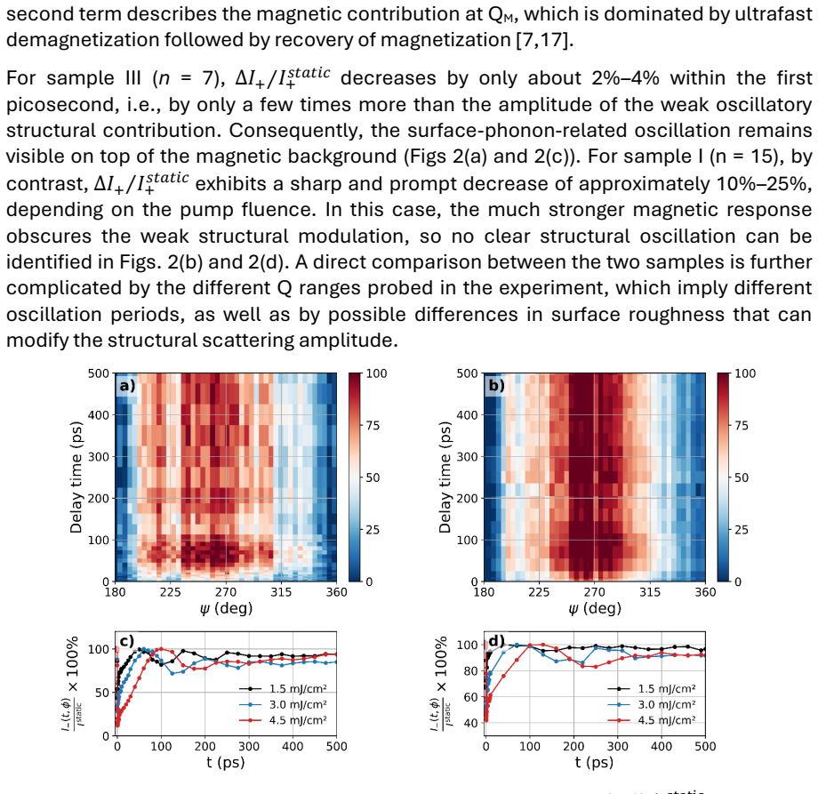
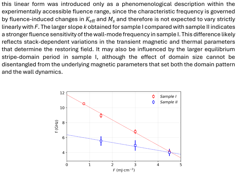
At the stripe-domain wavevector the helicity-summed XRMS signal exhibits ultrafast demagnetization and a weak phonon oscillation, while the dichroic signal shows a strong oscillatory response whose frequency decreases with pump fluence. The authors attribute the latter to pump-induced changes in effective anisotropy and saturation magnetization modifying the restoring field of an internal domain-wall mode. Time-resolved XRMS thus provides access to internal domain-wall motion in disordered stripe textures on picosecond time scales.
What carries the argument
The dichroic component of the XRMS signal, which isolates the in-plane magnetization inside the Néel-type domain walls and thereby probes their internal mode.
If this is right
- The oscillation frequency in the dichroic channel decreases as pump fluence is increased.
- Pump light modifies the effective anisotropy and saturation magnetization, thereby altering the restoring field of the internal domain-wall mode.
- The helicity-summed signal is dominated by demagnetization and coherent surface phonons rather than wall motion.
- This technique isolates wall-specific dynamics on picosecond timescales in labyrinthine stripe domains.
Where Pith is reading between the lines
- The same approach might be applied to other chiral magnetic structures to observe their internal modes.
- Fluence-dependent frequency shifts could serve as a real-time probe of local magnetic parameters in operating devices.
- Extensions to different materials could test whether the softening is universal or depends on the specific anisotropy landscape.
Load-bearing premise
The strong oscillations appear only in the dichroic channel because they come from an internal domain-wall mode whose restoring field is set by the effective anisotropy and saturation magnetization.
What would settle it
A calculation that predicts the mode frequency from measured anisotropy and magnetization values and finds no match to the observed fluence-dependent frequencies would falsify the attribution to the internal wall mode.
Figures
read the original abstract
We measure picosecond dynamics of labyrinthine stripe domains with chiral N\'eel-type domain walls using time-resolved x-ray resonant magnetic scattering (XRMS). At the stripe-domain wavevector, the helicity-summed signal shows ultrafast demagnetization, recovery, and a weak oscillatory contribution that we identify as a signature of laser-launched coherent surface phonons. In contrast, the dichroic signal, which is sensitive to the in-plane magnetization inside the N\'eel walls, shows a strong oscillatory response whose frequency decreases with increasing pump fluence. We attribute this softening to pump-induced changes in the effective anisotropy and saturation magnetization, which modify the restoring field of an internal domain-wall mode. Time-resolved XRMS thus isolates wall-specific dynamics and provides access to internal domain-wall motion in disordered stripe textures on picosecond time scales.
Editorial analysis
A structured set of objections, weighed in public.
Referee Report
Summary. The manuscript reports time-resolved XRMS measurements on labyrinthine stripe domains with chiral Néel-type domain walls. The helicity-summed signal exhibits ultrafast demagnetization, recovery, and weak oscillations identified as laser-launched coherent surface phonons. The dichroic signal, sensitive to in-plane magnetization within the walls, shows strong oscillatory response whose frequency decreases with increasing pump fluence; this is attributed to pump-induced softening of an internal domain-wall mode via changes in effective anisotropy and saturation magnetization.
Significance. If the mode attribution is substantiated, the work demonstrates isolation of wall-specific picosecond dynamics in disordered textures using XRMS, offering a probe of internal Néel-wall motion that could inform ultrafast chiral magnetism studies. The experimental distinction between summed and dichroic channels is a clear strength.
major comments (2)
- [Abstract] Abstract: The central claim attributes the fluence-dependent frequency softening in the dichroic channel to modification of the restoring field of an internal domain-wall mode by pump-altered Keff and Ms. No dispersion relation, numerical estimate, or direct comparison (e.g., measured f versus calculated sqrt(Keff·Ms) after subtracting demagnetization/phonon contributions) is supplied to confirm the observed frequencies fall within the expected range.
- [Abstract] Abstract and results: Alternative sources of oscillatory dichroic contrast (transient in-plane canting, helicity-dependent scattering artifacts, or residual phonon coupling) are not quantitatively excluded; the attribution therefore rests on the untested assumption that the signal arises specifically from the internal DW mode.
minor comments (2)
- [Abstract] Abstract: Fluence values, error bars on frequencies, and details of the oscillatory fits are not reported, limiting assessment of the softening trend.
- The manuscript would benefit from explicit statement of the expected frequency range for the internal mode under the reported pump conditions.
Simulated Author's Rebuttal
We thank the referee for the careful reading of our manuscript and the constructive comments. We respond to each major comment below.
read point-by-point responses
-
Referee: [Abstract] Abstract: The central claim attributes the fluence-dependent frequency softening in the dichroic channel to modification of the restoring field of an internal domain-wall mode by pump-altered Keff and Ms. No dispersion relation, numerical estimate, or direct comparison (e.g., measured f versus calculated sqrt(Keff·Ms) after subtracting demagnetization/phonon contributions) is supplied to confirm the observed frequencies fall within the expected range.
Authors: We agree that the manuscript does not contain an explicit dispersion relation or numerical estimate of the domain-wall mode frequency. The attribution relies on the observed fluence dependence combined with the channel-specific response, but a direct comparison would strengthen the claim. In the revised manuscript we will add a brief analytical estimate based on the expected sqrt(K_eff * M_s) scaling (after subtracting the measured demagnetization and phonon contributions) and compare it to the observed frequencies. revision: yes
-
Referee: [Abstract] Abstract and results: Alternative sources of oscillatory dichroic contrast (transient in-plane canting, helicity-dependent scattering artifacts, or residual phonon coupling) are not quantitatively excluded; the attribution therefore rests on the untested assumption that the signal arises specifically from the internal DW mode.
Authors: The manuscript highlights the clear experimental distinction: the helicity-summed channel shows only weak phonon oscillations while the dichroic channel exhibits strong, fluence-dependent oscillations. This difference makes residual phonon coupling or helicity-independent artifacts unlikely. Transient in-plane canting is inconsistent with the recovery dynamics reported. Nevertheless, we acknowledge that a more quantitative discussion of these alternatives is absent. We will expand the results and discussion sections to address each alternative explicitly. revision: partial
Circularity Check
No circularity: experimental observation with direct attribution, no self-referential reduction
full rationale
The paper reports measured oscillatory dichroic XRMS signals whose frequency decreases with fluence and attributes this to pump-induced changes in Keff and Ms modifying the restoring field of an internal Néel-wall mode. No equations, dispersion relations, or self-citations are invoked that would derive or fit the observed frequency from the same data by construction; the link remains an interpretive statement. The result is self-contained as an experimental finding against external benchmarks, with no load-bearing steps matching the enumerated circularity patterns.
Axiom & Free-Parameter Ledger
Reference graph
Works this paper leans on
-
[1]
The stripe -domain periodicity was determined from the radius of the magnetic scattering ring
All multilayers exhibited PMA and formed labyrinthine stripe domains in the remanent state at room temperature (~300 K). The stripe -domain periodicity was determined from the radius of the magnetic scattering ring. The domain wave vectors were QM ≈ 0.010 nm−1 (sample I), QM ≈ 0.0145 nm−1 (sample II), and QM ≈ 0.021 nm−1 (sample III), corresponding to str...
2030
-
[2]
A. Thiaville, S. Rohart, É. Jué, V. Cros, and A. Fert, Europhys. Lett. 100, 57002 (2012). DOI: https://doi.org/10.1209/0295-5075/100/57002
-
[3]
S. Emori, U. Bauer, S. -M. Ahn, E. Martinez, and G. S. D. Beach, Nat. Mater. 12, 611 –616 (2013). DOI: https://doi.org/10.1038/nmat3675
-
[4]
K. -S. Ryu, L. Thomas, S. -H. Yang, and S. Parkin, Nat. Nanotechnol. 8, 527 –533 (2013). DOI: https://doi.org/10.1038/nnano.2013.102
-
[5]
C. Moreau-Luchaire, C. Moutafis, N. Reyren, J. Sampaio, C. A. F. Vaz, N. Van Horne, K. Bouzehouane, K. Garcia, C. Deranlot, P. Warnicke, P. Wohlhüter, J. -M. George, M. Weigand, J. Raabe, V. Cros, and A. Fert, Nat. Nanotechnol. 11, 444–448 (2016). DOI: https://doi.org/10.1038/nnano.2015.313
-
[6]
W. Legrand, J.-Y. Chauleau, D. Maccariello, N. Reyren, S. Collin, K. Bouzehouane, N. Jaouen, V. Cros, and A. Fert, Sci. Adv. 4, eaat0415 (2018). DOI: https://doi.org/10.1126/sciadv.aat0415
-
[7]
U. Atxitia, D. Hinzke, O. Chubykalo -Fesenko, U. Nowak, H. Kachkachi, O. N. Mryasov, R. F. Evans, and R. W. Chantrell, Phys. Rev. B 82, 134440 (2010). DOI: https://doi.org/10.1103/PhysRevB.82.134440
-
[8]
B. Koopmans, G. Malinowski, F. Dalla Longa, D. Steiauf, M. Fähnle, T. Roth, M. Cinchetti, and M. Aeschlimann, Nat. Mater. 9, 259–265 (2010). DOI: https://doi.org/10.1038/nmat2593
-
[9]
J. P. Hannon, G. T. Trammell, M. Blume, and D. Gibbs, Phys. Rev. Lett. 61, 1245 (1988). DOI: https://doi.org/10.1103/PhysRevLett.61.1245
-
[10]
J.-Y. Chauleau, W. Legrand, N. Reyren, D. Maccariello, S. Collin, H. Popescu, K. Bouzehouane, V. Cros, N. Jaouen, A. Fert. Phys. Rev. Lett. 120, 037202 (2018). DOI: https://doi.org/10.1103/PhysRevLett.120.037202
-
[11]
S. Flewett, E. Burgos -Parra, M. Garrido Strelow, Y. Sassi, C. Léveillé, F. Ajejas, N. Reyren, and N. Jaouen, Phys. Rev. B 103, 184401 (2021). DOI: https://doi.org/10.1103/PhysRevB.103.184401
-
[12]
N. Kerber, D. Ksenzov, F. Freimuth, F. Capotondi, E. Pedersoli, I. Lopez-Quintas, B. Seng, J. Cramer, K. Litzius, D. Lacour, H. Zabel, Y. Mokrousov, M. Kläui, and C. Gutt, Nat. Commun. 11, 6304 (2020). DOI: https://doi.org/10.1038/s41467-020-19613-z 9
-
[13]
C. Léveillé, E. Burgos-Parra, Y. Sassi, F. Ajejas, V. Chardonnet, E. Pedersoli, F. Capotondi, G. De Ninno, F. Maccherozzi, S. Dhesi, D. M. Burn, G. van der Laan, O. S. Latcham, A. V. Shytov, V. V. Kruglyak, E. Jal, V. Cros, J. -Y. Chauleau, N. Reyren, M. Viret, and N. Jaouen, Nat. Commun. 13, 1412 (2022). DOI: https://doi.org/10.1038/s41467-022-28899-0
-
[14]
N. L. Schryer and L. R. Walker, J. Appl. Phys. 45, 5406 –5421 (1974). DOI: https://doi.org/10.1063/1.1663252
-
[15]
F. Capotondi, E. Pedersoli, N. Mahne, R. H. Menk, G. Passos, L. Raimondi, C. Svetina, G. Sandrin, M. Zangrando, M. Kiskinova, S. Bajt, M. Barthelmess, H. Fleckenstein, H. N. Chapman, J. Schulz, J. Bach, R. Frömter, S. Schleitzer, L. Müller, C. Gutt, and G. Grübel. Rev. Sci. Instrum. 84, 051301 (2013). DOI: https://doi.org/10.1063/1.4807157
-
[16]
F. Capotondi, E. Pedersoli, F. Bencivenga, M. Manfredda , N. Mahne, L. Raimondi, C. Svetina, M. Zangrando, A. Demidovich, I. Nikolov, M. Danailov, C. Masciovecchio, and M. Kiskinova, J. Synchrotron Radiat. 22, 544–552 (2015). DOI: https://doi.org/10.1107/S1600577515004919
-
[17]
E. Pedersoli, F. Capotondi, D. Cocco, M. Zangrando, B. Kaulich, R. H. Menk, A. Locatelli, T. O. Mentes, C. Spezzani, G. Sandrin, D. M. Bacescu, M. Kiskinova, S. Bajt, M. Barthelmess, A. Barty, J. Schulz, L. Gumprecht, H. N. Chapman, A. J. Nelson, M. Frank, M. J. Pivovaroff, B. W. Woods, M. J. Bogan, and J. Hajdu, Rev. Sci. Instrum. 82, 043711 (2011). DOI:...
-
[18]
E. Beaurepaire, J. -C. Merle, A. Daunois, and J. -Y. Bigot, Phys. Rev. Lett. 76, 4250 (1996). DOI: https://doi.org/10.1103/PhysRevLett.76.4250
-
[19]
O. Hellwig, G. P. Denbeaux, J. B. Kortright, and E. E. Fullerton, Physica B 336, 136 –144 (2003). DOI: https://doi.org/10.1016/S0921-4526(03)00282-5
-
[20]
K. Fallon, S. McVitie, W. Legrand, F. Ajejas, D. Maccariello, S. Collin, V. Cros, and N. Reyren, Phys. Rev. B 100, 214431 (2019). DOI: https://doi.org/10.1103/PhysRevB.100.214431
-
[21]
F. Capotondi, A. A. Maznev, F. Bencivenga, S. Bonetti, D. Engel, D. Fainozzi, D. Fausti, L. Foglia, C. Gutt, N. Jaouen, D. Ksenzov, C. Masciovecchio, K. A. Nelson, I. Nikolov, M. Pancaldi, E. Pedersoli, B. Pfau, L. Raimondi, F. Romanelli, R. Totani, a nd M. Trigo, Phys. Rev. Lett. 135, 266101 (2025). DOI: https://doi.org/10.1103/dq8w-62bm
-
[22]
I. Lemesh, F. Büttner, and G. S. D. Beach, Phys. Rev. B 95, 174423 (2017). DOI: https://doi.org/10.1103/PhysRevB.95.174423
-
[23]
B. Pfau, S. Schaffert, L. Müller, C. Gutt, A. Al -Shemmary, F. Büttner, R. Delaunay, S. Düsterer, S. Flewett, R. Frömter, J. Geilhufe, E. Guehrs, C. M. Günther, R. Hawaldar, M. Hille, N. Jaouen, A. Kobs, K. Li, J. Mohanty, H. Redlin, W. F. Schlotter, D. Stickler, R. Treusch, B. Vodungbo, M. Kläui, H. P. Oepen, J. Lüning, G. Grübel, and S. Eisebitt, Nat. C...
-
[24]
C. Gutt, E. M. Allaria, E. Burgos -Parra, F. Capotondi, D. de Angelis, N. Jaouen, N. Kerber, D. Ksenzov, C. Léveillé, M. Pancaldi, and E. Pedersoli (2024). 20199086 [Data set]. Elettra Sincrotrone Trieste. https://doi.org/10.34965/I8821
discussion (0)
Sign in with ORCID, Apple, or X to comment. Anyone can read and Pith papers without signing in.