Compute-Optimal Network Design for Echocardiography Myocardial Segmentation and Perfusion Quantification using Neural Scaling Laws
Pith reviewed 2026-06-27 23:00 UTC · model grok-4.3
The pith
Neural scaling laws fitted on data subsets predict full-dataset performance and select compact networks that match expert cardiologist results on myocardial perfusion quantification.
A machine-rendered reading of the paper's core claim, the machinery that carries it, and where it could break.
Core claim
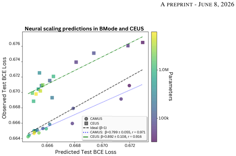
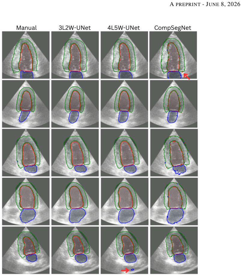
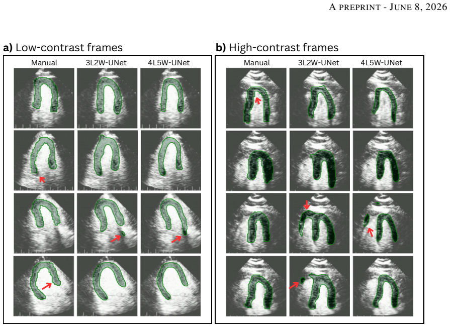
Extrapolation based on the scaling law is predictive of test loss at the full dataset size, allowing selection of two networks that obtained state-of-the-art performance on CAMUS with a 240-fold reduction in parameter count. The gradient of the scaling law transfers from CAMUS to the CEUS dataset with a bias in the predicted losses. The automatically segmented masks perform equivalently to a senior cardiologist in myocardial perfusion quantification.
What carries the argument
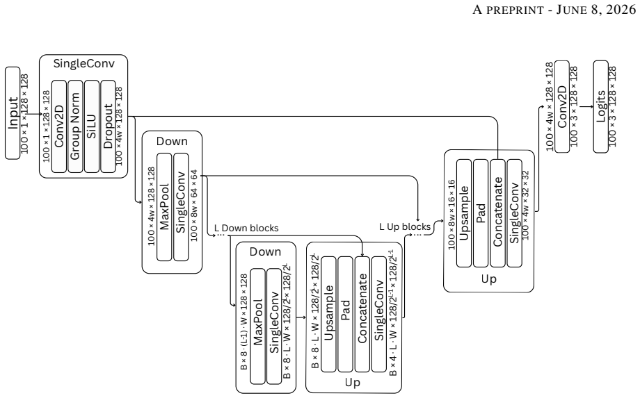
Neural scaling laws that relate test loss to model parameter count, fitted on performance from data subsets to extrapolate optimal network sizes for myocardial segmentation.
Load-bearing premise
The scaling law fitted on performance from data subsets will accurately extrapolate to the full dataset size and its gradient will transfer to the CEUS dataset with only a bias that does not invalidate optimal model selection.
What would settle it
Train the two extrapolated optimal networks on the complete CAMUS training set and check whether their measured test loss matches the scaling-law prediction or whether substantially larger networks achieve lower loss.
Figures
read the original abstract
Myocardial perfusion quantification using contrast-enhanced ultrasound offers a bedside non-ionizing alternative to nuclear imaging modalities. However, its clinical adoption is hindered by time-consuming manual labelling. Automated segmentation has proved challenging due to a paucity of in-domain training data. Adapting strategies currently used to optimise large language models for large datasets, we apply neural scaling laws to predict network performance for myocardial segmentation. We extrapolate performance on subsets of the data to determine optimal network size on the CAMUS echocardiography dataset and a 25-patient contrast-enhanced ultrasound (CEUS) dataset. Finally, we validate the clinical utility of our models by comparing the final myocardial perfusion parameters with those obtained by a senior cardiologist. Extrapolation based on the scaling law is predictive of test loss at the full dataset size, allowing us to select two networks that obtained state-of-the-art performance on CAMUS with a 240-fold reduction in parameter count. We observe the gradient of the scaling law transfers from CAMUS to the CEUS dataset with a bias in the predicted losses. The automatically segmented masks perform equivalently to a senior cardiologist in myocardial perfusion quantification. These results establish neural scaling laws as a practical tool for data-driven compute-optimal model design for small imaging datasets.
Editorial analysis
A structured set of objections, weighed in public.
Referee Report
Summary. The paper claims that neural scaling laws fitted to test losses on data subsets of the CAMUS echocardiography dataset can be extrapolated to predict performance at full dataset size. This enables selection of two networks achieving state-of-the-art myocardial segmentation on CAMUS with a 240-fold reduction in parameter count. The scaling-law gradient transfers to a 25-patient CEUS dataset after constant bias correction, and the resulting automatic segmentations produce myocardial perfusion quantification parameters equivalent to those from a senior cardiologist.
Significance. If the subset-based extrapolation reliably identifies compute-optimal models that generalize and match clinical performance, the work would offer a practical, data-driven method for model-size selection in data-scarce medical imaging tasks, potentially reducing compute demands without sacrificing accuracy.
major comments (2)
- [Abstract] Abstract and scaling-law section: the central claim that extrapolation from subset performances is predictive of full-dataset test loss and enables optimal model selection rests on the power-law fit being robust; with high run-to-run variance typical in echocardiography segmentation and typically few subset sizes available for fitting, sensitivity of the fitted coefficients to exact subset choices or noise could invalidate both the CAMUS model selection and the CEUS transfer claim.
- [Results] CEUS transfer paragraph: the assumption that only a constant bias correction is needed for the scaling-law gradient to transfer from CAMUS to the 25-patient CEUS set is load-bearing for claiming the same networks remain optimal; without an explicit check that the bias-adjusted predictions correctly rank models on held-out CEUS performance, the transfer step risks circularity.
minor comments (2)
- Clarify the exact number of subset sizes used to fit the scaling law and report the uncertainty or variance in the fitted exponents.
- Provide the precise definition of the scaling law functional form (e.g., the equation relating loss to model size and data size) in the methods.
Simulated Author's Rebuttal
We thank the referee for their constructive comments, which highlight important aspects of robustness and validation in our scaling-law approach. We address each major comment below with point-by-point responses. Where the concerns identify areas for clarification or additional checks, we indicate that revisions will be made.
read point-by-point responses
-
Referee: [Abstract] Abstract and scaling-law section: the central claim that extrapolation from subset performances is predictive of full-dataset test loss and enables optimal model selection rests on the power-law fit being robust; with high run-to-run variance typical in echocardiography segmentation and typically few subset sizes available for fitting, sensitivity of the fitted coefficients to exact subset choices or noise could invalidate both the CAMUS model selection and the CEUS transfer claim.
Authors: We agree that sensitivity to subset choice and run variance is a valid concern for power-law fits with limited points. However, the manuscript already validates the extrapolation by showing that predictions from subset fits closely match the measured test loss on the full CAMUS dataset for the selected models (see scaling-law section and Figure 3). This empirical match provides evidence that the fit is sufficiently robust for model selection in this setting. To further address the referee's point, we will add a sensitivity analysis in the revision, including fits across different random subset selections and reporting the resulting variation in predicted optimal model sizes. revision: yes
-
Referee: [Results] CEUS transfer paragraph: the assumption that only a constant bias correction is needed for the scaling-law gradient to transfer from CAMUS to the 25-patient CEUS set is load-bearing for claiming the same networks remain optimal; without an explicit check that the bias-adjusted predictions correctly rank models on held-out CEUS performance, the transfer step risks circularity.
Authors: The manuscript reports that the scaling-law gradient observed on CAMUS transfers to CEUS after a constant bias correction, and the same networks selected via CAMUS scaling laws achieve strong performance on the CEUS task. We acknowledge that an explicit verification that the bias-adjusted predictions preserve model ranking on held-out CEUS data would strengthen the transfer claim and reduce any perception of circularity. We will add this check in the revision by training a small set of models on CEUS subsets, applying the bias correction, and confirming the ranking matches actual held-out performance. revision: yes
Circularity Check
No significant circularity in scaling-law extrapolation
full rationale
The paper fits scaling laws to measured test losses on data subsets of CAMUS, extrapolates to predict full-dataset performance, selects two architectures on that basis, and then reports that the selected models achieve SOTA when trained on the full set. This is a standard predictive use of scaling laws rather than a reduction by construction; the full-dataset losses serve as an independent check. No load-bearing self-citations, no fitted parameters renamed as predictions, and no ansatz or uniqueness claim imported from the authors' prior work appear in the provided text. The CEUS transfer with bias correction is an empirical observation, not a definitional loop.
Axiom & Free-Parameter Ledger
free parameters (1)
- scaling law coefficients
axioms (1)
- domain assumption Performance on data subsets follows a predictable scaling law that can be extrapolated to larger data sizes
Reference graph
Works this paper leans on
-
[1]
Marc Dewey, Maria Siebes, Marc Kachelrieß, Klaus F. Kofoed, Pál Maurovich-Horvat, Konstantin Nikolaou, Wenjia Bai, Andreas Kofler, Robert Manka, Sebastian Kozerke, Amedeo Chiribiri, Tobias Schaeffter, Florian Michallek, Frank Bengel, Stephan Nekolla, Paul Knaapen, Mark Lubberink, Roxy Senior, Meng-Xing Tang, Jan J. Piek, Tim van de Hoef, Johannes Martens,...
2020
-
[2]
Diagnostic Value of Extracellular V olume Quantification and Myocardial Perfusion Analysis at CT in Cardiac Amyloidosis.Radiology, 300(2):326–335, August 2021
Jean-François Deux, Refaat Nouri, Vania Tacher, Amira Zaroui, Haytham Derbel, Islem Sifaoui, Virgile Chevance, Fourat Ridouani, Arnault Galat, Mounira Kharoubi, Silvia Oghina, Soulef Guendouz, Etienne Audureau, Em- manuel Teiger, Hicham Kobeiter, and Thibaud Damy. Diagnostic Value of Extracellular V olume Quantification and Myocardial Perfusion Analysis a...
2021
-
[3]
Ultrasound study of right ventricular myocardial perfusion and functional changes in hypertrophic cardiomyopathy.BMC Cardiovascular Disorders, 24(1):63, January 2024
Shan Cao, Lingjie Yang, Liyun Liu, Yuming Mu, and Lina Guan. Ultrasound study of right ventricular myocardial perfusion and functional changes in hypertrophic cardiomyopathy.BMC Cardiovascular Disorders, 24(1):63, January 2024
2024
-
[4]
Einstein, Shepard D
Andrew J. Einstein, Shepard D. Weiner, Adam Bernheim, Michal Kulon, Sabahat Bokhari, Lynne L. Johnson, Jeffrey W. Moses, and Stephen Balter. Multiple Testing, Cumulative Radiation Dose, and Clinical Indications in Patients Undergoing Myocardial Perfusion Imaging.JAMA : the journal of the American Medical Association, 304(19):2137–2144, November 2010
2010
-
[5]
Myocardial perfusion scans: projected population cancer risks from current levels of use in the United States.Circulation, 122(23):2403–2410, December 2010
Amy Berrington de Gonzalez, Kwang-Pyo Kim, Rebecca Smith-Bindman, and Dorothea McAreavey. Myocardial perfusion scans: projected population cancer risks from current levels of use in the United States.Circulation, 122(23):2403–2410, December 2010
2010
-
[6]
Jerome, Peter L
Scott D. Jerome, Peter L. Tilkemeier, Mary B. Farrell, and Leslee J. Shaw. Nationwide Laboratory Adherence to Myocardial Perfusion Imaging Radiation Dose Reduction Practices.JACC: Cardiovascular Imaging, 8(10):1170– 1176, October 2015
2015
-
[7]
Scannell, and Anedeo Chiribiri
Ebraham Alskaf, Utkarsh Dutta, Cian M. Scannell, and Anedeo Chiribiri. Deep learning applications in myocardial perfusion imaging, a systematic review and meta-analysis.Informatics in Medicine Unlocked, 32:101055, January 2022. 12 APREPRINT- JUNE8, 2026
2022
-
[8]
Kasprzak
Roxy Senior, Antonella Moreo, Nicola Gaibazzi, Luciano Agati, Klaus Tiemann, Bharati Shivalkar, Stephan von Bardeleben, Leonarda Galiuto, Hervé Lardoux, Giuseppe Trocino, Ignasi Carrió, Dominique Le Guludec, Gianmario Sambuceti, Harald Becher, Paolo Colonna, Folkert Ten Cate, Ezio Bramucci, Ariel Cohen, Gianpaolo Bezante, Costantina Aggeli, and Jaroslaw D...
2013
-
[9]
Mina Amiri, Rupert Brooks, and Hassan Rivaz. Fine-Tuning U-Net for Ultrasound Image Segmentation: Dif- ferent Layers, Different Outcomes.IEEE Transactions on Ultrasonics, Ferroelectrics, and Frequency Control, 67(12):2510–2518, December 2020
2020
-
[10]
Deep Neural Architectures for Contrast Enhanced Ultrasound (CEUS) Focal Liver Lesions Automated Diagnosis.Sensors, 21(12):4126, January 2021
C˘at˘alin Daniel C˘aleanu, Cristina Laura Sîrbu, and Georgiana Simion. Deep Neural Architectures for Contrast Enhanced Ultrasound (CEUS) Focal Liver Lesions Automated Diagnosis.Sensors, 21(12):4126, January 2021. Number: 12
2021
-
[11]
Joint Segmentation and Differential Diagnosis of Thyroid Nodule in Contrast-Enhanced Ultrasound Images.IEEE Transactions on Biomedical Engineering, 70(9):2722–2732, September 2023
Fang Chen, Haojie Han, Peng Wan, Hongen Liao, Chunrui Liu, and Daoqiang Zhang. Joint Segmentation and Differential Diagnosis of Thyroid Nodule in Contrast-Enhanced Ultrasound Images.IEEE Transactions on Biomedical Engineering, 70(9):2722–2732, September 2023
2023
-
[12]
Semantic segmentation method for myocardial contrast echocardiogram based on DeepLabV3+ deep learning architecture.Mathematical Biosciences and Engineering, 20(2):2081–2093, 2023
Huan Cheng, Jucheng Zhang, Yinglan Gong, Zhaoxia Pu, Jun Jiang, Yonghua Chu, Ling Xia, Huan Cheng, Jucheng Zhang, Yinglan Gong, Zhaoxia Pu, Jun Jiang, Yonghua Chu, and Ling Xia. Semantic segmentation method for myocardial contrast echocardiogram based on DeepLabV3+ deep learning architecture.Mathematical Biosciences and Engineering, 20(2):2081–2093, 2023....
2081
-
[13]
Lee R. Dice. Measures of the Amount of Ecologic Association Between Species.Ecology, 26(3):297–302, 1945. _eprint: https://esajournals.onlinelibrary.wiley.com/doi/pdf/10.2307/1932409
-
[14]
Left-Ventricular V olume Estimation in Contrast-Enhanced Echocardiography Using Deep Learning
Jieyu Hu, Erik Smistad, Bjørnar Grenne, Espen Holte, Håvard Dalen, and Lasse Lovstakken. Left-Ventricular V olume Estimation in Contrast-Enhanced Echocardiography Using Deep Learning. In2023 IEEE International Ultrasonics Symposium (IUS), pages 1–4, September 2023
2023
-
[15]
Scaling Laws for Neural Language Models
Jared Kaplan, Sam McCandlish, Tom Henighan, Tom B. Brown, Benjamin Chess, Rewon Child, Scott Gray, Alec Radford, Jeffrey Wu, and Dario Amodei. Scaling Laws for Neural Language Models, January 2020. arXiv:2001.08361 [cs]
work page internal anchor Pith review Pith/arXiv arXiv 2020
-
[16]
Training Compute-Optimal Large Language Models
Jordan Hoffmann, Sebastian Borgeaud, Arthur Mensch, Elena Buchatskaya, Trevor Cai, Eliza Rutherford, Diego de Las Casas, Lisa Anne Hendricks, Johannes Welbl, Aidan Clark, Tom Hennigan, Eric Noland, Katie Millican, George van den Driessche, Bogdan Damoc, Aurelia Guy, Simon Osindero, Karen Simonyan, Erich Elsen, Jack W. Rae, Oriol Vinyals, and Laurent Sifre...
work page internal anchor Pith review Pith/arXiv arXiv 2022
-
[17]
Scaling Laws For Deep Learning Based Image Reconstruction, February 2023
Tobit Klug and Reinhard Heckel. Scaling Laws For Deep Learning Based Image Reconstruction, February 2023. arXiv:2209.13435 [eess]
-
[18]
Manmatha, Ashwin Swaminathan, Zhuowen Tu, Stefano Ermon, and Stefano Soatto
Hao Li, Yang Zou, Ying Wang, Orchid Majumder, Yusheng Xie, R. Manmatha, Ashwin Swaminathan, Zhuowen Tu, Stefano Ermon, and Stefano Soatto. On the Scalability of Diffusion-based Text-to-Image Generation. In2024 IEEE/CVF Conference on Computer Vision and Pattern Recognition (CVPR), pages 9400–9409, Seattle, WA, USA, June 2024. IEEE
2024
-
[19]
Revisiting model scaling with a U-net benchmark for 3D medical image segmentation.Scientific Reports, 15(1):29795, August 2025
Ziyan Huang, Jin Ye, Haoyu Wang, Zhongying Deng, Zhikai Yang, Yanzhou Su, Jie Liu, Tianbin Li, Yun Gu, Shaoting Zhang, Yu Qiao, Lixu Gu, and Junjun He. Revisiting model scaling with a U-net benchmark for 3D medical image segmentation.Scientific Reports, 15(1):29795, August 2025
2025
-
[20]
Fubao Zhu, Longxi Li, Jinyu Zhao, Chen Zhao, Shaojie Tang, Jiaofen Nan, Yanting Li, Zhongqiang Zhao, Jianzhou Shi, Zenghong Chen, Chuang Han, Zhixin Jiang, and Weihua Zhou. A new method incorporating deep learning with shape priors for left ventricular segmentation in myocardial perfusion SPECT images.Computers in Biology and Medicine, 160:106954, June 2023
2023
-
[21]
Seyed Mohammad Entezarmahdi, Reza Faghihi, Mehran Yazdi, Negar Shahamiri, Parham Geramifar, and Mahdi Haghighatafshar. QCard-NM: Developing a semiautomatic segmentation method for quantitative analysis of the right ventricle in non-gated myocardial perfusion SPECT imaging.EJNMMI Physics, 10(1):21, March 2023
2023
-
[22]
How to perform an ultrasound contrast myocardial perfusion examination?European Heart Journal - Cardiovascular Imaging, 23(6):727–729, June 2022
Bernard Cosyns, Andreas Helfen, Howard Leong-Poi, and Roxy Senior. How to perform an ultrasound contrast myocardial perfusion examination?European Heart Journal - Cardiovascular Imaging, 23(6):727–729, June 2022. 13 APREPRINT- JUNE8, 2026
2022
-
[23]
Image Segmentation Using Deep Learning: A Survey.IEEE Transactions on Pattern Analysis and Machine Intelligence, 44(7):3523–3542, July 2022
Shervin Minaee, Yuri Boykov, Fatih Porikli, Antonio Plaza, Nasser Kehtarnavaz, and Demetri Terzopoulos. Image Segmentation Using Deep Learning: A Survey.IEEE Transactions on Pattern Analysis and Machine Intelligence, 44(7):3523–3542, July 2022
2022
-
[24]
Narinder Singh Punn and Sonali Agarwal. Modality specific U-Net variants for biomedical image segmentation: A survey.Artificial Intelligence Review, 55(7):5845–5889, October 2022. arXiv:2107.04537 [eess]
-
[25]
UNet 3+: A Full-Scale Connected UNet for Medical Image Segmentation, April 2020
Huimin Huang, Lanfen Lin, Ruofeng Tong, Hongjie Hu, Qiaowei Zhang, Yutaro Iwamoto, Xianhua Han, Yen-Wei Chen, and Jian Wu. UNet 3+: A Full-Scale Connected UNet for Medical Image Segmentation, April 2020. arXiv:2004.08790 [eess]
-
[26]
Medical Image Segmentation Review: The success of U-Net, November 2022
Reza Azad, Ehsan Khodapanah Aghdam, Amelie Rauland, Yiwei Jia, Atlas Haddadi Avval, Afshin Bozorgpour, Sanaz Karimijafarbigloo, Joseph Paul Cohen, Ehsan Adeli, and Dorit Merhof. Medical Image Segmentation Review: The success of U-Net, November 2022. arXiv:2211.14830 [eess]
-
[27]
Deep Learning for Segmentation Using an Open Large-Scale Dataset in 2D Echocardiography.IEEE transactions on medical imaging, 38(9):2198–2210, September 2019
Sarah Leclerc, Erik Smistad, Joao Pedrosa, Andreas Ostvik, Frederic Cervenansky, Florian Espinosa, Torvald Espeland, Erik Andreas Rye Berg, Pierre-Marc Jodoin, Thomas Grenier, Carole Lartizien, Jan Dhooge, Lasse Lovstakken, and Olivier Bernard. Deep Learning for Segmentation Using an Open Large-Scale Dataset in 2D Echocardiography.IEEE transactions on med...
2019
-
[28]
Myocardial Segmentation of Contrast Echocardiograms Using Random Forests Guided by Shape Model
Yuanwei Li, Chin Pang Ho, Navtej Chahal, Roxy Senior, and Meng-Xing Tang. Myocardial Segmentation of Contrast Echocardiograms Using Random Forests Guided by Shape Model, June 2018. arXiv:1806.07490 [cs]
work page internal anchor Pith review Pith/arXiv arXiv 2018
-
[29]
Vaitkus, Marek Belohlavek, Vinayak Krishnamurthy, and Iman Borazjani
Taeouk Kim, Mohammadali Hedayat, Veronica V . Vaitkus, Marek Belohlavek, Vinayak Krishnamurthy, and Iman Borazjani. Automatic segmentation of the left ventricle in echocardiographic images using convolutional neural networks.Quantitative Imaging in Medicine and Surgery, 11(5):1763–1781, May 2021
2021
-
[30]
Segmentation of Anatomical Structures of the Left Heart from Echocardiographic Images Using Deep Learning
Mhd Jafar Mortada, Selene Tomassini, Haidar Anbar, Micaela Morettini, Laura Burattini, and Agnese Sbrollini. Segmentation of Anatomical Structures of the Left Heart from Echocardiographic Images Using Deep Learning. Diagnostics (Basel, Switzerland), 13(10):1683, May 2023
2023
-
[31]
Compositional Segmentation of Cardiac Images Leveraging Metadata, October 2024
Abbas Khan, Muhammad Asad, Martin Benning, Caroline Roney, and Gregory Slabaugh. Compositional Segmentation of Cardiac Images Leveraging Metadata, October 2024. arXiv:2410.23130 [eess]
-
[32]
CoST-UNet: Convolution and swin transformer based deep learning architecture for cardiac segmentation.Biomedical Signal Processing and Control, 96:106633, October 2024
Md Rabiul Islam, Marwa Qaraqe, and Erchin Serpedin. CoST-UNet: Convolution and swin transformer based deep learning architecture for cardiac segmentation.Biomedical Signal Processing and Control, 96:106633, October 2024
2024
-
[33]
Anatomically accurate cardiac segmentation using Dense Associative Networks
Zahid Ullah and Jihie Kim. Anatomically accurate cardiac segmentation using Dense Associative Networks. Engineering Applications of Artificial Intelligence, 162:112742, December 2025
2025
-
[34]
DAF- Mamba: Dynamic Selective and Adaptive Fused Mamba for Cardiac Image Segmentation.Pattern Recognition, page 112664, November 2025
Yonglin Chen, Yixiang Wang, Zhongyuan Liu, Yuyan Weng, Chihui Long, Yalong Yang, and Jinhui Tang. DAF- Mamba: Dynamic Selective and Adaptive Fused Mamba for Cardiac Image Segmentation.Pattern Recognition, page 112664, November 2025
2025
-
[35]
Diego D. B. Carvalho, Zeynettin Akkus, Stijn C. H. van den Oord, Arend F. L. Schinkel, Antonius F. W. van der Steen, Wiro J. Niessen, Johan G. Bosch, and Stefan Klein. Lumen Segmentation and Motion Estimation in B-Mode and Contrast-Enhanced Ultrasound Images of the Carotid Artery in Patients With Atherosclerotic Plaque. IEEE Transactions on Medical Imagin...
2015
-
[36]
Deep-learning-based washout classification for decision support in contrast-enhanced ultrasound examinations of the liver.Journal of Medical Imaging, 12(4):044502, July 2025
Hannah Strohm, Sven Rothlübbers, Jürgen Jenne, Dirk-André Clevert, Thomas Fischer, Niklas Hitschrich, Bernhard Mumm, Paul Spiesecke, and Matthias Günther. Deep-learning-based washout classification for decision support in contrast-enhanced ultrasound examinations of the liver.Journal of Medical Imaging, 12(4):044502, July 2025
2025
-
[37]
CEUSegNet: A Cross-Modality Lesion Segmentation Network for Contrast-Enhanced Ultrasound
Zheling Meng, Yangyang Zhu, Xiao Fan, Jie Tian, Fang Nie, and Kun Wang. CEUSegNet: A Cross-Modality Lesion Segmentation Network for Contrast-Enhanced Ultrasound. In2022 IEEE 19th International Symposium on Biomedical Imaging (ISBI), pages 1–5, March 2022
2022
-
[38]
CEUS-SAM: Cross-Modal Prompt-Based SAM Network for Breast CEUS Image Segmentation
Xutao Li, Min Xu, Ximiao Zhang, Sihua Niu, Jiaan Zhu, and Xiuzhuang Zhou. CEUS-SAM: Cross-Modal Prompt-Based SAM Network for Breast CEUS Image Segmentation. In2024 IEEE International Conference on Bioinformatics and Biomedicine (BIBM), pages 994–999, December 2024
2024
-
[39]
Yuxiang Duan, Jili Long, Shunyi Zhao, Hao Wang, and Yuriy S. Shmaliy. AI-Enhanced High-Precision Segmenta- tion and Perfusion Analysis in Myocardial Contrast Echocardiography.IEEE Transactions on Cybernetics, pages 1–13, 2026
2026
-
[40]
Deep Learning Scaling is Predictable, Empirically
Joel Hestness, Sharan Narang, Newsha Ardalani, Gregory Diamos, Heewoo Jun, Hassan Kianinejad, Md Mostofa Ali Patwary, Yang Yang, and Yanqi Zhou. Deep Learning Scaling is Predictable, Empirically, December 2017. arXiv:1712.00409 [cs]. 14 APREPRINT- JUNE8, 2026
work page internal anchor Pith review Pith/arXiv arXiv 2017
-
[41]
Rosenfeld, Amir Rosenfeld, Yonatan Belinkov, and Nir Shavit
Jonathan S. Rosenfeld, Amir Rosenfeld, Yonatan Belinkov, and Nir Shavit. A Constructive Prediction of the Generalization Error Across Scales, December 2019. arXiv:1909.12673 [cs]
-
[42]
Scaling Vision Transformers
Xiaohua Zhai, Alexander Kolesnikov, Neil Houlsby, and Lucas Beyer. Scaling Vision Transformers. In2022 IEEE/CVF Conference on Computer Vision and Pattern Recognition (CVPR), pages 1204–1213, New Orleans, LA, USA, June 2022. IEEE
2022
-
[43]
Anna L. Emanuel, Rick I. Meijer, Erik van Poelgeest, Pien Spoor, Erik H. Serné, and Etto C. Eringa. Contrast- enhanced ultrasound for quantification of tissue perfusion in humans.Microcirculation, 27(1):e12588, 2020. _eprint: https://onlinelibrary.wiley.com/doi/pdf/10.1111/micc.12588
-
[44]
G. Hommel. A Stagewise Rejective Multiple Test Procedure Based on a Modified Bonferroni Test.Biometrika, 75(2):383–386, 1988. 15
1988
discussion (0)
Sign in with ORCID, Apple, or X to comment. Anyone can read and Pith papers without signing in.